. 2007 May;81(10):5246-56.
doi: 10.1128/JVI.02778-06. Epub 2007 Feb 28.

A shared transcription termination signal on negative and ambisense RNA genome segments of Rift Valley fever, sandfly fever Sicilian, and Toscana viruses

Affiliations

A shared transcription termination signal on negative and ambisense RNA genome segments of Rift Valley fever, sandfly fever Sicilian, and Toscana viruses

César G Albariño et al. J Virol. 2007 May.

Abstract

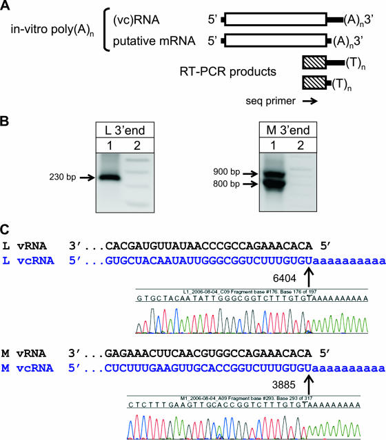
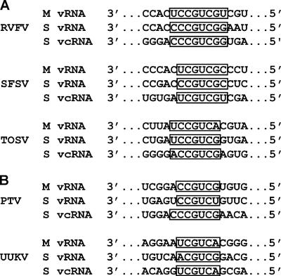
The Phlebovirus genus (family Bunyaviridae) is composed of a diverse group of arboviruses that cause disease syndromes ranging from mild febrile illness to hemorrhagic fever with high fatality. Although antigenically similar, these viruses differ by approximately 25% at the genome level, and their ecologies, including geographic ranges, preferred vector species, and hosts, vary considerably. In contrast to other ambisense viruses, where RNA hairpin structures which serve as transcription termination signals are frequently found separating the opposite-sense open reading frames, no evidence of predicted high-energy hairpin structures was found at the ambisense junctions of phlebovirus S RNA segments. However, a conserved sequence motif was identified on both negative and ambisense genome segments that functions as a transcription termination signal for the N, NSs, and GPC mRNAs in three diverse phleboviruses, namely, Rift Valley fever, sandfly Sicilian, and Toscana viruses. The exact termination of nascent virus mRNA molecules was determined by 3' rapid amplification of cDNA ends. Surprisingly, analysis of the termini of mRNAs from both S and M segments of these three viruses revealed that transcription termination occurred immediately upstream of a conserved sequence motif with the general features 3'-C(1-3)GUCG/A-5'. In contrast, no corresponding sequence motif was found in the L segments, and analysis indicated a "runoff" transcript approach to L mRNA termination. The absolute requirement of the identified transcription termination motif was demonstrated by using a highly efficient Rift Valley fever virus reverse genetics system to generate live recombinant viruses with S segments lacking the termination signal motif for the NP or NSs mRNA and showing that these recombinant viruses generated mRNAs that failed to terminate correctly.


Figures

FIG. 1.
FIG. 1.
(A) Diagram depicting the 3′RACE strategy employed throughout these studies for detection of L and M segment mRNAs. (B) Agarose gel results depicting the 3′RACE amplification of RVF virus L (left) and M (right) segment RNA species. Lane 1, virus-specific amplicons; lane 2, size marker. Full-length replication products (vcRNA) were detected for both L and M segments. However, in contrast to the case with the M segment, no smaller fragments corresponding to L segment mRNA species were observed. Similar results were obtained with both TOS and SFS viruses (data not shown). (C) Chromatogram sequence data indicating the exact sites of in vitro polyadenylation of L and M segment full-length vcRNA replication products, as indicated by arrows. Note that polyadenylation of the RVF virus L segment occurred only after the last genomic nucleotide at position 6404, indicating the lack of upstream mRNA termination. All nucleotide numbering is relative to the virus GenBank entry.
FIG. 2.
FIG. 2.
(A) Alignment of virus-sense M segment genome and 3′RACE-detected mRNA species of RVF, SFS, and TOS viruses. The initial position of in vitro polyadenylation is indicated by a bold arrow and can be visualized directly in each respective 3′RACE amplification product sequence chromatogram. The putative M segment transcription signal motifs are boxed and depicted in red. (B) Diagram depicting the nucleotide positions of the glycoprotein precursor molecule translation initiation and stop codons (boxed area), the site of transcription termination (bold arrow), and the last genomic nucleotide (thin arrow) on the M segments of RVF, SFS, and TOS viruses. All nucleotide numbering is relative to the virus GenBank entry.
FIG. 3.
FIG. 3.
(A) Schematic of the 3′RACE strategy employed to detect ambisense S segment vcRNA and mRNA species for NSs and N. (B) Agarose gel results depicting 3′RACE amplification of RVF virus S segment full-length vRNA (left panel, left lane) and full-length vcRNA (left panel, right lane) and NSs and N mRNAs (right panel). Lane L, size marker. (C) Chromatogram sequence data indicating the nucleotide positions of NSs (902) and N (850) mRNA termination. Underlined nucleotides indicate the positions of stop codons for N (915) and NSs (832), with the thin blunt arrows indicating the relative position and direction of each ORF. Note that in each panel the experimentally identified transcription termination signal is boxed and that both NSs and N mRNA species terminate prior to the opposite ambisense ORF. All nucleotide numbering is relative to the virus GenBank entry.
FIG. 4.
FIG. 4.
(A) Alignment of SFS virus S segment vcRNA or vRNA and the respective N or NSs mRNA species. Sequence chromatograms indicate the sites for transcription termination of N and NSs mRNAs, at positions 838 and 952, respectively. (B) Alignment of TOS virus S segment vcRNA or vRNA and the respective NSs or N mRNA species. Sequence chromatograms indicate the sites for transcription termination of N and NSs mRNAs, at positions 983 and 1062, respectively. Note that in each panel the putative transcription termination signal is boxed, underlined nucleotides indicate the relative positions of stop codons, and a thin blunt arrow indicates the relative position and direction of each respective ORF. All nucleotide numbering is relative to the virus GenBank entry.
FIG. 5.
FIG. 5.
(A) Summary alignment of M and S segment vRNA and vcRNA genomes of RVF, SFS, and TOS viruses. Note that in each panel, the experimentally identified putative transcription termination signal is boxed. (B) Summary alignment of Punta Toro (PT) and Uukuniemi (UUK) virus M and S segment vRNA and vcRNA genomes indicating the presence of predicted sequence motifs (boxed) found in appropriate genomic locations downstream of each respective stop codon that may play a role in mRNA transcription termination. Note the high nucleotide sequence identity of these predicted motifs with experimentally determined putative transcription termination signals found in RVF, SFS, and TOS viruses. All nucleotide numbering is relative to the virus GenBank entry.
FIG. 6.
FIG. 6.
(A) Schematic drawing of the wt RVF virus S segment depicting NSs mRNA (left) and N mRNA (right) and the relative position of each putative transcription termination signal. (B) Diagram depicting the deletion of each transcription termination signal, with S-mut 1 containing a deletion of the putative NSs signal s1 and S-mut 2 containing a deletion of the putative N signal s2. (C) Agarose gel results depicting the 3′RACE amplification of vRNA (top left), vcRNA (top right), NSs mRNA (bottom left), and N mRNA (bottom right). Lanes wt, m1, and m2 contain amplification products from wt, S-mut 1, and S-mut 2 viruses, respectively; lane L, size marker. The top right panel indicates that both mutant viruses were able to complete full-length viral complementary S segment replication despite the fact that each putative transcription termination signal was individually removed. Note that the S-mut 1 virus did not generate discrete amplification products for NSs mRNA (bottom left, lane m1) and that the S-mut 2 virus did not generate discrete amplification products for N mRNA (bottom right, lane m2).
FIG. 7.
FIG. 7.
(A) Schematic drawing of wt RVF virus M segment depicting the relative locations of the GPC ORF, stop codon, poly(C) region, and transcription termination signal and the resulting M segment mRNA molecule. (B) Schematic drawings of RVF virus M segment cDNA plasmid mutants. The M-mut1 (upper panel) construct contained a deletion of the 14-nt C-rich region (ΔCn) located immediately upstream of the putative transcription termination signal only. The M-mut2 (lower panel) construct contained a deletion of the 6-nt putative transcription termination signal (Δs [3′-CCGUCG-5′]) only. (C) Results of 3′RACE amplification to detect vcRNA replication products and mRNA species of RVF virus M segment wt, M-mut 1 (m1), and M-mut 2 (m2) constructs. Note that deletion of the 14-nt C-rich region alone (m1) did not disrupt authentic transcription termination compared to that in the wt, whereas deletion of the 6-nt putative signal alone (m2) abolished the production of shorter mRNA species.

Similar articles

Cited by

References

    1. Barr, J. N., J. W. Rodgers, and G. W. Wertz. 2006. Identification of the Bunyamwera bunyavirus transcription termination signal. J. Gen. Virol. 87:189-198. - PubMed
    1. Bird, B. H., C. G. Albarino, and S. T. Nichol. Rift Valley fever virus lacking the NSm proteins retains high virulence in vivo and may provide a model of human delayed onset neurologic disease. Virology, in press. - PubMed
    1. Bird, B. H., M. L. Khristova, P. E. Rollin, T. G. Ksiazek, and S. T. Nichol. 2007. Complete genome analysis of 33 ecologically and biologically diverse Rift Valley fever virus strains reveals widespread virus movement and low genetic diversity due to recent common ancestry. J. Virol. 81:2805-2816. - PMC - PubMed
    1. Bridgen, A., and R. M. Elliott. 1996. Rescue of a segmented negative-strand RNA virus entirely from cloned complementary DNAs. Proc. Natl. Acad. Sci. USA 93:15400-15404. - PMC - PubMed
    1. Buchmeier, M. J., M. D. Bowen, and C. J. Peters. 2001. Arenaviridae: the viruses and their replication, p. 1635-1668. In D. M. Knipe, P. M. Howley, D. E. Griffin, R. A. Lamb, M. A. Martin, B. Roizman, and S. E. Straus (ed.), Fields virology, 4th ed. Lippincott Williams & Wilkins, Philadelphia, PA.

Publication types

MeSH terms

LinkOut - more resources