. 2007 May;81(10):5024-35.
doi: 10.1128/JVI.02444-06. Epub 2007 Feb 28.

Antiviral antibodies are necessary for control of simian immunodeficiency virus replication

Affiliations

Antiviral antibodies are necessary for control of simian immunodeficiency virus replication

Christopher J Miller et al. J Virol. 2007 May.

Abstract

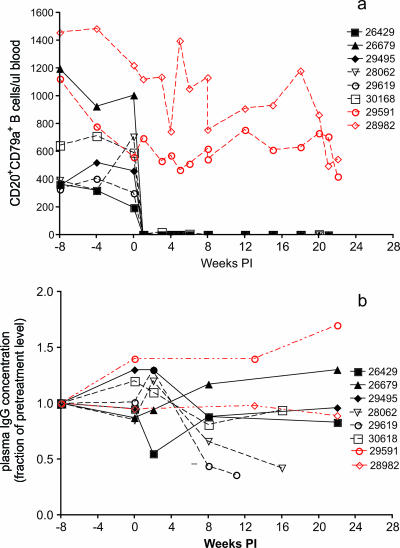
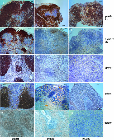
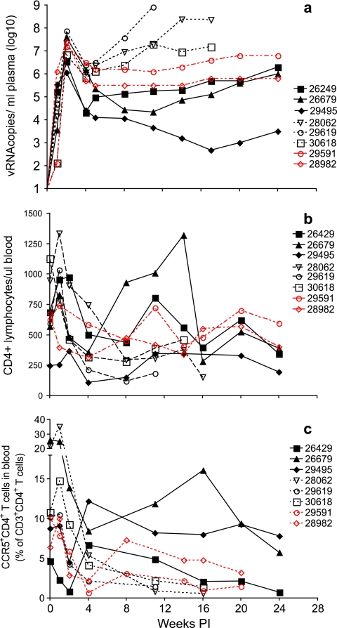
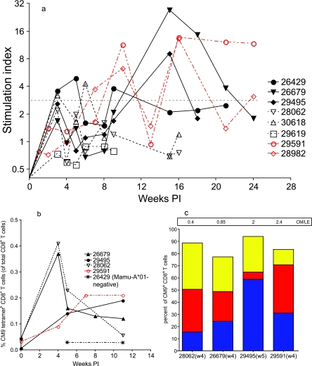
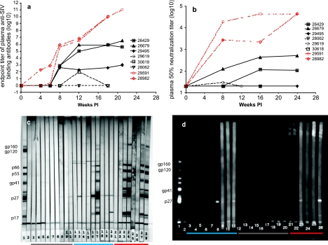
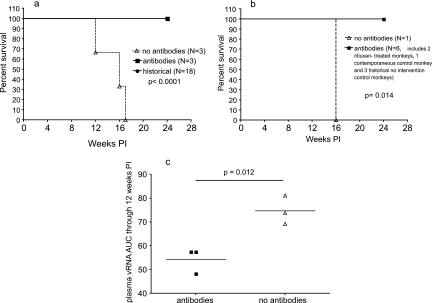
To better define the role of B cells in the control of pathogenic simian immunodeficiency virus (SIV) replication, six rhesus monkeys were depleted of B cells by intravenous infusion of rituximab (anti-CD20) 28 days and 7 days before intravaginal SIVmac239 inoculation and every 21 days thereafter until AIDS developed. Although the blood and tissues were similarly depleted of B cells, anti-SIV immunoglobulin G (IgG) antibody responses were completely blocked in only three of the six animals. In all six animals, levels of viral RNA (vRNA) in plasma peaked at 2 weeks and declined by 4 weeks postinoculation (PI). However, the three animals prevented from making an anti-SIV antibody response had significantly higher plasma vRNA levels through 12 weeks PI (P = 0.012). The remaining three B-cell-depleted animals made moderate anti-SIV IgG antibody responses, maintained moderate plasma SIV loads, and showed an expected rate of disease progression, surviving to 24 weeks PI without developing AIDS. In contrast, all three of the B-cell-depleted animals prevented from making anti-SIV IgG responses developed AIDS by 16 weeks PI (P = 0.0001). These observations indicate that antiviral antibody responses are critical in maintaining effective control of SIV replication at early time points postinfection.


Figures

FIG. 1.
FIG. 1.
Effect of rituximab infusion on concentrations of CD20+ CD79a+ B cells in blood (a) and on plasma IgG concentrations (b). Black symbols and lines denote the rituximab-treated monkeys. The dashed black lines denote monkeys in group A (28062, 29619, and 30618) that survived AIDS free for a shorter period of time than those in group B (26429, 26679, and 29495; solid black lines) (see the text). The red lines and symbols indicate the gamma globulin-infused monkeys. Day 0 is the day of SIV inoculation. Rituximab was infused every 21 days beginning 4 weeks prior to SIV inoculation.
FIG. 2.
FIG. 2.
Effect of rituximab infusion on the frequencies of CD20+ and CD79a+ B cells in tissues. Panels a, d, g, j, and m correspond to human gamma globulin-infused monkey 29591; panels b, e, h, k, and n correspond to rituximab-infused monkey 28062; and panels c, f, i, l, and o correspond to rituximab-infused monkey 29495. (a to c) CD20+ B cells in pretreatment (pre-TX) lymph node (Ln) biopsy specimens. (d to f) CD20+ B cells in lymph node (LN) biopsy specimens collected 6 weeks after initial rituximab treatment (2 weeks PI with SIV). (g to i) CD20+ B cells in spleen necropsy samples. (j to l) CD20+ B cells in colon necropsy samples. (m to o) CD79a+ B cells in spleen necropsy samples.
FIG. 3.
FIG. 3.
Plasma vRNA levels and concentrations of CD4+ T cells in blood. Shown are plasma SIV RNA levels (a), absolute numbers of CD3+ CD4+ T cells per microliter of blood (b), and frequencies of circulating CCR5+ CD3+ CD4+ T cells (c). Black symbols and lines denote the rituximab-treated monkeys; the solid black lines denote the group B monkeys. The red symbols and lines indicate the human gamma globulin-infused monkeys. Day 0 is the day of SIV inoculation. Rituximab was infused every 21 days beginning 4 weeks prior to SIV inoculation.
FIG. 4.
FIG. 4.
SIV-specific T-cell proliferation and the kinetics and phenotypes of Gag-specific CD8+-T-cell responses in Mamu-A*01-positive monkeys. (a) Levels of SIV-specific T-cell proliferation; (b) frequencies of CM9 tetramer-positive CD8+ T cells (percentages of total CD8+ T cells) in blood from four Mamu-A*01-positive monkeys and one Mamu-A*01-negative monkey (negative control) between 0 and 11 weeks PI; (c) frequencies of the CM9 tetramer-positive CD8+-T-cell phenotype in four Mamu-A*01-positive monkeys at 4 to 5 weeks PI. CM9 tetramer-positive, CD8+-T-cell phenotypes are defined as central memory (blue; C45RA, CD28+), intermediate effector memory (red; C45RA, CD28), and late effector memory (yellow; C45RA+, CD28). The ratios of central-memory to late-effector-memory (CM/LE) CM9-specific CD8+ T cells in the samples are indicated in the box at the top of the graph. w4, week 4; w5, week 5.
FIG. 5.
FIG. 5.
Effect of rituximab infusion on systemic antiviral antibody responses. (a) End point titers of anti-SIV binding antibodies in plasma. (b) 50% SIVmac251-neutralizing antibody titers in plasma. (c) SIVmac239 immunoblots. Lanes 3 to 20, plasma samples collected prior to inoculation, 8 weeks PI, and approximately 1 week prior to necropsy or at 20 weeks PI. Lanes 21 to 28, plasma samples collected prior to gamma globulin infusion, after gamma globulin infusion but prior to SIV inoculation, and at 8 and 20 weeks PI. Lanes: 1, positive control; 2, negative control; 3 to 5; monkey 28062; 6 to 8, monkey 29619; 9 to 11, monkey 30618; 12 to 14, monkey 26429; 15 to 17, monkey 26679; 18 to 20, monkey 29495; 21 to 24, monkey 29591; and 25 to 28, monkey 28982. (d) SIVmac 239 chemiluminescent immunoblots. Plasma samples collected from each animal prior to inoculation and 2 and 4 weeks PI were analyzed (lanes 3 to 26). Lanes: 1, positive control; 2, negative control; 3 to 5, monkey 26429; 6 to 8, monkey 26679; 9 to 11, monkey 29495; 12 to 14, monkey 28062; 15 to 17, monkey 29619; 18 to 20, monkey 30618; 21 to 23, monkey 29591; 24 to 26, monkey 28982.
FIG. 6.
FIG. 6.
Relationship between antiviral antibody responses, survival, and plasma SIV levels. RNA levels in rituximab-treated monkeys were determined. (a) Survival through 24 weeks PI. Based on a Kaplan-Meier analysis, significantly more rituximab-treated monkeys that made antiviral antibody responses (n = 3) and treatment-naïve monkeys (n = 18) than B-cell-depleted monkeys that were prevented from making antiviral antibody responses (n = 3) survived to 24 weeks PI (P < 0.0001). (b) Survival through 24 weeks PI of seven Mamu-A*01-positive monkeys. Based on a Kaplan-Meier analysis, the rituximab-treated Mamu-A*01-positive monkeys that made antiviral antibody responses (n = 2), the human gamma globulin-treated Mamu-A*01-positive monkey that made an antiviral antibody response (n = 1), and the historically treatment-naïve Mamu-A*01-positive monkeys that made antiviral antibody responses (n = 3) survived longer than the B-cell-depleted Mamu-A*-01-positive monkey that was prevented from making an antiviral antibody response (n = 1) (P = 0.014). (c) AUCs corresponding to the plasma vRNA levels in the six B-cell-depleted monkeys through 12 weeks PI. The mean plasma vRNA levels through 12 weeks PI (the time point when the first animal was necropsied) were transformed into AUCs. In a two-tailed paired t test, the mean plasma vRNA AUC for B-cell-depleted monkeys that made antiviral antibody responses (n = 3; left horizontal line) was significantly lower than that for B-cell-depleted monkeys that were prevented from making antiviral antibody responses (n = 3; right horizontal line; P = 0.012).

References

    1. Abel, K., L. Compton, T. Rourke, D. Montefiori, D. Lu, K. Rothaeusler, L. Fritts, K. Bost, and C. J. Miller. 2003. Simian-human immunodeficiency virus SHIV89.6-induced protection against intravaginal challenge with pathogenic SIVmac239 is independent of the route of immunization and is associated with a combination of cytotoxic T-lymphocyte and alpha interferon responses. J. Virol. 77:3099-3118. - PMC - PubMed
    1. Allen, T. M., J. Sidney, M. F. del Guercio, R. L. Glickman, G. L. Lensmeyer, D. A. Wiebe, R. DeMars, C. D. Pauza, R. P. Johnson, A. Sette, and D. I. Watkins. 1998. Characterization of the peptide binding motif of a rhesus MHC class I molecule (Mamu-A*01) that binds an immunodominant CTL epitope from simian immunodeficiency virus. J. Immunol. 160:6062-6071. - PubMed
    1. Baba, T. W., V. Liska, R. Hofmann-Lehmann, J. Vlasak, W. Xu, S. Ayehunie, L. A. Cavacini, M. R. Posner, H. Katinger, G. Stiegler, B. J. Bernacky, T. A. Rizvi, R. Schmidt, L. R. Hill, M. E. Keeling, Y. Lu, J. E. Wright, T. C. Chou, and R. M. Ruprecht. 2000. Human neutralizing monoclonal antibodies of the IgG1 subtype protect against mucosal simian-human immunodeficiency virus infection. Nat. Med. 6:200-206. - PubMed
    1. Baskin, G. B., M. Murphey-Corb, E. A. Watson, and L. N. Martin. 1988. Necropsy findings in rhesus monkeys experimentally infected with cultured simian immunodeficiency virus (SIV)/delta. Vet. Pathol. 25:456-467. - PubMed
    1. Dailey, P. J., M. Zamroud, R. Kelso, J. Kolberg, and M. Urdea. 1995. Quantitation of simian immunodeficiency virus (SIV) RNA in plasma of acute and chronically infected rhesus macaques using a branched DNA (bDNA) signal amplification assay. J. Med. Primatol. 24:209.

Publication types

MeSH terms