. 2007 May;81(10):5046-57.
doi: 10.1128/JVI.02746-06. Epub 2007 Feb 28.

Development of Sindbis viruses encoding nsP2/GFP chimeric proteins and their application for studying nsP2 functioning

Affiliations

Development of Sindbis viruses encoding nsP2/GFP chimeric proteins and their application for studying nsP2 functioning

Svetlana Atasheva et al. J Virol. 2007 May.

Abstract

Sindbis virus (SINV) is one of almost 30 currently known alphaviruses. In infected cells, it produces only a few proteins that function in virus replication and interfere with the development of the antiviral response. One of the viral nonstructural proteins, nsP2, not only exhibits protease and RNA helicase activities that are directly involved in viral RNA replication but also plays critical roles in the development of transcriptional and translational shutoffs in the SINV-infected cells. These multiple activities of nsP2 complicate investigations of this protein's functions and further understanding of its structure. Using a transposon-based approach, we generated a cDNA library of SINV genomes with a green fluorescent protein (GFP) gene randomly inserted into nsP2 and identified a number of sites that can be used for GFP cloning without a strong effect on virus replication. Recombinant SIN viruses encoding nsP2/GFP chimeric protein were capable of growth in tissue culture and interfering with cellular functions. SINV, expressing GFP in the nsP2, was used to isolate nsP2-specific protein complexes formed in the cytoplasm of the infected cells. These complexes contained viral nsPs, all of the cellular proteins that we previously coisolated with SINV nsP3, and some additional protein factors that were not found before in detectable concentrations. The random insertion library-based approach, followed by the selection of the viable variants expressing heterologous proteins, can be applied for mapping the domain structure of the viral nonstructural and structural proteins, cloning of peptide tags for isolation of the protein-specific complexes, and studying their formation by using live-cell imaging. This approach may also be applicable to presentation of additional antigens and retargeting of viruses to new receptors.


Figures

FIG. 1.
FIG. 1.
Selection of replicating SIN viruses encoding nsP2/GFP protein. A library of viral genomes having random GFP insertions in the nsP2 gene was generated as described in Materials and Methods. After transfection of the RNA pools, the genomes of the GFP-expressing viruses from the individual plaques were sequenced to identify the sites of the insertions. The numbers of amino acids, after which the insertions occurred, are indicated.
FIG. 2.
FIG. 2.
Recombinant viruses encoding nsP2/GFP and analysis of their replication in BHK-21 cells. (A) Schematic representation of wt SINV Toto1101 genome and genomes of SIN/nsP2GFP/8 and SIN/nsP2GFP/472, containing in-frame insertions of the entire GFP gene after aa 8 and 472 of nsP2, respectively. Arrows indicate the positions of the subgenomic promoter. (B) Infectivity of in vitro-synthesized viral RNAs and comparative plaque sizes determined in the infectious center assay (see Materials and Methods for details). Plaques were stained after 48 h of incubation under agarose cover. (C) BHK-21 cells were infected with the indicated viruses at an MOI of 10 PFU/cell. At the times indicated, media were replaced, and virus titers were determined by a plaque assay as described in Materials and Methods. The results of one of two reproducible experiments are presented.
FIG. 3.
FIG. 3.
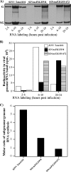
Synthesis of virus-specific RNAs in the cells infected with recombinant viruses. (A) 5 × 10⁵ BHK-21 cells in 6-well Costar plates were infected with SINV Toto1101, SIN/nsP2GFP/8, and SIN/nsP2GFP/472 at an MOI of 20 PFU/cell. At the indicated times, the medium in the wells was replaced by 0.8 ml of αMEM supplemented with 10% FBS, ActD (1 μg/ml), and [3H]uridine (20 μCi/ml). After 4 h of incubation at 37°C, RNAs were isolated and analyzed by agarose gel electrophoresis as described in Materials and Methods. (B) The synthesis of viral genome RNA was analyzed by excising the bands corresponding to 49S viral genome RNA from the gel shown in panel A, followed by measurement of the radioactivity by scintillation counting. (C) Bands corresponding to 26S subgenome RNA (at 6 h postinfection) were also excised from the gel, the radioactivity was measured by scintillation counting, and the molar ratio of subgenome to genome RNA synthesis was evaluated. G and SG indicate the positions of viral genomic and subgenomic RNAs. The results for one of two reproducible experiments are presented.
FIG. 4.
FIG. 4.
Processing and distribution of SINV nsPs in the cells infected with different recombinant viruses. (A) Schematic representation of the genomes of the viruses used in these experiments. SIN/2V/389 was previously described (14). It had GFP insertion after aa 389 of nsP3 and a single-point mutation leading to replacement of glycine by valine in the P2 position of the cleavage site between nsP2 and nsP3 (45). This nsP2 G806→V mutation blocked P2/3 cleavage during P123 processing. It also contained an additional mutation leading to the replacement of a stop codon between nsP3 and nsP4 by the cysteine-coding one. Other recombinant viruses are described in the legend to Fig. 2. (B) At 9 h postinfection, cells were harvested, and equal amounts of protein were used for gel electrophoresis. After transfer, the nitrocellulose membranes were processed by rabbit anti-nsP3 (upper panel) or anti-nsP2 (lower panel) antibodies and horseradish peroxidase-conjugated, secondary donkey anti-rabbit immunoglobulin G antibodies. (C) Analysis of the nonstructural polyprotein processing. At 3 h postinfection, proteins were labeled with [35S]methionine as described in Materials and Methods and chased for 15, 30, or 60 min. The positions of the processed and partially processed proteins are indicated. (D) BHK-21 cells were infected with SIN/nsP2GFP/8 and SIN/nsP2GFP/472 as described in Materials and Methods. At 8 h postinfection, the cells were fixed with 1% formaldehyde, and images were acquired on a Zeiss LSM510 META confocal microscope using a ×63 1.4NA oil immersion planapochromal lens.
FIG. 5.
FIG. 5.
Analysis of protein synthesis in the cells infected with recombinant viruses. 5 × 10⁵ BHK-21 cells were infected with SINV Toto1101, SIN/nsP2GFP/8, and SIN/nsP2GFP/472 at an MOI of 20 PFU/cell. At the indicated times, proteins were pulse-labeled with [35S]methionine for 30 min and analyzed on a sodium dodecyl sulfate-10% polyacrylamide gel. The gels were dried and autoradiographed (A) or analyzed on a Storm 860 PhosphorImager (B) as described in Materials and Methods. The levels of the synthesis of cellular proteins were determined by measuring radioactivity in the protein band corresponding to actin and normalized to radioactivity in the actin band in the lane representing the uninfected cells. The results from one of two reproducible experiments are presented.
FIG. 6.
FIG. 6.
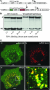
Replication of the SINV/nsP2GFP/nsP3Cherry virus expressing two fluorescent proteins fused with different nsPs and distribution of nsP2/GFP and nsP3/Cherry in the infected cells. (A) Schematic representation of wt SINV Toto1101 and SINV/nsP2GFP/nsP3Cherry genomes. (B) A total of 5 × 10⁵ BHK-21 cells in six-well Costar plates were infected with SINV Toto1101 and SINV/nsP2GFP/nsP3Cherry at an MOI of ∼20 PFU/cell. At the indicated times, the medium in the wells was replaced by 0.8 ml of αMEM supplemented with 10% FBS, ActD (1 μg/ml), and [3H]uridine (20 μCi/ml). After 4 h of incubation at 37°C, the RNAs were isolated and analyzed by agarose gel electrophoresis as described in Materials and Methods. (C) BHK-21 cells were infected with SINV/nsP2GFP/nsP3Cherry at an MOI of ∼20 PFU/cell. At 12 h postinfection, they were fixed with 1% formaldehyde, and the three-dimensional images were acquired on a Zeiss LSM510 META confocal microscope using a ×63 1.4NA oil immersion planapochromal lens. The enlarged cell fragment presented in panel d is indicated in panel c.
FIG. 7.
FIG. 7.
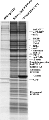
Analysis of proteins coisolated with nsP2/GFP from the replicon- or virus-infected cells. BHK-21 cells were infected with packaged SINrep/nsP2GFP/472/Pac virus and SINrep/GFP or SIN/nsP2GFP/472 virus at an MOI of 20 infectious units or PFU per cell. Proteins were isolated by using GFP-specific antibodies as described in Materials and Methods, and after elution from the affinity columns were separated on sodium dodecyl sulfate-10% polyacrylamide gels and stained with Coomassie brilliant blue R-250. Coomassie blue-stained bands were excised from the gels, and the proteins were identified by MALDI-TOF analysis. The names of the identified proteins are indicated. An open box indicates a GFP-containing band present in the lysate of SINrep/GFP replicon-infected cells. The results for one of five reproducible experiments are presented.
FIG. 8.
FIG. 8.
Summary of the data about the structure and functional domains in alphavirus nsP2. (A) Positions of RNA helicase, protease, and putative S-adenosyl-l-methionine-dependent RNA methyltransferase domains. (B) Positions of GFP insertions that do not cause deleterious effect on virus replication. (C) Positions of adaptive point mutations in the putative methyltransferase domains of SINV, SFV, and VEEV that have a strong effect on both RNA replication and the ability of replicating alphaviruses or replicons to cause CPE (13, 37, 38). (D) Positions of the compensatory mutations found in the SINV and VEEV genomes with a mutated replication enhancer, the 51-nt CSE (11). (E) Positions of deletion in SFV nsP2 (51) and GFP insertions in SINV nsP2 that make these proteins incapable of P23 polyprotein processing.

References

    1. Agapov, E. A., K. E. Reed, and C. M. Rice. 1998. Use of the vaccinia virus expression system for the study of HCV protein processing, p. 303-314. In J. Y. N. Lau (ed.), Methods in molecular medicine, vol. 19. Humana Press, New York, NY. - PubMed
    1. Ahola, T., P. Kujala, M. Tuittila, T. Blom, P. Laakkonen, A. Hinkkanen, and P. Auvinen. 2000. Effects of palmitoylation of replicase protein nsP1 on alphavirus infection. J. Virol. 74:6725-6733. - PMC - PubMed
    1. Ahola, T., A. Lampio, P. Auvinen, and L. Kaariainen. 1999. Semliki Forest virus mRNA capping enzyme requires association with anionic membrane phospholipids for activity. EMBO J. 18:3164-3172. - PMC - PubMed
    1. Barton, D. J., S. G. Sawicki, and D. L. Sawicki. 1991. Solubilization and immunoprecipitation of alphavirus replication complexes. J. Virol. 65:1496-1506. - PMC - PubMed
    1. Bredenbeek, P. J., I. Frolov, C. M. Rice, and S. Schlesinger. 1993. Sindbis virus expression vectors: packaging of RNA replicons by using defective helper RNAs. J. Virol. 67:6439-6446. - PMC - PubMed

Publication types

MeSH terms

LinkOut - more resources