. 2007 Feb 28;27(9):2155-62.
doi: 10.1523/JNEUROSCI.4945-06.2007.

Neurosteroid synthesis-mediated regulation of GABA(A) receptors: relevance to the ovarian cycle and stress

Affiliations

Neurosteroid synthesis-mediated regulation of GABA(A) receptors: relevance to the ovarian cycle and stress

Jamie Maguire et al. J Neurosci. .

Abstract

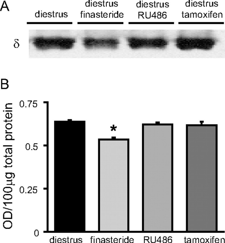
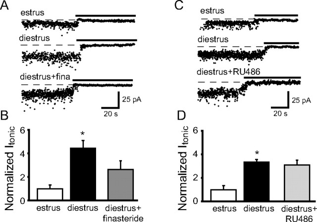
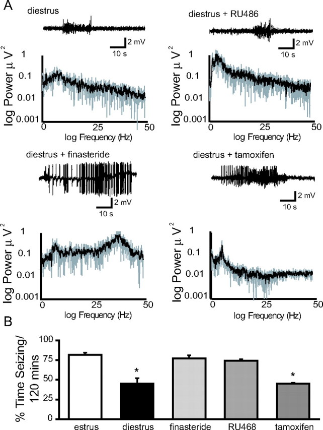
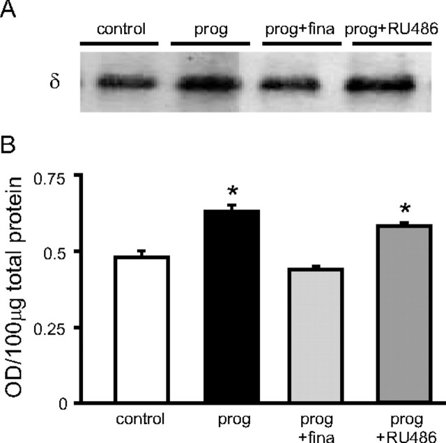
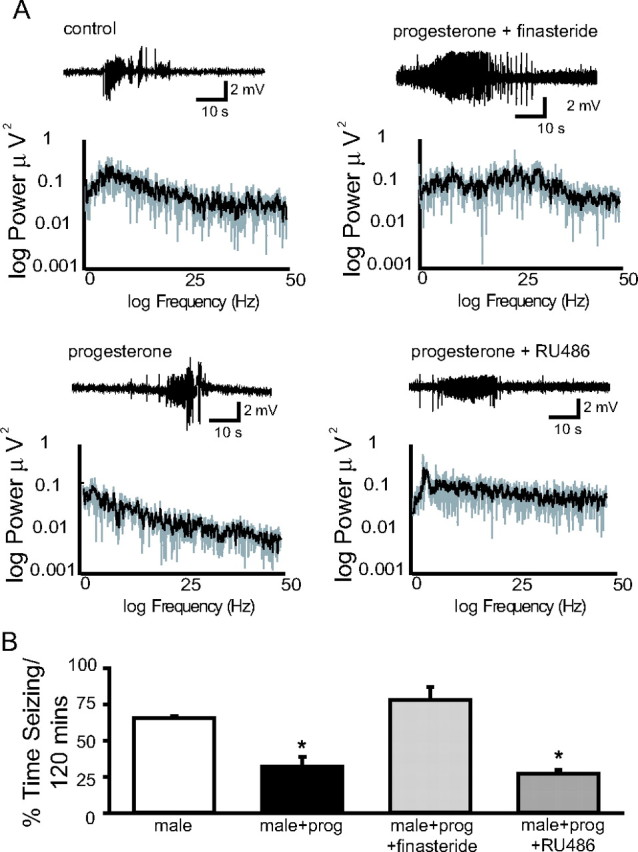
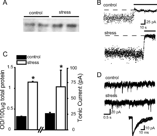
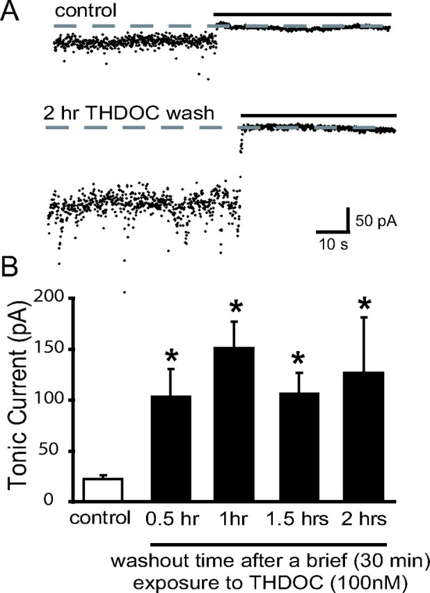
Recently, we demonstrated cyclic alterations in GABA(A) receptor (GABA(A)R) subunit composition over the ovarian cycle correlated with fluctuations in progesterone levels. However, it remains unclear whether this physiological regulation of GABA(A)Rs is directly mediated by hormones. Here, we show that both ovarian and stress hormones are capable of reorganizing GABA(A)Rs by actions through neurosteroid metabolites. The cyclic alterations in GABA(A)Rs demonstrated in female mice can be mimicked with exogenous progesterone treatment in males or in ovariectomized females. Progesterone (5 mg/kg, twice daily) upregulates the expression of GABA(A)R delta subunits and enhances the tonic inhibition mediated by these receptors in dentate gyrus granule cells (DGGCs). These changes in males as well as ovarian cycle-induced changes in females can be blocked by finasteride, an antagonist of neurosteroid synthesis from progesterone. The altered GABA(A)R expression is unaffected by the progesterone receptor antagonist RU486 [mifepristone (11beta-[p-(dimethylamino)phenyl]-17beta-hydroxy-17-(1-propynyl)estra-4,9-dien-3-one)], suggesting that neurosteroid synthesis and not progesterone receptor activation underlies the hormone-mediated effects on GABA(A)R expression. Neurosteroids can alter GABA(A)R expression on a rapid timescale, because GABA(A)R upregulation can be induced in brain slices maintained in vitro after a short (30 min) treatment with the neurosteroid 3alpha,5alpha-tetrahydrodeoxycorticosterone (THDOC) (100 nM). Consistent with these rapid alterations, acute stress, a condition known to quickly raise THDOC levels, within 30 min induces upregulation of GABA(A)R delta subunit expression and increase tonic inhibition in DGGCs. These results reveal that several physiological conditions characterized by elevations in neurosteroid levels induce a reorganization of GABA(A)Rs through the action of neurosteroids.


Figures

Figure 1.
Figure 1.
Neurosteroid synthesis is required for ovarian cycle-related changes in GABAARs. A, Representative immunoblots for GABAAR δ subunits of total membrane hippocampal protein from untreated diestrous mice or diestrous mice treated with either finasteride (100 mg/kg), RU486 (50 mg/kg), or tamoxifen (1 mg/kg) from estrus throughout diestrus. B, The histogram of average optical density of Western blots highlights the blocking effect of finasteride, unlike RU486 or tamoxifen, on the upregulation of the GABAAR δ subunit at diestrus. The asterisk denotes significance (p < 0.05). Error bars indicate SEM.
Figure 2.
Figure 2.
Neurosteroid synthesis is required to induce the increased tonic inhibition during diestrus. A, Representative recordings from control estrous and diestrous mice and mice treated with finasteride from estrus through diestrus. The dashed lines indicate the basal current in the presence of saturating concentrations of the GABAAR antagonist SR95531 that was perfused during the time indicated by the horizontal black bars. B, The bar graphs of the average tonic current normalized to the average tonic current recorded during estrus in DGGCs show a significant increase in diestrous mice, which is blocked with finasteride treatment. The asterisk denotes significance (p < 0.05, t test). C, Representative recordings from control estrous and diestrous mice and mice treated with RU486 from estrus through diestrus. D, Histogram of the average tonic current normalized to the average tonic current recorded during estrus in DGGCs demonstrates a significant increase in diestrous mice, which is unaffected by RU486 treatment. The asterisk denotes significance (p < 0.05, t test). Error bars indicate SEM.
Figure 3.
Figure 3.
The decrease in excitability during diestrus is mediated by neurosteroid synthesis. A, Representative EEG recordings from control diestrous mice and diestrous mice treated with RU486, finasteride, or tamoxifen. Finasteride blocks the decrease in excitability during diestrus. Fast Fourier transform analysis demonstrates the increased power of the EEG in diestrous mice treated with finasteride. B, The histograms of the percentage time seizing per 2 h recording session show the decrease in seizure activity during diestrus, which is blocked by finasteride treatment. The asterisk denotes significance (p < 0.05). Error bars indicate SEM.
Figure 4.
Figure 4.
Progesterone mediates the changes in GABAARs related to the ovarian cycle. A, Representative immunoblots of total membrane hippocampal protein from untreated male mice or male mice treated with 5 mg/kg progesterone twice daily for 2 d alone or in combination with either finasteride or RU486. B, The histogram of average optical density of Western blots demonstrates that progesterone administration increases GABAAR δ subunit expression, which can be blocked with finasteride treatment but not with RU486. The asterisk denotes significance (p < 0.05). Error bars indicate SEM. prog, Progesterone; fina, finasteride.
Figure 5.
Figure 5.
Neurosteroid synthesis from exogenous progesterone administration decreased excitability. A, Representative EEG recordings from control male mice and male mice treated twice daily for 2 d with 5 mg/kg progesterone alone or in combination with finasteride or RU486. Finasteride blocks the decrease in excitability in progesterone-treated males. Fast Fourier transform analysis demonstrates the increase in power in diestrous mice treated with finasteride. B, The histograms of the percentage time seizing per 2 h recording session demonstrate the decrease in excitability in progesterone-treated males, which is blocked only by finasteride not by RU486. The asterisk denotes significance (p < 0.05). Error bars indicate SEM. prog, Progesterone.
Figure 6.
Figure 6.
Acute stress increases GABAAR δ subunit expression and tonic inhibition. A, Representative immunoblots with antibodies to GABAAR δ subunits of total membrane hippocampal protein from two unstressed control male mice and two mice male mice exposed to 2 min of CO2 stress. B, Representative recordings from control unstressed male mice and male mice subjected to 2 min of CO2 stress. C, The bar graphs of average optical densities of Western blots and average tonic current demonstrate the upregulation of the GABAAR δ subunit expression and increased tonic inhibition after acute stress compared with control. The asterisk denotes significance (p < 0.05). Error bars indicate SEM. D, Representative recordings from control unstressed males and males exposed to 2 min of CO2 stress. The inset shows superimposed averaged sIPSCs recorded in DGGCs from control unstressed and males exposed to 2 min of CO2 stress.
Figure 7.
Figure 7.
Acute THDOC treatment in vitro induces a long-lasting increase in tonic inhibition. A, Representative recordings in DGGCs from control slices and slices treated for 30 min with THDOC, and then washed out for 2 h. Note the increase in tonic current in slices treated with THDOC despite the 2 h washout, suggesting long-lasting changes in tonic inhibition. B, Bar graphs show the average tonic current in DGGCs from control slices or slices acutely treated with THDOC after increasing periods of washout. Acute THDOC treatment significantly enhances tonic current after various lengths of washout from 30 min to 2 h. The asterisk denotes significance (p < 0.05, t test). Error bars indicate SEM.

Similar articles

Cited by

References

    1. Agis-Balboa RC, Pinna G, Zhubi A, Maloku E, Veldic M, Costa E, Guidotti A. Characterization of brain neurons that express enzymes mediating neurosteroid biosynthesis. Proc Natl Acad Sci USA. 2006;103:14602–14607. - PMC - PubMed
    1. Akinci MK, Johnston GA. Sex differences in acute swim stress-induced changes in the binding of MK-801 to the NMDA subclass of glutamate receptors in mouse forebrain. J Neurochem. 1993;61:2290–2293. - PubMed
    1. Barbaccia ML, Roscetti G, Trabucchi M, Cuccheddu T, Concas A, Biggio G. Neurosteroids in the brain of handling-habituated and naive rats: effect of CO2 inhalation. Eur J Pharmacol. 1994;261:317–320. - PubMed
    1. Barbaccia ML, Roscetti G, Trabucchi M, Mostallino MC, Concas A, Purdy RH, Biggio G. Time-dependent changes in rat brain neuroactive steroid concentrations and GABA(A) receptor function after acute stress. Neuroendocrinology. 1996a;63:166–172. - PubMed
    1. Barbaccia ML, Concas A, Roscetti G, Bolacchi F, Mostallino MC, Purdy RH, Biggio G. Stress-induced increase in brain neuroactive steroids: antagonism by abecarnil. Pharmacol Biochem Behav. 1996b;54:205–210. - PubMed

Publication types

MeSH terms