. 2007 Feb 28;27(9):2176-85.
doi: 10.1523/JNEUROSCI.5176-06.2007.

Functional axonal regeneration through astrocytic scar genetically modified to digest chondroitin sulfate proteoglycans

Affiliations

Functional axonal regeneration through astrocytic scar genetically modified to digest chondroitin sulfate proteoglycans

William B J Cafferty et al. J Neurosci. .

Abstract

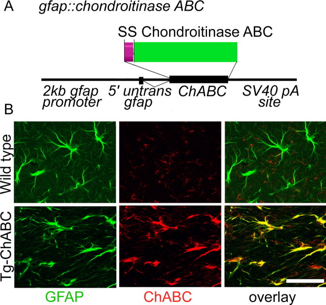
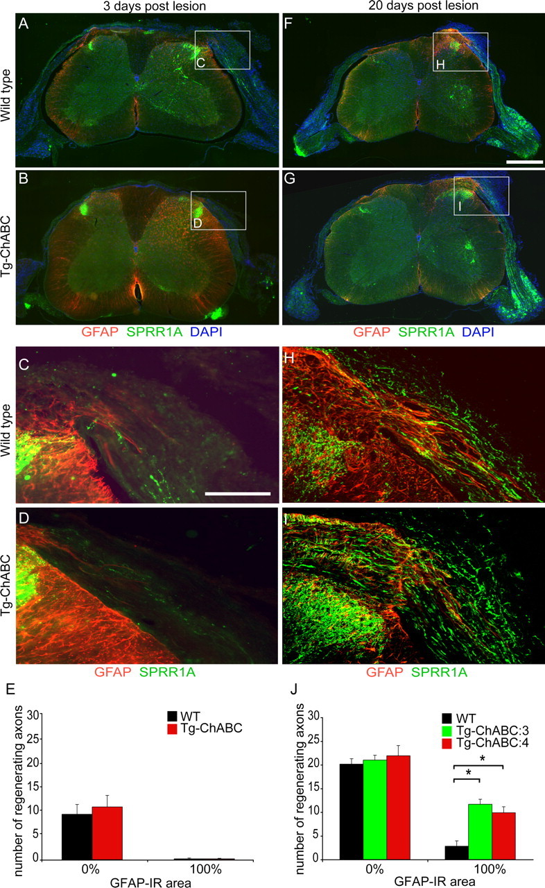
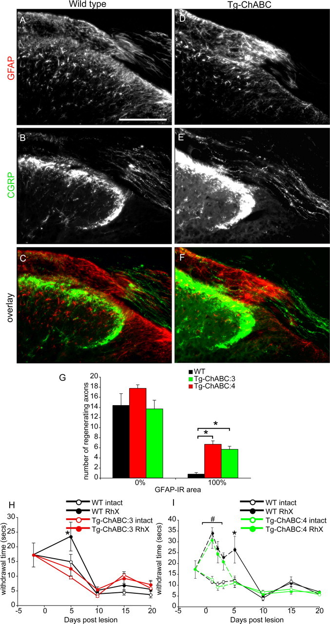
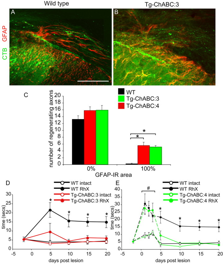
Axotomized neurons within the damaged CNS are thought to be prevented from functional regeneration by inhibitory molecules such as chondroitin sulfate proteoglycans (CSPGs) and myelin-associated inhibitors. Here, we provide a transgenic test of the role of CSPGs in limiting regeneration, using the gfap promotor to express a CSPG-degrading enzyme chondroitinase ABC (ChABC) in astrocytes. Corticospinal axons extend within the lesion site, but not caudal to it, after dorsal hemisection in the transgenic mice. The presence of the gfap-ChABC transgene yields no significant improvement in motor function recovery in this model. In contrast, functionally significant sensory axon regeneration is observed after dorsal rhizotomy in transgenic mice. These transgenic studies confirm a local efficacy for reduced CSPG to enhance CNS axon growth after traumatic injury. CSPGs appear to function in a spatially distinct role from myelin inhibitors, implying that combination-based therapy will be especially advantageous for CNS injuries.


Figures

Figure 1.
Figure 1.
Transgenic expression of chondroitinase ABC. A, A schematic illustrates the DNA vector providing expression of ChABC under the control of the murine gfap promoter. Photomicrographs illustrate sections of motor cortex 2 weeks after stab lesion. Astrocytes in both wild-type and ChABC:3 and Tg-ChABC:4 mice upregulate expression of GFAP (green). B, An antibody that recognizes ChABC (red) shows 100% overlap with GFAP (yellow) in transgenic mice. SV40, Simian virus 40.
Figure 2.
Figure 2.
Transgenic expression of functional ChABC after dorsal hemisection. Photomicrographs of sagittal sections from intact wild-type (A), Tg-ChABC:3 (F), and Tg-ChABC:4 (K) mice show normal chondroitin sulfate proteoglycan (CS56-IR) deposition in spinal gray and white matter. Dorsal hemisection increases deposition of CS56-IR CSPGs at the lesion site of wild-type mice (B, C). Chondroitin-4-sulfate immunoreactivity reflects GAG degradation and is entirely absent from the lesion site (D, E). Dorsal hemisection in Tg-ChABC:3 (G, H) and Tg-ChABC:4 (L, M) shows no increase in CS56-IR CSPGs at the lesion site. Instead, there is robust C4S immunoreactivity of hydrolyzed CSPGs within the lesion site in Tg-ChABC:3 (I, J) and Tg-ChABC:4 (N, O) mice. Scale bar, 500 μm.
Figure 3.
Figure 3.
Transgenic expression of functional ChABC after dorsal rhizotomy. A–P, Photomicrographs show the DREZ from intact (A, B) and rhizotomized (C, D) wild-type (WT) plus intact (I, J) and rhizotomized (K, L) Tg-ChABC mice. GFAP-IR astrocytes (red) extend into the damaged dorsal root in both lesioned wild-type (C) and Tg-ChABC (L) mice. C4S-IR CSPG stubs are present in the damaged GFAP-rich DREZ of Tg-ChABC mice (K, L) but are entirely absent from wild-type mice (C, D). Sections from intact (E, F) and rhizotomized (G, H) wild-type and intact (M, N) and rhizotomized (O, P) Tg-ChABC mice were treated in vitro with ChABC enzyme to digest all CSPGs and then stained with C4S to reveal maximum stub concentration. The optical density of C4S immunoreactivity in three areas (the CNS adjacent to the DREZ, the DREZ, and adjacent PNS; A) of rhizotomized and intact wild-type and Tg-ChABC mice with and without in vitro ChABC treatment compares the relative efficacy of transgenic ChABC expression versus subsequent digestion of CSPGs in vivo. Q, No significant difference in optical density of C4S immunoreactivity was observed within the CNS, DREZ, or PNS zones between wild-type and Tg-ChABC mice without in vitro ChABC treatment. ChABC treatment increased the density of stubs 10-fold in both wild-type and Tg-ChABC mice equally (Q) (data are mean ± SEM). R, On the rhizotomized side, the optical density of stub staining is significantly higher in the CNS, DREZ, and PNS zones of Tg-ChABC mice compared with wild-type mice (data are mean ± SEM; p < 0.001, ANOVA). In vitro digestion of Drx sections reveals that transgenic expression of ChABC in vivo is highly efficient in digesting CSPGs near the DREZ (R). Scale bar, 200 μm.
Figure 4.
Figure 4.
Transgenic ChABC increases the number of CST axons in astrocytic scar of a SCI. A–F, Photomicrographs illustrate sagittal sections of wild-type (WT) (A, B), Tg-ChABC:3 (C, D), and Tg-ChABC:4 (E, F) mice 4 weeks after bilateral dorsal over-hemisection. BDA-IR CST (green) axons can be seen approaching the lesion site (delineated by GFAP-IR astrocytes; red). Insets (B, D, F) show regenerating CST axons stopping >1 mm away from the lesion epicenter (denoted by asterisk) in wild-type mice (B); CST axons can be seen coursing throughout the lesion in Tg-ChABC:3 (B) and Tg-ChABC:4 (F) mice. G, Assessment of astrocytic process orientation revealed an identical profile of directionality in both genotypes (data represent mean ± SEM), with process alignment changing from a longitudinal orientation (Long.) distal from the lesion to a transverse orientation (Trans.) at the lesion center. H, Quantification of CST axon growth illustrates that significantly more axons approach the lesion site in Tg-ChABC:3 and Tg-ChABC:4 mice compared with wild-type controls. Data represent mean ± SEM CST axon index (*p < 0.05, one-way ANOVA). I, Wild-type, Tg-ChABC:3, and Tg-ChABC:4 mice underwent open-field locomotion assessment after dorsal over-hemisection; there was no significant difference in hindlimb movement between the groups. Scale bars: A, C, E, 500 μm; B, D, F, 200 μm.
Figure 5.
Figure 5.
Transgenic expression of ChABC promotes regeneration of sensory neurons after dorsal rhizotomy. A–D, F–I, Photomicrographs illustrate transverse section of cervical spinal cord from adult wild-type (WT) (A, F) and Tg-ChABC (B, G) mice that had undergone unilateral rhizotomy from C5–C8 3 d (A–D) or 20 d (F–I) after lesion (intact root left side, rhizotomized root right side; red, GFAP; green, SPRR1A; blue, DAPI). E, Insets show high-power photomicrographs of the rhizotomized DREZ. Three days after lesion, modest numbers of SPRR1A-IR (green) regenerating axons can be seen approaching the damaged DREZ (delineated by GFAP-IR astrocytes; red) in both wild-type (C) and Tg-ChABC (D) mice; no SPRR1A-IR regenerating axons were seen central to the DREZ (E) (data represent mean ± SEM axons; x-axis demarcates PNS from CNS by GFAP staining; 0% GFAP-IR refers to the PNS; 100% GFAP-IR refers to the CNS). F, H, J, Twenty days after lesion, SPRR1A-IR axons can be seen throughout the dorsal root in wild-type mice (F, H), but few axons penetrate the DREZ and enter the spinal cord (H, J). In contrast, significant numbers of SPRR1A-IR axons can be seen growing up to and past the DREZ and entering the spinal cord in Tg-ChABC:3 and Tg-ChABC:4 mice (I, J) (data represent mean ± SEM; *p < 0.001, one-way ANOVA). Scale bars: A, B, F, G, 500 μm; C, D, H, I, 100 μm.
Figure 6.
Figure 6.
Transgenic expression of ChABC restores nociceptive function after dorsal rhizotomy. A–F, Photomicrographs illustrate transverse section of ipsilateral C7 spinal cord from wild-type (WT) (A–C) and Tg-ChABC (D–F) 21 d after dorsal rhizotomy. GFAP-IR astrocytes bulge into the PNS in both wild-type (A, C) and Tg-ChABC:3 (D, F) mice. Regenerating CGRP-IR axons abruptly stop growing when they reach the damaged DREZ in wild-type mice (B, C). G, CGRP-IR axons traverse the damaged DREZ in Tg-ChABC mice (E, F) and reenter the spinal cord. Quantification of the number of CGRP-IR axons at the DREZ (x-axis demarcates PNS from CNS by GFAP staining; 0% GFAP-IR refers to the PNS; 100% GFAP-IR refers to the CNS) shows Tg-ChABC:3 and Tg-ChABC:4 to have significantly more axons entering the spinal cord compared with wild-type control mice (data represent mean ± SEM; *p < 0.001, one-way ANOVA). H, I, Thermal withdrawal latencies were recorded from wild-type (black lines), Tg-ChABC:3 (H, red lines), and Tg-ChABC:4 (I, green lines) mice to assess return of nociceptive function after rhizotomy. Withdrawal latency was significantly elevated ipsilateral to the lesion after rhizotomy in wild-type (filled black circles) and Tg-ChABC (filled red and green circles) mice compared with their contralateral intact sides (open circles, H, I) (data represent mean ± SEM withdrawal time; # p < 0.05, one-way ANOVA). Wild-type mice recovered thermal sensation within 10 d of lesion. Tg-ChABC:3 and Tg-ChABC:4 recovered significant thermal sensory function by 5 d after lesion (*p < 0.05, one-way ANOVA) 5 d earlier than wild-type controls. Scale bar, 100 μm.
Figure 7.
Figure 7.
Transgenic expression of ChABC restores cutaneous mechanosensation after dorsal rhizotomy. A, B, Photomicrographs illustrate transverse sections of ipsilateral C7 spinal cord from wild-type (WT) (A) and Tg-ChABC (B) 21 d after dorsal rhizotomy. The DREZ is demarcated by the presence of GFAP-IR reactive astrocytes (red); regenerating CTB-IR axons abruptly stop growing when they reach the damaged DREZ in wild-type mice (green, A). CTB-IR axons traverse the damaged DREZ in Tg-ChABC mice and reenter the spinal cord (green, B). C, Quantification of the number of CTB-IR axons at the DREZ (C) (x-axis demarcates PNS from CNS by GFAP staining; 0% GFAP-IR refers to the PNS; 100% GFAP-IR refers to the CNS) shows Tg-ChABC:3 and Tg-ChABC:4 to have significantly more axons entering the spinal cord compared with wild-type control mice (data represent mean ± SEM; *p < 0.001, one-way ANOVA). Mechanosensation of injured mice was assessed using a modified tape removal task. D, E, Sense assessment was significantly elevated ipsilateral to the lesion after rhizotomy in wild-type (filled black circles) and Tg-ChABC (filled red and green circles) mice compared with their contralateral intact sides (open circles) (data represent mean ± SEM withdrawal time; # p < 0.05, one-way ANOVA). Wild-type mice maintained a deficit throughout the testing period, but Tg-ChABC:3 and Tg-ChABC:4 mice recovered significant function by 5 d after lesion (*p < 0.05, one-way ANOVA) compared with rhizotomized wild-type mice. Scale bar, 100 μm.

Similar articles

Cited by

References

    1. Abad F, Feria M, Boada J. Chronic amitriptyline decreases autotomy following dorsal rhizotomy in rats. Neurosci Lett. 1989;99:187–190. - PubMed
    1. Aldskogius H, Kozlova EN. Central neuron-glial and glial-glial interactions following axon injury. Prog Neurobiol. 1998;55:1–26. - PubMed
    1. Asher RA, Morgenstern DA, Fidler PS, Adcock KH, Oohira A, Braistead JE, Levine JM, Margolis RU, Rogers JH, Fawcett JW. Neurocan is upregulated in injured brain and in cytokine-treated astrocytes. J Neurosci. 2000;20:2427–2438. - PMC - PubMed
    1. Bareyre FM, Kerschensteiner M, Raineteau O, Mettenleiter TC, Weinmann O, Schwab ME. The injured spinal cord spontaneously forms a new intraspinal circuit in adult rats. Nat Neurosci. 2004;7:269–277. - PubMed
    1. Barritt AW, Davies M, Marchand F, Hartley R, Grist J, Yip P, McMahon SB, Bradbury EJ. Chondroitinase ABC promotes sprouting of intact and injured spinal systems after spinal cord injury. J Neurosci. 2006;26:10856–10867. - PMC - PubMed

Publication types

MeSH terms

Substances