. 2007 Feb 28;27(9):2272-82.
doi: 10.1523/JNEUROSCI.3470-06.2007.

Response anticipation and response conflict: an event-related potential and functional magnetic resonance imaging study

Affiliations

Response anticipation and response conflict: an event-related potential and functional magnetic resonance imaging study

Jin Fan et al. J Neurosci. .

Abstract

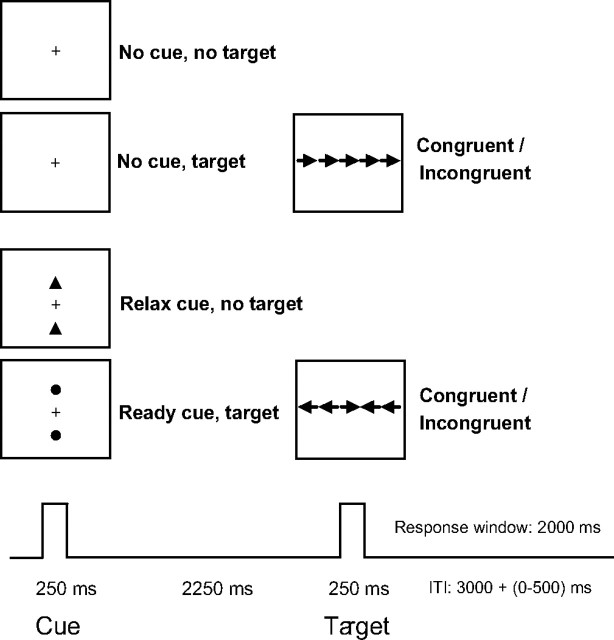
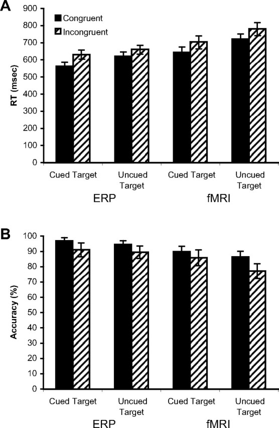
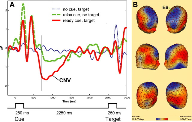
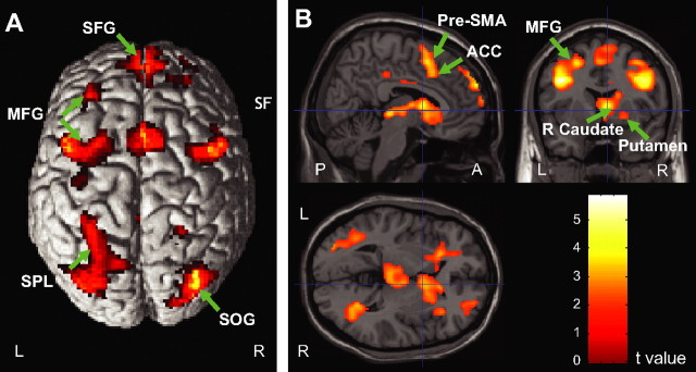
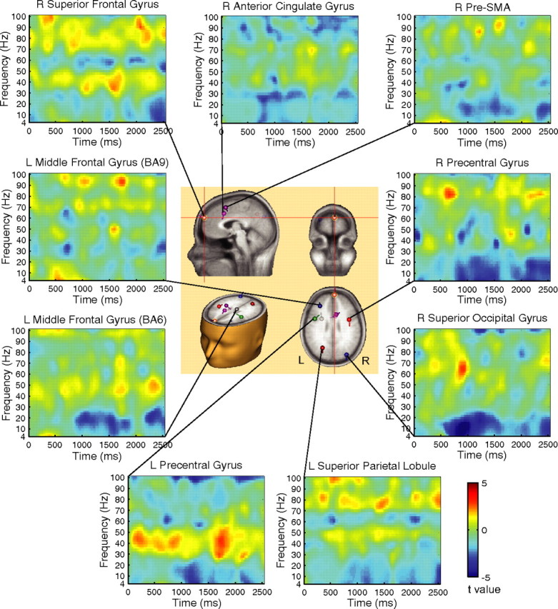
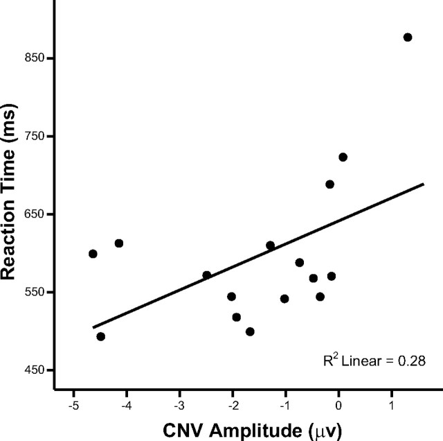
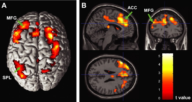
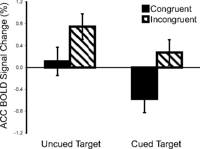
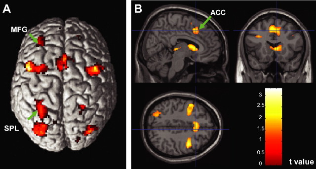
Response anticipation and response conflict processes are supported by executive control. However, few neuroimaging studies have attempted to study the relationship between these two processes in the same experimental session. In this study, we isolated brain activity associated with response anticipation (after a cue to prepare vs relax) and with response conflict (responding to a target with incongruent vs congruent flankers) and examined the independence and interaction of brain networks supporting these processes using event-related potentials (ERPs) and functional magnetic resonance imaging. Response anticipation generated a contingent negative variation ERP that correlated with shorter reaction times, and was associated with activation of a thalamo-cortico-striatal network, as well as increased gamma band power in frontal and parietal regions, and decreased spectral power in theta, alpha, and beta bands in most regions. Response conflict was associated with increased activation in the anterior cingulate cortex (ACC) and prefrontal cortex of the executive control network, with an overlap in activation with response anticipation in regions including the middle frontal gyrus, ACC, and superior parietal lobule. Although the executive control network showed increased activation in response to unanticipated versus anticipated targets, the response conflict effect was not altered by response anticipation. These results suggest that common regions of a dorsal frontoparietal network and the ACC are engaged in the flexible control of a wide range of executive processes, and that response anticipation modulates overall activity in the executive control network but does not interact with response conflict processing.


Figures

Figure 1.
Figure 1.
Schematic of the task for eliciting response anticipation and response conflict. In each trial, a 250 ms cue period was followed by an imperative 250 ms target stimulus (the center arrow in a row of 5 arrows), with a 2250 ms cue–target interval and a 2000 ms response window. The intertrial interval was jittered between 3000 and 3500 ms with an average duration of 3250 ms. There were two cue conditions: uncued trials and cued trials, including relax and ready cues. There were two target conditions: targets with congruent or incongruent flankers. The participants' task was to indicate the direction of the target.
Figure 2.
Figure 2.
A, B, Behavioral results corresponding to cue and target conditions during ERP recording and fMRI scanning for RT (A) and accuracy (B). For responses to cued versus uncued targets, RTs were shorter during both ERP and fMRI testing, and accuracy was greater during fMRI testing. In addition, RTs were longer and accuracy was reduced for responses to incongruent versus congruent targets. There was no significant interaction between cue and target conditions. Error bars represent ±1 SEM.
Figure 3.
Figure 3.
A, ERP waveforms as a function of cue type. The ERP amplitude was greater in the ready cue than in the relax cue condition at ∼700 ms after the cue onset. B, Scalp topography of the voltage difference of ready cue minus relax cue at 699 ms. The time point corresponds to the vertical line in A. E6 indicates the location of the electrode with the maximum CNV amplitude.
Figure 4.
Figure 4.
Brain activation associated with response anticipation shown on the surface (A) and cross views (B). The superior frontal gyrus (SFG), pre-SMA extending to ACC, middle frontal gyrus (MFG), superior parietal lobule (SPL) of the dorsal parietal cortex along the intraparietal sulcus (IPs), superior occipital gyrus (SOG), and right caudate nucleus and putamen showed greater activation in the ready versus relax cue comparison. For a full list of activated regions, see Table 1.
Figure 5.
Figure 5.
Power change difference diagrams for the ready minus relax cue contrast as a function of time–frequency and the dipoles of ERP data. The center panel shows the dipole locations. The small balls and bars represent the locations and orientations of the dipoles, respectively. The right superior frontal gyrus showed greater gamma (>30 Hz) power maintained over the cue–target interval, whereas the right superior occipital gyrus showed power decrease in theta (4–8 Hz), alpha (8–12 Hz), and beta (12–30 Hz) bands. The cue onset is at 0 ms, and the target onset is at 2500 ms.
Figure 6.
Figure 6.
Scattergram showing the relationship between mean CNV amplitudes and mean RTs in the ready cue condition across participants.
Figure 7.
Figure 7.
Brain activation associated with response conflict (incongruent vs congruent) shown on the surface (A) and cross views (B). MFG, Middle frontal gyrus; SPL, superior parietal lobule.
Figure 8.
Figure 8.
ACC activation under different cue and target conditions. Greater activation was observed during the response to uncued versus cued targets, and to targets with incongruent versus congruent flankers. However, the conflict effects did not differ between these two cue conditions. Error bars represent ±1 SEM for each condition.
Figure 9.
Figure 9.
A, B, Regions showing consistent activation in response anticipation and response conflict processes shown on the surface (A) and cross views (B). MFG, Middle frontal gyrus; SPL, superior parietal lobule.

Similar articles

Cited by

References

    1. Alexander GE, Crutcher MD. Preparation for movement: neural representations of intended direction in three motor areas of the monkey. J Neurophysiol. 1990;64:133–150. - PubMed
    1. Andrew C, Pfurtscheller G. Event-related coherence as a tool for studying dynamic interaction of brain regions. Electroencephalogr Clin Neurophysiol. 1996;98:144–148. - PubMed
    1. Badler JB, Heinen SJ. Anticipatory movement timing using prediction and external cues. J Neurosci. 2006;26:4519–4525. - PMC - PubMed
    1. Barcelo F, Suwazono S, Knight RT. Prefrontal modulation of visual processing in humans. Nat Neurosci. 2000;3:399–403. - PubMed
    1. Bastiaansen MC, Brunia CH. Anticipatory attention: an event-related desynchronization approach. Int J Psychophysiol. 2001;43:91–107. - PubMed

Publication types

LinkOut - more resources