. 2007 Feb 28;27(9):2298-308.
doi: 10.1523/JNEUROSCI.5175-06.2007.

NMDA receptor function and NMDA receptor-dependent phosphorylation of huntingtin is altered by the endocytic protein HIP1

Affiliations

NMDA receptor function and NMDA receptor-dependent phosphorylation of huntingtin is altered by the endocytic protein HIP1

Martina Metzler et al. J Neurosci. .

Abstract

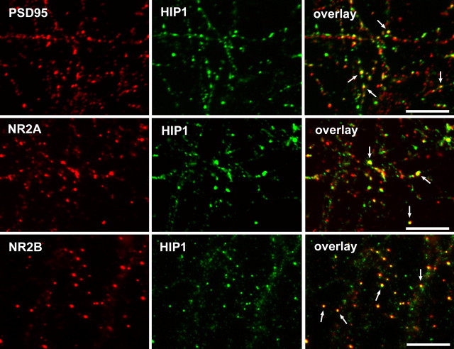
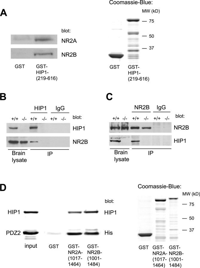
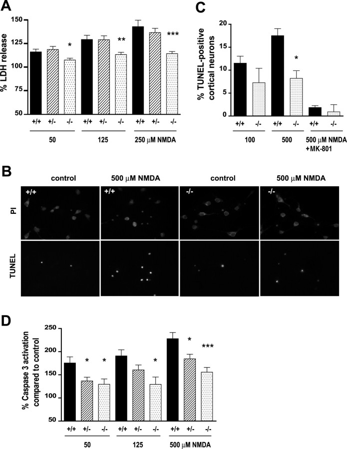
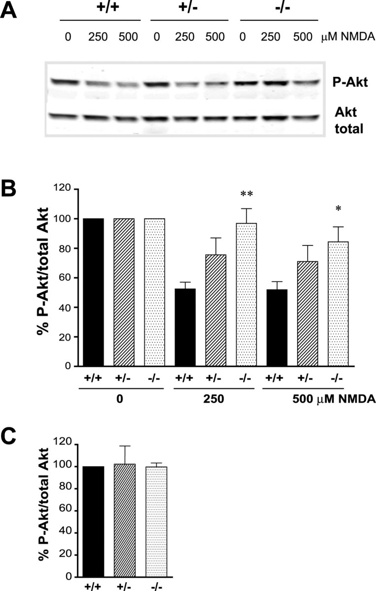
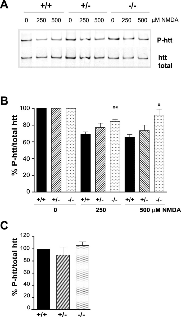
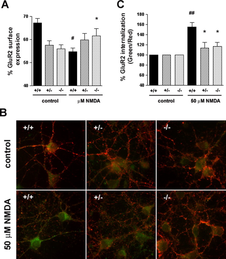
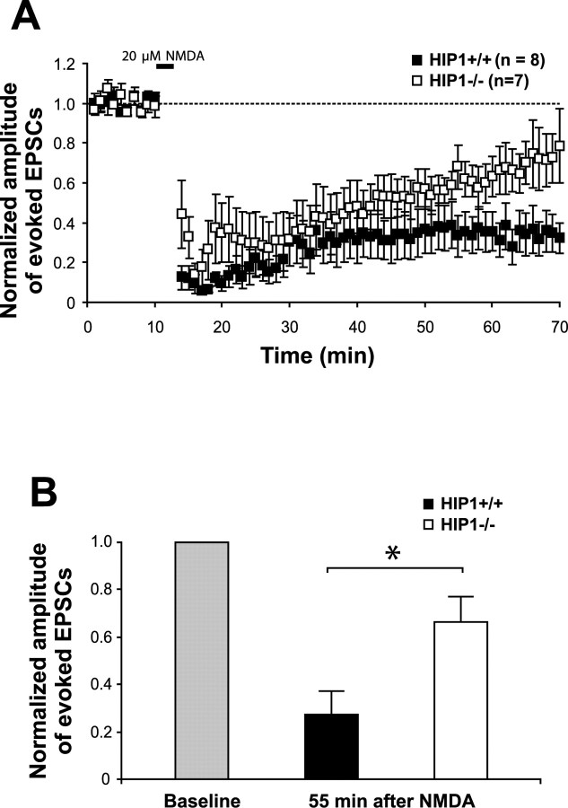
Huntingtin-interacting protein 1 (HIP1) is an endocytic adaptor protein that plays a role in clathrin-mediated endocytosis and the ligand-induced internalization of AMPA receptors (AMPARs) (Metzler et al., 2003). In the present study, we investigated the role of HIP1 in NMDA receptor (NMDAR) function by analyzing NMDA-dependent transport and NMDA-induced excitotoxicity in neurons from HIP1-/- mice. HIP1 colocalizes with NMDARs in hippocampal and cortical neurons and affinity purifies with NMDARs by GST (glutathione S-transferase) pull down and coimmunoprecipitation. A profound decrease in NMDA-induced AMPAR internalization of 75% occurs in neurons from HIP1-/- mice compared with wild type, using a quantitative single-cell-based internalization assay. This defect in NMDA-dependent removal of surface AMPARs is in agreement with the observed defect in long-term depression induction in hippocampal brain slices of HIP1-/- mice and supports a role of HIP1 in AMPAR internalization in vivo. HIP1-/- neurons are partially protected from NMDA-induced excitotoxicity as assessed by LDH (lactate dehydrogenase) release, TUNEL (terminal deoxynucleotidyl transferase-mediated biotinylated dUTP nick end labeling) and caspase-3 activation assays, which points to a role of HIP1 in NMDA-induced cell death. Interestingly, phosphorylation of Akt and its substrate huntingtin (htt) decreases during NMDA-induced excitotoxicity by 48 and 31%, respectively. This decrease is significantly modulated by HIP1, resulting in 94 and 48% changes in P-Akt and P-htt levels in HIP1-/- neurons, respectively. In summary, we have shown that HIP1 influences important NMDAR functions and that both HIP1 and htt participate in NMDA-induced cell death. These findings may provide novel insights into the cellular mechanisms underlying enhanced NMDA-induced excitotoxicity in Huntington's disease.


Figures

Figure 1.
Figure 1.
HIP1 colocalizes with NMDARs. HIP1 expression was analyzed by double immunofluorescence in primary hippocampal neurons after 14 DIV. HIP1 immunostaining was either detected with the mAb HIP1#9 (top and middle) or the pAb HIP1FP (bottom) shown in green. Expression of PSD-95, NR2A, and NR2B is shown in red. HIP1 localizes to synapses, at which it colocalizes with PSD-95. HIP1 also shows partial colocalization with NR2A (middle) and NR2B (bottom). Arrows indicate colocalization. Scale bars, 10 μm.
Figure 2.
Figure 2.
HIP1 interacts with NMDARs in vitro and in brain lysate. A, Soluble proteins from brain lysate were affinity purified with either GST alone or GST-HIP1-(219–616) bound to glutathione-Sepharose beads. The input amount of GST alone and GST fusion protein was verified by Coomassie blue staining, and their sizes were compared with a molecular-weight (MW) marker. Bound proteins were eluted and analyzed by Western blot and probed with anti-NR2A and anti-NR2B Abs as indicated. B, Brains from 3-month-old wild-type and HIP1−/− mice were homogenized and solubilized in the presence of 0.2% SDS and 0.8% Triton X-100. Proteins were immunoprecipitated with mAb HIP1#9 or normal IgG, separated by Western blot, and probed with Abs against HIP1 and NR2B. The same amount of sample was loaded on gels stained for NR2B and HIP1. C, Proteins were immunoprecipitated as described in B with an Ab against NR2B and normal IgG. D, Recombinant HIP1-(219–616)-His6 and recombinant PSD-95-PDZ2-His6 were expressed and purified from bacterial lysate. Twenty-five micrograms of purified HIP1 or PDZ2 were incubated with GST alone, GST-NR2A-(1017–1464), and GST-NR2B-(1001–1484) bound to glutathione-Sepharose beads. The input amount of GST alone and GST fusion proteins was verified by Coomassie blue staining, and their sizes were compared with a molecular-weight marker. The amount of specifically bound protein was eluted, immunoblotted, and probed with mAb HIP1#9 or an anti-His Ab, demonstrating direct interaction between HIP1 and NMDARs and PDZ2 and NMDARs, respectively. IP, Immunoprecipitate.
Figure 3.
Figure 3.
Reduced NMDA-induced excitotoxicity in HIP1−/− neurons. A, Cortical neurons at 12 DIV were treated with different doses of NMDA and 30 μm glycine for 10 min. After drug removal, neurons were cultured for 14 h, and release of LDH into the cell culture supernatant was measured. Duplicate samples for each condition and genotype in each of six independent experiments were determined. B, Striatal neurons from wild-type and HIP1−/− mice were treated with 500 μm NMDA and 30 μm glycine for 5 min at 9 DIV. After drug removal, neurons were cultured for 20 h and stained for TUNEL-positive neurons (green) followed by PI counterstaining (red). C, Cortical neurons at 12 DIV from wild-type and HIP1−/− mice were treated with 100 or 500 μm NMDA and 30 μm glycine for 5 min in the presence or absence of 50 μm MK-801 and processed as described in B. The number of TUNEL-positive neurons was determined and expressed relative to the number of total neurons. For each sample, 200 neurons were counted for each condition and genotype in each of three independent experiments. D, Cortical neurons at 12 DIV were treated with varying doses of NMDA and 30 μm glycine for 10 min. After drug removal, neurons were cultured for 3 h, and caspase-3 activation was measured fluorometrically by cleavage of Ac-DEVD-AFC. Duplicate samples for each condition and genotype in each of seven independent experiments were determined. *p < 0.05; **p < 0.005; ***p < 0.0005 compared with wild type; t test. Error bars denote ±SEM.
Figure 4.
Figure 4.
HIP1 promotes dephosphorylation of Akt during NMDA-induced excitotoxicity. A, Cortical neurons from wild-type and HIP1−/− mice were treated with varying doses of NMDA and 30 μm glycine for 10 min at 12 DIV. After drug removal, neurons were cultured for 90 min and processed for Western blotting. Membranes were probed with Abs against P-Akt-S473 and total Akt. B, The ratio between P-Akt-S473 and total Akt was determined by densitometry and expressed relative to controls. Duplicate samples for each condition and genotype in each of eight independent experiments were determined (*p < 0.05; **p < 0.005 compared with wild type; t test). C, The absolute ratio of P-Akt-S473 and total Akt was determined by densitometry under control conditions for all three genotypes and expressed relative to wild type. Error bars denote ±SEM.
Figure 5.
Figure 5.
HIP1 promotes dephosphorylation of htt during NMDA-induced excitotoxicity. A, Cortical neurons from wild-type and HIP1−/− mice were treated with varying doses of NMDA and 30 μm glycine for 10 min at 12 DIV. After drug removal, neurons were cultured for 90 min and processed for immunoprecipitation of htt. Precipitated protein was separated by Western blot, and membranes were probed with Abs against P-htt-S421 and total htt (mAb 2166). B, The ratio between P-htt-S421 and total htt was determined by densitometry and expressed relative to controls. Ratios for each condition and genotype in each of six independent experiments were determined (*p < 0.05; **p < 0.005 compared with wild type; t test). C, The absolute ratio between P-htt-S421 and total htt was determined by densitometry under control conditions for all three genotypes and expressed relative to wild type (n = 4). Error bars denote ±SEM.
Figure 6.
Figure 6.
Reduced NMDA-induced AMPAR internalization in HIP1−/− neurons. A, HIP1 modulates NMDA-induced GluR2 trafficking in cortical neurons. Cortical neurons were established from wild-type (+/+), heterozygous (+/−), and homozygous (−/−) littermates. After 12–15 DIV, the percentage of surface-expressed GluR2 was determined in a quantitative colorimetric assay in control and 100 μm NMDA-stimulated cultures under nonpermeant and permeant conditions. Results were averaged for each group in each of three independent experiments (#p < 0.05 for within-genotype, between-treatment comparison; *p < 0.05 for within-treatment, between-genotype comparison; t test). B, HIP1 modulates NMDA-induced GluR2 internalization in striatal neurons. Striatal neurons were cultured from wild-type (+/+), heterozygous (+/−), or homozygous (−/−) littermates and live-labeled with an Ab recognizing the extracellular domain of GluR2, followed by treatment with either control solution (ECS alone) or 50 μm NMDA, 10 μm glycine, and 5 μm strychnine for 5 min at 37°C. Cultures were washed and incubated for 20 min at 37°C to allow internalization. Surface GluR2 was then visualized using a red secondary Ab, whereas internalized GluR2 was labeled with a green secondary Ab. C, Internalization was quantified as the green-to-red signal ratio, which represents the ratio of the internalized GluR2 to the GluR2 remaining on the cell surface after different treatments (see Materials and Methods). Green-to-red ratios were averaged for each group in each of three independent experiments (##p < 0.005 for within-genotype, between-treatment comparison; *p < 0.05 for within-treatment, between-genotype comparison; t test). Error bars denote ±SEM.
Figure 7.
Figure 7.
LTD is reduced in hippocampal slices of HIP1−/− mice. A, Chemically induced LTD was established in hippocampal slices of wild-type mice (+/+; n = 8) and compared with HIP1−/− littermates (−/−; n = 7). B, The normalized amplitude of evoked EPSCs 55 min after bath application of NMDA is significantly reduced in hippocampal brain slices from wild-type mice compared with HIP1−/− littermates (*p < 0.05; t test). Error bars denote ±SEM.

References

    1. Alloway PG, Howard L, Dolph PJ. The formation of stable rhodopsin-arrestin complexes induces apoptosis and photoreceptor cell degeneration. Neuron. 2000;28:129–138. - PubMed
    1. Beal MF, Kowall NW, Ellison DW, Mazurek MF, Swartz KJ, Martin JB. Replication of the neurochemical characteristics of Huntington's disease by quinolinic acid. Nature. 1986;321:168–171. - PubMed
    1. Beattie EC, Carroll RC, Yu X, Morishita W, Yasuda H, von Zastrow M, Malenka RC. Regulation of AMPA receptor endocytosis by a signaling mechanism shared with LTD. Nat Neurosci. 2000;3:1291–1300. - PubMed
    1. Bonfoco E, Krainc D, Ankarcrona M, Nicotera P, Lipton SA. Apoptosis and necrosis: two distinct events induced, respectively, by mild and intense insults with N-methyl-d-aspartate or nitric oxide/superoxide in cortical cell cultures. Proc Natl Acad Sci USA. 1995;92:7162–7166. - PMC - PubMed
    1. Carroll RC, Lissin DV, von Zastrow M, Nicoll RA, Malenka RC. Rapid redistribution of glutamate receptors contributes to long-term depression in hippocampal cultures. Nat Neurosci. 1999a;2:454–460. - PubMed

Publication types