. 2007 Feb 28;27(9):2309-17.
doi: 10.1523/JNEUROSCI.4565-06.2007.

Development of hydrocephalus in mice expressing the G(i)-coupled GPCR Ro1 RASSL receptor in astrocytes

Affiliations

Development of hydrocephalus in mice expressing the G(i)-coupled GPCR Ro1 RASSL receptor in astrocytes

Elizabeth J Sweger et al. J Neurosci. .

Abstract

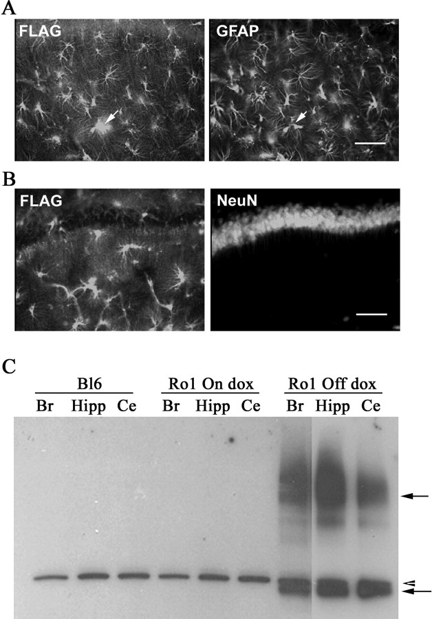
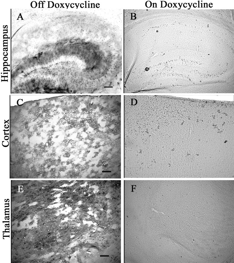
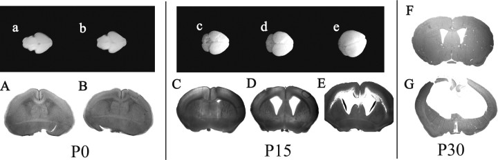
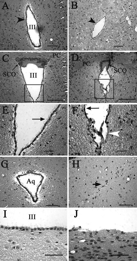
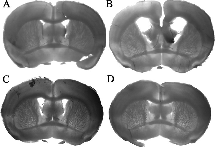
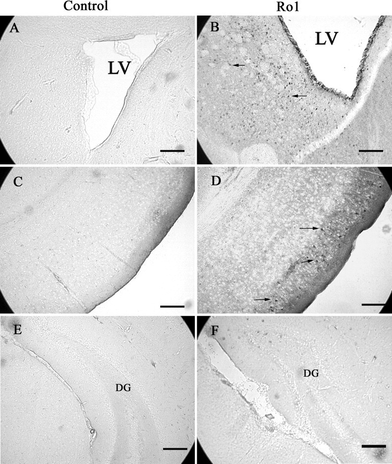
We developed a transgenic mouse line that expresses the G(i)-coupled RASSL (receptor activated solely by synthetic ligand) Ro1 in astrocytes to study astrocyte-neuronal communication. Surprisingly, we found that all transgenics expressing Ro1 developed hydrocephalus. We analyzed these mice in an effort to develop a new model of hydrocephalus that will further our understanding of the pathophysiology of the disease. Expression of Ro1 was restricted to astrocytes by crossing the transgenic hGFAP-tTA (tet transactivator behind the human glial fibrillary acidic protein promoter) mouse line with the transgenic tetO-Ro1/tetO-LacZ mouse line. This cross produced double-transgenic mice that expressed Ro1 in astrocytes. All double transgenics developed hydrocephalus by postnatal day 15, whereas single-transgenic littermate controls appeared normal. Hydrocephalic Ro1 mice displayed enlarged ventricles, partial denudation of the ependymal cell layer, altered subcommissural organ morphology, and obliteration of the cerebral aqueduct. Severely hydrocephalic mice also had increased levels of phospho-Erk and GFAP expression. Administration of doxycycline to breeding pairs suppressed Ro1 expression and the onset of hydrocephalus in double-transgenic offspring. Ro1 animals maintained on dox did not develop hydrocephalus; however, if taken off doxycycline at weaning, double-transgenic mice developed enlarged ventricles within 7 weeks, indicating that Ro1 expression also induces hydrocephalus in adults. This study discovered a new model of hydrocephalus in which the rate of pathogenesis can be controlled enabling the study of the pathogenesis of both juvenile and adult onset hydrocephalus.


Figures

Figure 1.
Figure 1.
Ro1 structure [from Coward et al. (1998)]. To create Ro1, the second extracellular loop of the κ-opioid receptor was replaced with the second extracellular loop of the δ-opioid receptor. A FLAG tag was added to the N terminus for detection. Ro1 has a greatly reduced affinity for endogenous ligands but still responds to the small molecule drug spiradoline.
Figure 2.
Figure 2.
Ro1 expression is restricted to astrocytes and is regulated by dox. A, B, Hippocampal brain sections were double-stained for FLAG and GFAP (astrocyte marker) or FLAG and NeuN (neuronal marker). A, Positive FLAG staining (left, arrow) colocalized with GFAP (right, arrow) but not with NeuN (B). The arrows point to the same cell. Scale bars, 100 μm. C, Brain lysates from a C57BL/6 control mouse, Ro1 mouse on dox (25 μg/ml), and Ro1 mouse off dox were immunoprecipitated with FLAG monoclonal antibody, and the blots were probed with FLAG polyclonal antibody. Only the Ro1 mouse off dox had positive bands (solid arrows). A nonspecific band was found in all samples (arrowhead). Br, Brain; Hipp, hippocampus; Ce, cerebellum.
Figure 3.
Figure 3.
LacZ expression is regulated by dox. Brain sections from Ro1 mice on dox (25 μg/ml) (B, D, F) and Ro1 mice off dox (A, C, E) were stained by Xgal histochemistry. Ro1 animals on dox had no detectable positive cells, whereas robust staining was seen in Ro1 animals off dox, indicating that dox is effectively controlling expression. Scale bars: A, B, 150 μm; C–F, 50 μm.
Figure 4.
Figure 4.
Ro1 mice maintained off dox develop hydrocephalus. B, C, All Ro1 pups from breeding pairs maintained off dox (B) develop overt hydrocephalus, and >50% die by 12 weeks of age (C). A, Ro1 mice maintained on dox (25 μg/ml) appear normal. Mice shown are P45.
Figure 5.
Figure 5.
Ro1 mice develop progressive enlargement of the lateral ventricles. At birth, whole brains and coronal sections of Ro1 mice (b, B) and single-transgenic control littermates (a, A) are indistinguishable. By P15, brains from Ro1 mice (d, e, D, E) are larger than controls (c, C) and have pronounced enlargement of the lateral ventricles. The hydrocephalus in Ro1 mice can be classified as mild (D) (n = 4), moderate (E) (n = 6), or severe (data not shown) (n = 2) cases based on ventricle-to-brain ratio. By P30, Ro1 mice (G) have severe hydrocephalus characterized by greatly enlarged lateral ventricles and extremely thin cortex. Control mice at P30 (F) appear normal. All mice were maintained off dox.
Figure 6.
Figure 6.
Alterations in the ventricular system of hydrocephalic Ro1 mice. Hematoxylin and eosin staining of coronal sections from single-transgenic control (P45, left column) and Ro1 (P33, right column) mice. A, B, In Ro1 mice (B), the third ventricle at the level of the periventricular hypothalamic nucleus is reduced in size and the ependymal cell layer appears thinner compared with controls (A). D, F, H, J, The subcommissural organ is disorganized in Ro1 mice (D), and there is a partial denudation of ependymal cells lining the ventricular walls (F, arrowhead; J). The remaining ependymal cells have fewer cilia (F, arrow), and the aqueduct is obliterated with no apparent ependymal cell layer (H). C, E, G, I, In control mice, the subcommissural organ (C) and aqueduct (G) show normal morphology. The ependymal layer is intact with multiple cilia per cell (E, arrow; I). E and F are enlargements of the boxes in C and D, respectively. Mice were maintained off dox. Aq, Aqueduct; SCO, subcommissural organ; III, third ventricle; PC, posterior commissure. Scale bars: A, B, G, H, I, J, 50 μm; C, D, 100 μm; E, F, 30 μm.
Figure 7.
Figure 7.
Ro1 mice will develop enlarged ventricles if taken off dox at weaning. A, B, In litters taken off dox at weaning (P21), Ro1 mice (B) develop enlarged ventricles when compared with single-transgenic littermate controls (A). C, D, Ro1 mice (D) have normal ventricles when mice were kept on dox continuously compared with single-transgenic littermate controls (C). All mice were processed at P60. A, n = 2; B, n = 4; C, n = 7; D, n = 8.
Figure 8.
Figure 8.
GFAP expression is upregulated in hydrocephalic Ro1 mice. Brain lysates from two Ro1 mice with severe hydrocephalus and one littermate control (P22) were separated by SDS-PAGE and probed with anti-GFAP antibody (A). Ro1 mice have increased GFAP levels in the hippocampus and cortex. Brain sections from control (B) and Ro1 (C) mice were stained for GFAP to show increased expression. Cortical sections are shown; pial surface is on the top right corner. Scale bar, 50 μm.
Figure 9.
Figure 9.
Phospho-Erk p44/42 levels are elevated in hydrocephalic Ro1 mice. Increased phospho-Erk p44/42 staining is observed around the lateral ventricles (B) and in the entorhinal cortex (D) of hydrocephalic Ro1 mice but not in littermate single-transgenic controls (P21) (A, C). No increases in phospho-Erk were observed in the hippocampus (F). Sections shown in A and B are at the level of the anterior commissure and striatum; C–F are at the level of the subcommissural organ. Scale bars, 150 μm. LV, Lateral ventricle; DG, dentate gyrus.

References

    1. Araque A, Li N, Doyle RT, Haydon PG. SNARE protein-dependent glutamate release from astrocytes. J Neurosci. 2000;20:666–673. - PMC - PubMed
    1. Araque A, Carmignoto G, Haydon PG. Dynamic signaling between astrocytes and neurons. Annu Rev Physiol. 2001;63:795–813. - PubMed
    1. Araque A, Martin ED, Perea G, Arellano JI, Buno W. Synaptically released acetylcholine evokes Ca2+ elevations in astrocytes in hippocampal slices. J Neurosci. 2002;22:2443–2450. - PMC - PubMed
    1. Bach A, Lallemand Y, Nicola MA, Ramos C, Mathis L, Maufras M, Robert B. Msx1 is required for dorsal diencephalon patterning. Development. 2003;130:4025–4036. - PubMed
    1. Blackshear PJ, Graves JP, Stumpo DJ, Cobos I, Rubenstein JL, Zeldin DC. Graded phenotypic response to partial and complete deficiency of a brain-specific transcript variant of the winged helix transcription factor RFX4. Development. 2003;130:4539–4552. - PubMed

Publication types