. 2007 Feb 28;27(9):2318-30.
doi: 10.1523/JNEUROSCI.4322-06.2007.

Transgenic mouse lines subdivide medial vestibular nucleus neurons into discrete, neurochemically distinct populations

Affiliations

Transgenic mouse lines subdivide medial vestibular nucleus neurons into discrete, neurochemically distinct populations

Martha W Bagnall et al. J Neurosci. .

Abstract

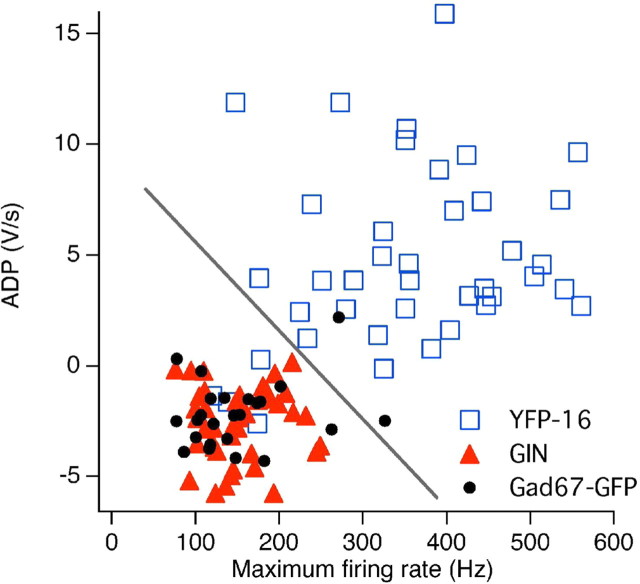
The identification of neuron types within circuits is fundamental to understanding their relevance to behavior. In the vestibular nuclei, several classes of neurons have been defined in vivo on the basis of their activity during behavior, but it is unclear how those types correspond to neurons identified in slice preparations. By targeting recordings to neurons labeled in transgenic mouse lines, this study reveals that the continuous distribution of intrinsic parameters observed in medial vestibular nucleus (MVN) neurons can be neatly subdivided into two populations of neurons, one of which is GABAergic and the other of which is exclusively glycinergic or glutamatergic. In slice recordings, GABAergic neurons labeled in the EGFP (enhanced green fluorescent protein)-expressing inhibitory neuron (GIN) line displayed lower maximum firing rates (<250 Hz) than glycinergic and glutamatergic neurons labeled in the yellow fluorescent protein-16 (YFP-16) line (up to 500 Hz). In contrast to cortical and hippocampal interneurons, GABAergic MVN neurons exhibited wider action potentials than glutamatergic (and glycinergic) neurons. Responses to current injection differed between the neurons labeled in the two lines, with GIN neurons modulating their firing rates over a smaller input range, adapting less during steady depolarization, and exhibiting less rebound firing than YFP-16 neurons. These results provide a scheme for robust classification of unidentified MVN neurons by their physiological properties. Finally, dye labeling in slices shows that both GABAergic and glycinergic neurons project to the contralateral vestibular nuclei, indicating that commissural inhibition is accomplished through at least two processing streams with differential input and output properties.


Figures

Figure 1.
Figure 1.
Distribution and morphology of YFP-16 and GIN neurons in the MVN. Coronal sections are from the YFP-16 (A) and GIN (B) lines. MVNp, Parvocellular MVN; MVNm, magnocellular MVN; NPH, nucleus prepositus hypoglossi; IV, fourth ventricle (darkened for contrast). Scale bar: A (for A, B), 200 μm. C, D, Magnified view of YFP-16 neurons (C) and GIN neurons in MVN (D). Scale bar: D (for C, D), 10 μm. E, Histogram of soma size, measured as the product of length and width. YFP-16 neurons are significantly larger than GIN neurons (p < 0.001). F, Histogram of number of processes extending from cell body. On average, YFP-16 neurons extend significantly more primary dendrites than GIN neurons (p < 0.0001). n = 108 (YFP-16) and 160 (GIN) neurons for both graphs.
Figure 2.
Figure 2.
Single-cell RT-PCR indicates that YFP-16 neurons are glutamatergic or glycinergic, whereas GIN neurons are GABAergic. RT-PCR was performed on YFP-16 and GIN neurons from slice recordings. Neurons were assayed for five indicators of transmitter content: VGluT1, VGluT2, GlyT2, GAD65, and GAD67. Two neurons are shown from each line. A, YFP-16 neuron A is VGluT2+, whereas neuron B is GlyT2+. B, GIN neuron A expressed the GABAergic markers GAD67 and GAD65, as well as the glycinergic marker GlyT2; GIN neuron B expressed only GAD67 and GAD65. Right, RNA controls were 0.5 ng RNA extracts from whole mouse brain processed alongside single cells.
Figure 3.
Figure 3.
Action potentials differ between YFP-16 and GIN neurons. A, Example action potentials from two YFP-16 neurons (black). B, Examples from two GIN neurons (gray). C, Relationship between ADP after spike repolarization and action potential width at half-height. Each point represents one neuron [YFP-16 (open squares), n = 49; GIN (gray triangles), n = 59; unidentified neurons from a separate study (dark gray dots), n = 61]. D, Maximum depth of the AHP in YFP-16 and GIN neurons plotted versus the time after the spike peak when that maximum is reached. Symbols and n are the same as those in C.
Figure 4.
Figure 4.
Input–ouput properties in YFP-16 and GIN neurons. Shown are examples of responses to depolarizing current steps in a YFP-16 neuron (A) and a GIN neuron (B). Steps of increasing amplitude were given until the neuron could no longer fire action potentials throughout the whole step. Bottom panels, Mean firing rate evoked during depolarization is plotted versus the amplitude of the current step for each neuron shown above. The break in line fit indicates the measurement of gain below and above 80 Hz for each neuron. C, Scatterplot of maximum firing rate (average over whole step) versus input resistance measured below spike threshold. Each point represents one neuron (YFP-16, n = 40; GIN, n = 43; unidentified neurons, n = 61). D, Maximum input current for which a neuron could sustain action potentials during 1 s of depolarization. YFP-16 neurons can respond to approximately threefold more current than GIN neurons (p < 0.0001) (Table 2). Bars indicate means. Three YFP-16 neurons whose maximum input current exceeded 4 nA are not shown here for graphical clarity. E, Histogram of adaptation ratio, defined as the firing rate at the end of the 1 s depolarization divided by firing rate at the beginning. YFP-16 neurons adapted significantly more than GIN neurons (p < 0.0001) (Table 2). F, Ratio of cellular gain in high range (>80 spikes per second) to gain in low range (<80 spikes per second). GIN neurons were significantly more linear than YFP-16 neurons (p < 0.0001) (Table 2).
Figure 5.
Figure 5.
Post-inhibitory rebound firing in YFP-16 and GIN neurons. A, Examples of rebound firing after 1 s of hyperpolarizing current injection in YFP-16 (open squares) and GIN (closed triangles) neurons. Neurons were forced to fire 10–20 Hz and hyperpolarized ∼30 mV to facilitate comparisons across cells. B, Data from individual neurons are plotted against their input resistance. YFP-16 neurons exhibit significantly more rebound firing than GIN neurons, although there is large heterogeneity (Table 2). Each symbol represents one neuron (YFP-16, n = 38; GIN, n = 24). C, Rebound firing and ADP are positively correlated across all neurons (n as in B). One neurons with rebound of 300 spikes per second was excluded from analyses. sp/s, Spikes per second.
Figure 6.
Figure 6.
Identification of MVN neurons that project commissurally. A, Diagram of dye-labeling method. Crystals of fluorescently labeled dextran (fluororuby or Texas Red) were deposited unilaterally into the ventral aspect of the MVN in live slices. The dye was taken up by cut axons and transported retrogradely to cell bodies of origin. B, Section from a GIN animal, showing axons at the midline. Left, GFP; middle, fluororuby; right, merge. Scale bar, 100 μm. C, D, Commissurally projecting YFP-16 (C) and GIN (D) neurons in the MVN contralateral to the injection. Scale bars, 50 μm.
Figure 7.
Figure 7.
Physiology of identified commissural YFP-16 and GIN neurons. A, Action potentials from a YFP-16 commissural neuron (left, blue) and a GIN commissural neuron (right, red). B, Data as in Figure 3C; the ADP is plotted versus the action potential half-width for each neuron. Commissural neurons in both lines display action potential waveforms similar to their parent populations [YFP-16 commissural (blue squares), n = 16; GIN commissural (red triangles), n = 17]. C, Reproduction of Figure 4C with data from commissural neurons overlaid (YFP-16, n = 17; GIN, n = 16). YFP-16 and GIN commissural neurons resemble the unidentified overall population of YFP-16 neurons. Slight differences may arise from developmental changes, because commissural recordings were made in younger animals (P14–P17 compared with P17–P28).
Figure 8.
Figure 8.
Measurements of intrinsic physiological characteristics successfully predict whether an MVN neuron is GABAergic. The action potential ADP value for each neuron is plotted versus its maximum firing rate; a line on the resulting graph accurately classifies 95% of YFP-16 and GIN neurons into their respective groups (see Results). The general utility of this dividing line was then evaluated by recording from GABAergic neurons labeled in the GAD67–GFP line, 92% of which are also classified correctly as GABAergic. YFP-16, n = 40; GIN, n = 43; GAD67–GFP, n = 24.

References

    1. Attwell PJ, Cooke SF, Yeo CH. Cerebellar function in consolidation of a motor memory. Neuron. 2002;34:1011–1020. - PubMed
    1. Autret L, Mechaly I, Scamps F, Valmier J, Lory P, Desmadryl G. The involvement of Cav3.2/alpha1H T-type calcium channels in excitability of mouse embryonic primary vestibular neurones. J Physiol (Lond) 2005;567:67–78. - PMC - PubMed
    1. Barmack NH, Fagerson M, Errico P. Cholinergic projection to the dorsal cap of the inferior olive of the rat, rabbit, and monkey. J Comp Neurol. 1993;328:263–281. - PubMed
    1. Beraneck M, Hachemaoui M, Idoux E, Ris L, Uno A, Godaux E, Vidal PP, Moore LE, Vibert N. Long-term plasticity of ipsilesional medial vestibular nucleus neurons after unilateral labyrinthectomy. J Neurophysiol. 2003;90:184–203. - PubMed
    1. Beraneck M, Idoux E, Uno A, Vidal PP, Moore LE, Vibert N. Unilateral labyrinthectomy modifies the membrane properties of contralesional vestibular neurons. J Neurophysiol. 2004;92:1668–1684. - PubMed

Publication types

LinkOut - more resources