. 2007 Feb 28;27(9):2369-76.
doi: 10.1523/JNEUROSCI.3565-06.2007.

Organization and regulation of small conductance Ca2+-activated K+ channel multiprotein complexes

Affiliations

Organization and regulation of small conductance Ca2+-activated K+ channel multiprotein complexes

Duane Allen et al. J Neurosci. .

Abstract

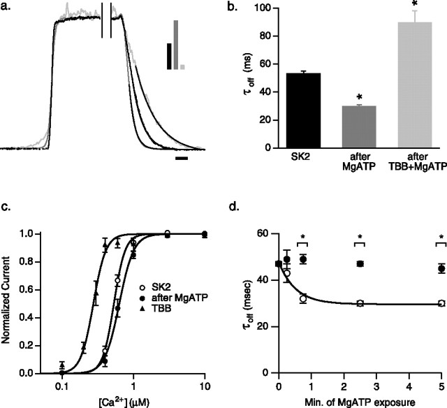
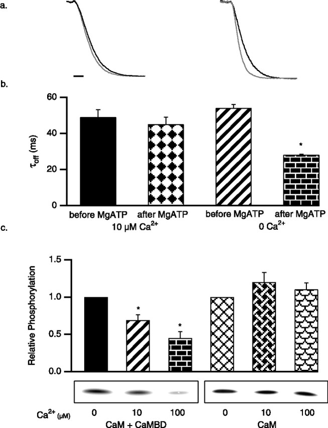
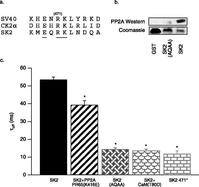
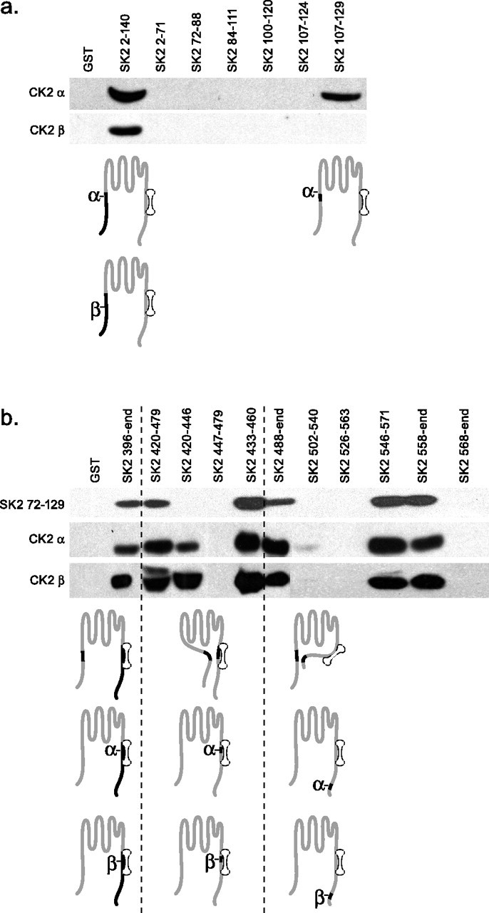
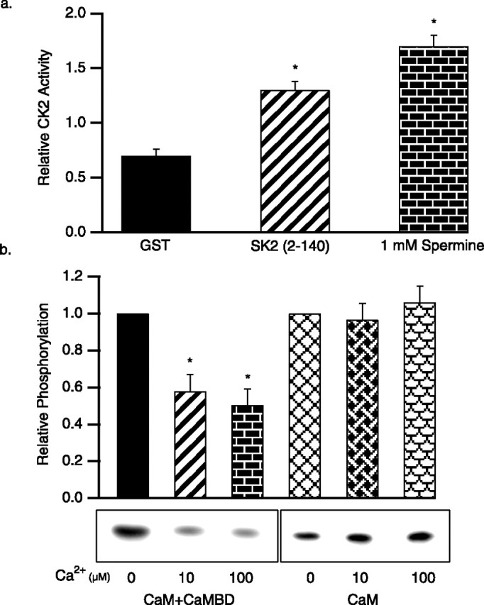
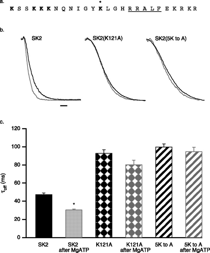
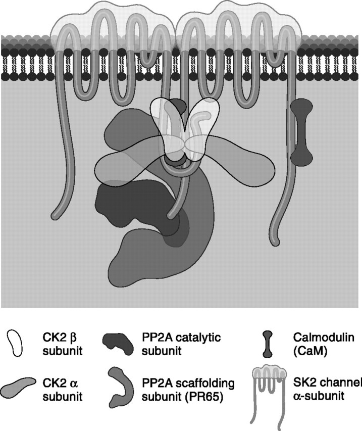
Small conductance Ca2+-activated K+ channels (SK channels) are complexes of four alpha pore-forming subunits each bound by calmodulin (CaM) that mediate Ca2+ gating. Proteomic analysis indicated that SK2 channels also bind protein kinase CK2 (CK2) and protein phosphatase 2A (PP2A). Coexpression of SK2 with the CaM phosphorylation surrogate CaM(T80D) suggested that the apparent Ca2+ sensitivity of SK2 channels is reduced by CK2 phosphorylation of SK2-bound CaM. By using 4,5,6,7-tetrabromo-2-azabenzimidazole, a CK2-specific inhibitor, we confirmed that SK2 channels coassemble with CK2. PP2A also binds to SK2 channels and counterbalances the effects of CK2, as shown by coexpression of a dominant-negative mutant PP2A as well as a mutant SK2 channel no longer able to bind PP2A. In vitro binding studies have revealed interactions between the N and C termini of the channel subunits as well as interactions among CK2 alpha and beta subunits, PP2A, and distinct domains of the channel. In the channel complex, lysine residue 121 within the N-terminal domain of the channel activates SK2-bound CK2, and phosphorylation of CaM is state dependent, occurring only when the channels are closed. The effects of CK2 and PP2A indicate that native SK2 channels are multiprotein complexes that contain constitutively associated CaM, both subunits of CK2, and at least two different subunits of PP2A. The results also show that the Ca2+ sensitivity of SK2 channels is regulated in a dynamic manner, directly through CK2 and PP2A, and indirectly by Ca2+ itself via the state dependence of CaM phosphorylation by CK2.


Figures

Figure 1.
Figure 1.
Activation of CK2 speeds SK2 channel deactivation and reduces the apparent Ca2+ sensitivity. a, Deactivation of normalized SK2 currents after rapid solution exchange from 10 μm to 0 Ca2+; black trace, control (τoff = 57.0 ms); dark gray trace, after a 2.5 min exposure to 5 mm MgATP in 0 Ca2+off = 29.3 ms); light gray trace, after 2.5 min exposure to 5 mm MgATP in 0 Ca2+ and TBB (τoff = 112.4 ms). Single exponential fits are added to traces. Bars in inset indicate relative current amplitudes. Calibration, 50 ms. b, Average SK2 deactivation time constants derived from single exponential fits for the indicated conditions. c, Normalized steady-state Ca2+ dose–response relationships for SK2 control (open circle), after application of MgATP (closed circle), and additionally in the presence of TBB (triangle). d, Time course of MgATP effect. Separate groups of patches were exposed to 5 mm MgATP for 0 min (n = 10), 0.25 min (n = 11), 0.75 min (n = 12), 2.5 min (n = 11), or 5 min (n = 8) to generate paired, before (black circles) and after (open circles) time points. The time constant of MgATP effect = 0.5 s.
Figure 2.
Figure 2.
CK2 activity for SK2-associated CaM is state dependent. SK2 current deactivation as in Figure 1, for control SK2 (black trace) and after application of 5 mm MgATP (gray trace; 45 s) in the presence of 10 μm (left) or 0 Ca2+ (right). Calibration, 50 ms. b, Average SK2 deactivation time constants derived from single exponential fits for the indicated conditions. c, In vitro CK2 phosphorylation reactions, stimulated by poly-l-lysine. Autoradiograph of phosphorylated CaM shows representative results for the conditions indicated, and the bar graph shows the average relative phosphorylation of CaM.
Figure 3.
Figure 3.
PP2A is coassembled with SK2 channels. a, Amino acid sequence comparison of PP2A binding sites on SV40 small T-antigen, CK2α, and SK2 (467–476). Mutated residues are underlined, and the position of the truncation, R471, is indicated. b, Top, Anti-His Western blot of proteins eluted from GST pull-down experiments with His-tagged PP2A PR65 as prey with the following baits: GST, SK2 396-end containing the mutation EQRK to AQAA, and SK2 396-end. Bottom, Coomassie blue-stained gel showing the input GST-fusion bait proteins. c, Average deactivation time constants derived from single exponential fits for control SK2, SK2 coexpressed with the dominant negative PP2A (PR65 K416E), the SK2 EQRK to AQAA triple mutant, SK2 coexpressed with CaM T80D, and the SK2 truncation at position 471.
Figure 4.
Figure 4.
Interactions between domains of SK2 and CK2. a, GST pull-down experiments used the indicated fragments of the N-terminal domain of SK2 as baits to detect interactions with N-terminally His-tagged CK2 α or CK2 β. Specifically bound prey proteins were detected by Western blotting with an anti-His antibody. b, Same as in a but with GST-fusion proteins of the C-terminal domain of SK2 as baits and additional testing for interactions with the membrane proximal portion of the SK2 N-terminal domain. Subunit schematics represent the interactions detected by the pull-down results.
Figure 5.
Figure 5.
The SK2 N-terminal domain activates CK2. a, Average CK2 phosphorylation of a prototypic CK2 substrate peptide stimulated by control GST, the SK2 N-terminal domain, or spermine. b, In vitro CK2 phosphorylation of CaM, either bound to the CaMBD (left) or alone (right), stimulated by the SK2 N-terminal domain. Autoradiograph of phosphorylated CaM shows representative results for the conditions indicated, and the bar graph shows the average relative phosphorylation of CaM.
Figure 6.
Figure 6.
K121 regulates CK2 coassembled with SK2 channels. a, Amino acid sequence (109–134) of the positively charged cluster in the SK2 N-terminal domain that includes the identified CK2 α binding site (RRALF; underlined). Asterisk indicates K121; the mutated lysine residues are in bold type. b, SK2 current deactivation for control SK2 (black traces) and after application of 5 mm MgATP (gray traces) for wild-type, SK2 K121A, and SK2 5KtoA channels. Calibration, 50 ms. c, Average deactivation time constants derived from single exponential fits for SK2, SK2 K121A, and SK2 5KtoA before and after 5 mm ATP application.
Figure 7.
Figure 7.
Model for coassembled SK2, CaM, CK2, and PP2A. The model, consistent with pull-down results, represents two of the four SK2 subunits and invokes an intersubunit interaction. CK2 and PP2A are drawn approximately to scale. In this configuration, the N-terminal domain of SK2 that includes K121 is positioned close to CK2 that binds to the adjacent sequence, RRALF.

Similar articles

Cited by

References

    1. Abel HJ, Lee JC, Callaway JC, Foehring RC. Relationships between intracellular calcium and afterhyperpolarizations in neocortical pyramidal neurons. J Neurophysiol. 2004;91:324–335. - PubMed
    1. Arrigoni G, Marin O, Pagano MA, Settimo L, Paolin B, Meggio F, Pinna LA. Phosphorylation of calmodulin fragments by protein kinase CK2: mechanistic aspects and structural consequences. Biochemistry. 2004;43:12788–12798. - PubMed
    1. Bildl W, Strassmaier T, Thurm H, Andersen J, Eble S, Oliver D, Knipper M, Mann M, Schulte U, Adelman JP, Fakler B. Protein kinase CK2 is coassembled with small conductance Ca(2+)-activated K+ channels and regulates channel gating. Neuron. 2004;43:847–858. - PubMed
    1. Bond CT, Herson PS, Strassmaier T, Hammond R, Stackman R, Maylie J, Adelman JP. Small conductance Ca2+-activated K+ channel knock-out mice reveal the identity of calcium-dependent afterhyperpolarization currents. J Neurosci. 2004;24:5301–5306. - PMC - PubMed
    1. Cai X, Liang CW, Muralidharan S, Kao JP, Tang CM, Thompson SM. Unique roles of SK and Kv4.2 potassium channels in dendritic integration. Neuron. 2004;44:351–364. - PubMed

Publication types

Substances

LinkOut - more resources