

Cannabinoid CB1 and CB2 receptors and fatty acid amide hydrolase are specific markers of plaque cell subtypes in human multiple sclerosis
- PMID: 17329437
- PMCID: PMC6673484
- DOI: 10.1523/JNEUROSCI.4814-06.2007
Cannabinoid CB1 and CB2 receptors and fatty acid amide hydrolase are specific markers of plaque cell subtypes in human multiple sclerosis
Abstract
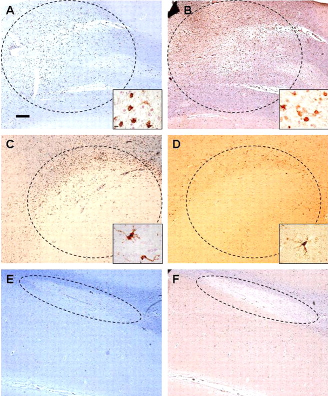
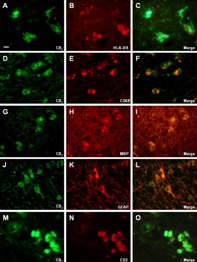
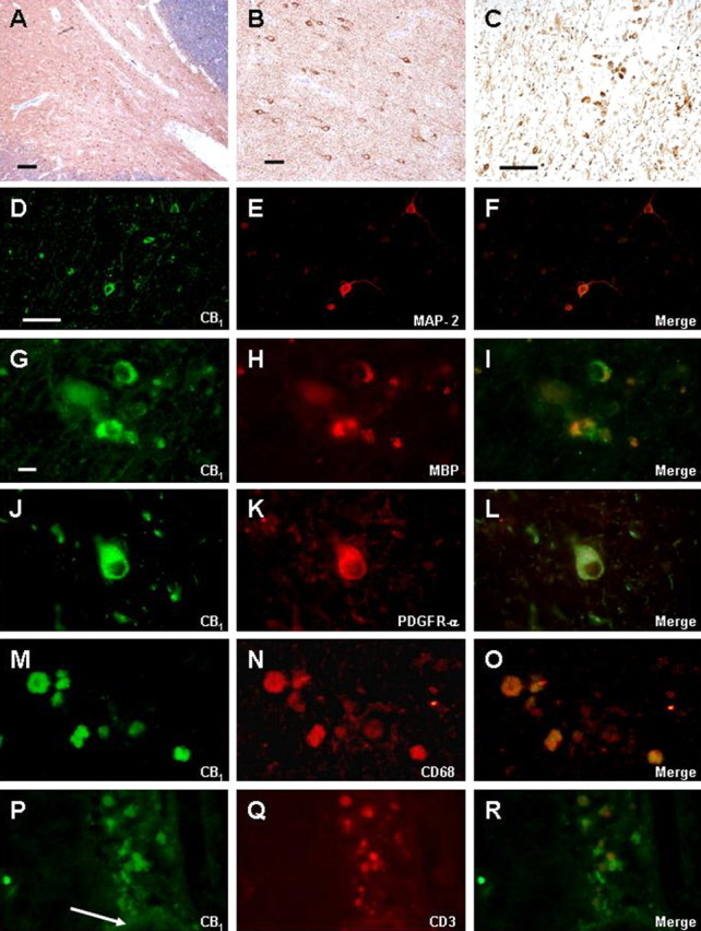
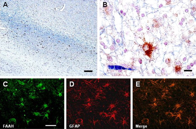
Increasing evidence supports the idea of a beneficial effect of cannabinoid compounds for the treatment of multiple sclerosis (MS). However, most experimental data come from animal models of MS. We investigated the status of cannabinoid CB1 and CB2 receptors and fatty acid amide hydrolase (FAAH) enzyme in brain tissue samples obtained from MS patients. Areas of demyelination were identified and classified as active, chronic, and inactive plaques. CB1 and CB2 receptors and FAAH densities and cellular sites of expression were examined using immunohistochemistry and immunofluorescence. In MS samples, cannabinoid CB1 receptors were expressed by cortical neurons, oligodendrocytes, and also oligodendrocyte precursor cells, demonstrated using double immunofluorescence with antibodies against the CB1 receptor with antibodies against type 2 microtubule-associated protein, myelin basic protein, and the platelet-derived growth factor receptor-alpha, respectively. CB1 receptors were also present in macrophages and infiltrated T-lymphocytes. Conversely, CB2 receptors were present in T-lymphocytes, astrocytes, and perivascular and reactive microglia (major histocompatibility complex class-II positive) in MS plaques. Specifically, CB2-positive microglial cells were evenly distributed within active plaques but were located in the periphery of chronic active plaques. FAAH expression was restricted to neurons and hypertrophic astrocytes. As seen for other neuroinflammatory conditions, selective glial expression of cannabinoid CB1 and CB2 receptors and FAAH enzyme is induced in MS, thus supporting a role for the endocannabinoid system in the pathogenesis and/or evolution of this disease.
Figures
References
-
- Baker D, Pryce G, Croxford JL, Brown P, Pertwee RG, Huffman JW, Layward L. Cannabinoids control spasticity and tremor in a multiple sclerosis model. Nature. 2000;404:84–87. - PubMed
-
- Bisogno T, Ligresti A, Di Marzo V. The endocannabinoid signalling system: biochemical aspects. Pharmacol Biochem Behav. 2005;81:224–238. - PubMed
Publication types
MeSH terms
Substances
LinkOut - more resources
Full Text Sources
Other Literature Sources
Medical
Research Materials