. 2007 Feb 28;2(2):e252.
doi: 10.1371/journal.pone.0000252.

The Pafah1b complex interacts with the reelin receptor VLDLR

Affiliations

The Pafah1b complex interacts with the reelin receptor VLDLR

Guangcheng Zhang et al. PLoS One. .

Abstract

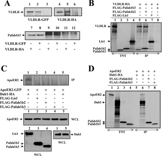
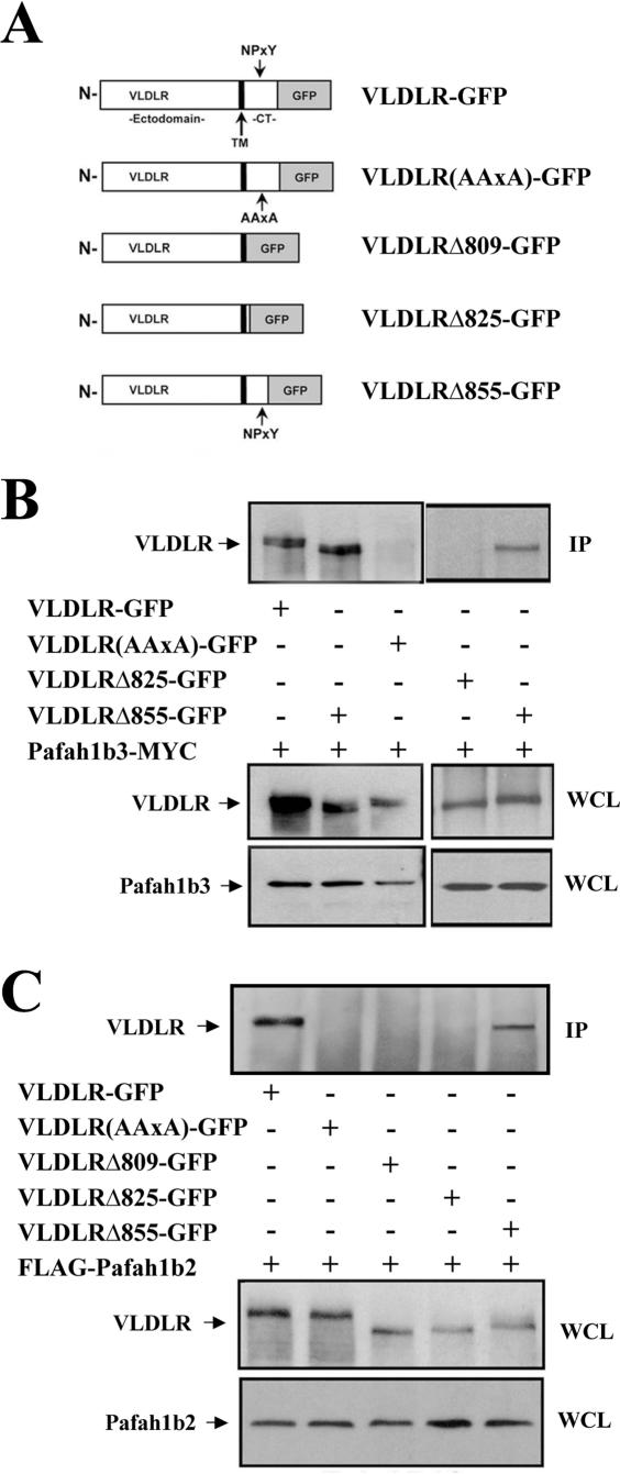
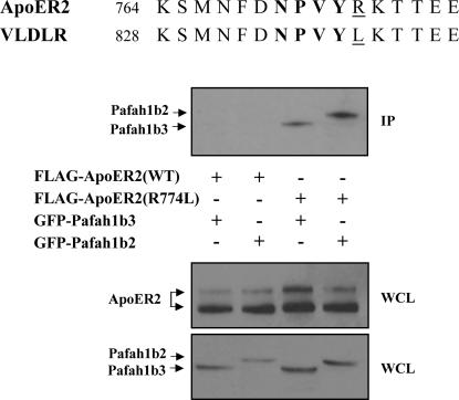
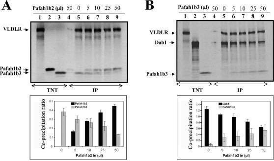
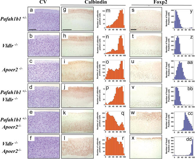
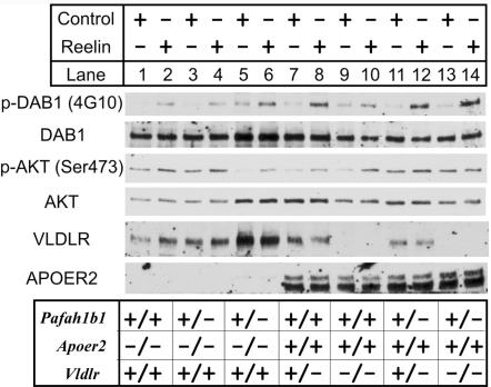
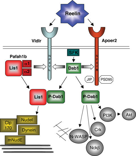
Reelin is an extracellular protein that directs the organization of cortical structures of the brain through the activation of two receptors, the very low-density lipoprotein receptor (VLDLR) and the apolipoprotein E receptor 2 (ApoER2), and the phosphorylation of Disabled-1 (Dab1). Lis1, the product of the Pafah1b1 gene, is a component of the brain platelet-activating factor acetylhydrolase 1b (Pafah1b) complex, and binds to phosphorylated Dab1 in response to Reelin. Here we investigated the involvement of the whole Pafah1b complex in Reelin signaling and cortical layer formation and found that catalytic subunits of the Pafah1b complex, Pafah1b2 and Pafah1b3, specifically bind to the NPxYL sequence of VLDLR, but not to ApoER2. Compound Pafah1b1(+/-);Apoer2(-/-) mutant mice exhibit a reeler-like phenotype in the forebrain consisting of the inversion of cortical layers and hippocampal disorganization, whereas double Pafah1b1(+/-);Vldlr(-/-) mutants do not. These results suggest that a cross-talk between the Pafah1b complex and Reelin occurs downstream of the VLDLR receptor.


Conflict of interest statement

Competing Interests: The authors have declared that no competing interests exist.

Figures

Figure 1
Figure 1
Pafah1b2 and Pafah1b3 bind VLDLR but not ApoER2. (A) Pafah1b3 binds VLDLR in transfected cells. Pafah1b3-MYC was co-expressed in 293T cells with GFP- (lanes 1–3) or HA-tagged VLDLR (lanes 4–6). VLDLR was co-precipitated with MYC antibodies directed against Pafah1b3 (lanes 2 and 5), but not with control antibodies (lanes 3 and 6). Conversely, Pafah1b3 was co-precipitated with HA (lane 9) or GFP (lane 10) directed against VLDLR, but not with control antibodies (lanes 11–12). Lanes 1 and 4 show VLDLR, and lanes 7–8 show Pafah1b3 in the WCL. Blots were probed with the GFP (lanes 1–3), HA (lanes 4–6) or Myc antibodies (lanes 7–12). (B) Pafah1b2 and Pafah1b3 bind VLDLR in a cell-free-system (TNT). In vitro translated proteins were radiolabeled with 35S and analyzed by SDS-PAGE (lanes 1–4). Individual Pafah1b subunits were combined with equal amounts of VLDLR and immunoprecipitated with FLAG (lanes 5–7) or a negative control antibody (lanes 8). Proteins were detected by autoradiography. Note that VLDLR co-precipitated with Pafah1b α subunits, but not with Lis1. (C) Pafah1b2 and Pafah1b3 do not bind ApoER2 in transfected cells. ApoER2-GFP was co-expressed in 293T cells with the indicated proteins, and co-immunoprecipitated with HA antibodies directed against Dab1 (lane 1). FLAG antibodies directed against the Pafah1b subunits (lanes 2–4) or a control antibody (lane 5) did not co-precipitate the receptor. Blots were probed with antibodies against GFP to detect co-precipitated ApoER2 (upper panel) or total ApoER2 expression in the corresponding WCLs (middle panel). FLAG or HA antibodies were used to detect Pafah1b subunits (lower left panel) or Dab1 (lower right panel) in the corresponding WCLs. ApoER2 co-precipitated exclusively with Dab1. (D) Pafah1b2 and Pafah1b3 do not bind ApoER2 in a cell-free-system (TNT). In vitro translated proteins were radiolabeled with 35S and analyzed by SDS-PAGE (lanes 1–4). Equal amounts of ApoER2 were combined with the indicated proteins and immunoprecipitated with a control antibody (lane 5), or with antibodies against Dab1 (lane 6), Pafah1b2 (lane 7) or Pafah1b3 (lane 8). Proteins were detected by autoradiography. ApoER2 again co-precipitated only with Dab1. IP, immunoprecipitate; WCL, whole cell lysate.
Figure 2
Figure 2
Pafah1b2 and Pafah1b3 interact specifically with the NPxY motif of VLDLR. (A) Diagram of the VLDLR expression constructs used in this study. TM = transmembrane region. CT = cytoplasmic tail. (B) The NPxY motif is required for VLDLR binding to Pafah1b3. Pafah1b3-MYC was co-expressed in 293T cells with the indicated GFP-tagged VLDLR constructs. Proteins were immunoprecipitated with Myc antibodies and the blot was probed with GFP antibodies to detect VLDLR receptors (upper panel). Only full-length VLDLR and the NPxY-containingVLDLR?855 co-precipitated with Pafah1b3. WCLs were probed with GFP (middle panels) or Myc (lower panels) antibodies to ensure that similar amounts of VLDLR or Pafah1b3 proteins were present in each sample. (C) The NPxY motif is required for VLDLR binding to Pafah1b2. FLAG-Pafah1b2 was co-expressed in 293T cells with GFP-tagged VLDLR constructs and immunoprecipitated with FLAG antibodies. Blots were probed with GFP antibodies to detect VLDLR proteins in the IP (upper panel) or WCLs (middle panel), or FLAG antibodies to detect Pafah1b2 in the WCLs (lower panel). Only full-length VLDLR and VLDLR?855 co-precipitated with Pafah1b2. IP, immunoprecipitate; WCL, whole cell lysate.
Figure 3
Figure 3
Substitution of Arg774 with a Leu in ApoER2 rescues binding to Pafah1b α subunits. (A) Amino acid sequence of VLDLR and ApoER2 near the NPxY motif in the cytoplasmic tail of each receptor. Unique amino acids R774 in ApoER2 and L838 in VLDLR are underlined. (B) A Leu residue following the NPxY motif is required for Pafah1b α subunit binding to lipoprotein receptors. FLAG-tagged intact ApoER2(WT) or a mutant receptor in which Arg774 was substituted by a Leu ApoER2(R774L) were co-expressed in COS7 cells with GFP-tagged Pafah1b α subunits. Proteins were immunoprecipitated with FLAG antibodies (upper panel) and immunoblotted with antibodies against GFP to detect co-precipitated α subunits. FLAG (middle panel) or GFP (lower panel) antibodies were used to detect ApoER2 receptors or the Pafah1b α subunits in the WCL. Pafah1b2 and Pafah1b2 co-precipitated with ApoER2(R774L), but not with intact ApoER2(WT). IP, immunoprecipitate; WCL, whole cell lysate.
Figure 4
Figure 4
Pafah1b α subunits compete with each other and with Dab1 for binding to VLDLR. (A) Pafah1b α subunits compete for VLDLR binding. Proteins were expressed in vitro using a cell-free-system (TNT) and radiolabeled with 35S (lanes 1–3). Equal amounts (50 μl) of VLDLR and Pafah1b3 were incubated with increasing amounts of Pafah1b2, as indicated. Proteins were immunoprecipitated with polyclonal antibodies against VLDLR (lanes 5–9) or a control antibody (lane 4) and detected by autoradiography. The plot represents the mean ratio of co-precipitated Pafah1b3 and Pafah1b2 normalized to the amount of precipitated VLDLR in each sample. Increasing amounts of Pafah1b2 reduced the amount of co-precipitated Pafah1b3. (B) Proteins were expressed in vitro (TNT) and radiolabeled with 35S (lanes 1–3). Equal amounts (50 μl) of VLDLR and Dab1 were incubated with increasing amounts of Pafah1b3, as indicated. Proteins were immunoprecipitated with polyclonal antibodies against VLDLR (lanes 5–9) or a control antibody (lane 4) and detected by autoradiography. The plot represents the mean ratio of co-precipitated Pafah1b3 and Dab1 normalized to the amount of precipitated VLDLR in each sample. Increasing amounts of Pafah1b3 reduced the amount of co-precipitated Dab1. Bars represent the standard error of the mean from triplicate experiments. IP, immunoprecipitate.
Figure 5
Figure 5
reeler-like disruption of cortical layers in Pafah1b1 +/− mice lacking ApoER2 but not VLDLR. Sagittal sections of the neocortex were obtained from adult mice of the indicated genotype. Adjacent sections were stained with cresyl violet (CV) (a-f) or subjected to immunohistochemistry with antibodies against calbindin to label cells in layers II–III (g-l), or Foxp2 to label cells in layer VI (s-x). Histograms represent the radial distribution of cells positive for calbindin (m-r) or Foxp2 (y-dd) from the bottom of the cortical plate (set as 0) to the pial surface (set as 100). Pafah1b1+/− and Vldlr −/− single or double mutants presented no obvious cortical layering defects. Apoer2 −/− single mutants exhibited some laminar dispersion of upper layer neurons. In contrast, Pafah1b1 +/−;Apoer2 −/− double mutants displayed a marked inversions of upper and lower layers (reeler-like phenotype) comparable to that seen in Apoer2 −/−;Vldlr −/− mice. Scale bars = 500 µm.
Figure 6
Figure 6
reeler-like disruption of hippocampal layers in Pafah1b1 +/−;Apoer2−/− double mutants. Comparable sagittal sections of the hippocampus obtained from adult mice of the indicated genotype were stained with cresyl violet. The hippocampus proper (HP) and the dentate gyrus (DG) of Apoer2+/− mice are normal, whereas a splitting of the pyramidal layers is evident in Pafah1b1+/− and in Apoer2−/− mice. Severe dyslamination of cellular layers is seen in double Pafah1b1+/−;Apoer2−/− (reeler-like). Scale bar = 500 µm.
Figure 7
Figure 7
Reelin induces Dab1 and Akt phosphorylation in Pafah1b1 +/−;Apoer2−/− double mutant neurons. Cortical neurons were cultured from mutant mice of the indicated genotype, and incubated with either control or Reelin-containing medium for 20 min. Lysates were analyzed by Western blot using the 4G10 antibody to detect Dab1 phosphorylation on tyrosine residues and a phospho-Akt antibody to detect Akt phosphorylation on serine 473. Blots were reprobed with antibodies against total Dab1 and Akt to ensure that similar amount of proteins were present in each sample, and with antibodies against VLDLR and ApoER2 to confirm the genotype of the mutants.
Figure 8
Figure 8
Integrated model of Reelin and Lis1 signaling. Reelin binds to VLDLR and ApoER2 and causes src-family kinase (SFK) activation and Dab1 phosphorylation. Dab1 binds to the NPxY motif of both, VLDLR and ApoER2. Upon Reelin stimulation, phosphoDab1 (P-Dab1) interacts with Lis1 and with other signal transduction molecules (grey circles). Lis1 also binds the catalytic subunits of the Pafah1b complex (α1 and α2) as well as components of the cytoplasmic dynein complex (yellow square). α1 and α2 also bind VLDLR at the NPxYL motif and compete with Dab1 for receptor occupancy. These subunits do not recognize the NPxYR motif of ApoER2. A unique domain of ApoER2 enables unique interactions with synaptic and trafficking proteins (white octagons). The binding of catalytic Pafah1b subunits to VLDLR may displace P-Dab1 and promote its interaction with Lis1. Signaling molecules downstream of Lis1 and Dab1 affect cytoskeleton dynamics by acting on microtubules (thick lines) or actin filaments (thin lines), thereby controlling neuronal migration and layer formation.

Similar articles

Cited by

References

    1. Reiner O, Carrozzo R, Shen Y, Wehnert M, Faustinella F, et al. Isolation of a Miller-Dieker lissencephaly gene containing G protein beta-subunit-like repeats. Nature. 1993;364:717–721. - PubMed
    1. Hong SE, Shugart YY, Huang DT, Al Shahwan S, Grant PE, et al. Autosomal recessive lissencephaly with cerebellar hypoplasia is associated with human RELN mutations. Nature Genet. 2000;26:93–96. - PubMed
    1. Rice DS, Curran T. Role of the Reelin signaling pathway in central nervous system development. Ann Rev Neurosci. 2001;24:1005–1039. - PubMed
    1. Tissir F, Goffinet AM. Reelin and brain development. Nat Rev Neurosci. 2003;4:496–505. - PubMed
    1. D'Arcangelo G. The reeler mouse: anatomy of a mutant. In: Dhossche DM, editor. International Review of Neurobiology. San Diego, CA: Elsevier Inc; 2005. pp. 383–417. - PubMed

Publication types

MeSH terms

Substances