. 2007 Apr;37(4):935-45.
doi: 10.1002/eji.200636542.

Arginase activity mediates reversible T cell hyporesponsiveness in human pregnancy

Affiliations

Arginase activity mediates reversible T cell hyporesponsiveness in human pregnancy

Pascale Kropf et al. Eur J Immunol. 2007 Apr.

Abstract

Complex regulation of T cell functions during pregnancy is required to ensure materno-fetal tolerance. Here we reveal a novel pathway for the temporary suppression of maternal T cell responses in uncomplicated human pregnancies. Our results show that arginase activity is significantly increased in the peripheral blood of pregnant women and remarkably high arginase activities are expressed in term placentae. High enzymatic activity results in high turnover of its substrate L-arginine and concomitant reduction of this amino acid in the microenvironment. Amino acid deprivation is emerging as a regulatory pathway of lymphocyte responses and we assessed the consequences of this enhanced arginase activity on T cell responses. Arginase-mediated L-arginine depletion induces down-regulation of CD3 zeta, the main signalling chain of the TCR, and functional T cell hyporesponsiveness. Importantly, this arginase-mediated T cell suppression was reversible, as inhibition of arginase activity or addition of exogenous L-arginine restored CD3 zeta chain expression and T cell proliferation. Thus, L-arginine metabolism constitutes a novel physiological mechanism contributing to the temporary suppression of the maternal immune response during human pregnancy.


Figures

Figure 1
Figure 1
Arginase is enhanced in pregnancy. (a) PlaC (n=9) and maternal PBMC (n=9) were isolated and arginase activity was measured in cell lysates (left panel). Arginase activities in maternal PBMC were compared to those found in PBMC from age-matched non-pregnant controls (right panel). Each symbol represents arginase activity of the cells from one individual and the horizontal bar represents the median. Statistical differences were determined using the Mann-Whitney test: left panel *p<0.0001, right panel *p=0.008. (b) Arginase protein expression was measured in cell lysates (30 μg/sample) from paired individuals and from controls by Western blot as previously described [24, 53]. hAI: recombinant human arginase I. (c) Arginase activity was measured in maternal (n=9; open circles) and non-pregnant control sera (n=9; open squares). Each symbol represents the urea content in the serum of one individual and the horizontal bar represents the median. Statistical differences were determined using the Mann-Whitney test: *p=0.006.
Figure 2
Figure 2
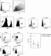
Identification of arginase expressing cells. (a) Identification of arginase I-expressing cells. The region containing cells expressing arginase I (A) was identified by back-gating ungated arginase I-positive cells (U) on the side scatter/forward scatter plot. (b) Frequency of arginase I-expressing cells. In region A, two populations of CD14+ cells were identified: CD14low (C) and CD14high (D)(CD14: B, dotted line: unstained cells). The frequency of arginase-expressing cells was determined in region B (CD14), C (CD14low) and D (CD14high). The markers of histograms and dot-plot were set according to the fluorescence of unstained cells and the value in parenthesis represents the percentage of cells positive for the isotype control. Data show the results of one representative experiment out of four independent experiments. (c) Neutrophils and alternatively activated macrophages express arginase I. Arginase I was expressed in 77.8% of CD15+CD14 cells (upper left), 90.8% of CD15+CD14low cells (upper right) and 25.3% of CD14lowCD206+ cells (lower left); the isotype control for arginase I in CD14lowCD206+ is shown in the lower right (1.7%). Data show the results of one representative experiment out of four independent experiments. (d) Arginase activity in peripheral neutrophils is increased during pregnancy. Neutrophils were isolated by double-density gradient centrifugation of Histopaque®-1119 and 1077 and arginase activity was measured as described in Material and methods. Each symbol represents arginase activity of the cells from one individual and the horizontal bar represents the median. Statistical differences were determined using the Mann-Whitney test: *p=0.02.
Figure 3
Figure 3
PlaC have the capacity to down-regulate CD3ζ chain and suppress proliferation of Jurkat cells in an arginase-/l-arginine-dependent manner. PlaC or maternal PBMC (2 × 10⁵ cells) were incubated with 1 × 10⁵ Jurkat cells in a final volume of 200 μL in the presence or in the absence of 10 μL 5.6 μM nor-NOHA or 2 μL 100 mM l-arginine. After 2 days, MFI of CD3ζ in TCR+ cells (a) or frequency of TCR+ cells incorporating BrdU (b) were determined. Jurkat cells were identified by increased side /forward scatter. A 100% response represents the value of the MFI of CD3ζ (a: MFI =4.6) or the percentage of proliferating Jurkat cells (b: 36.3%) in the cultures containing Jurkat cells alone. Data show the average ± SEM of three independent experiments.
Figure 4
Figure 4
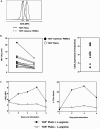
Down-regulation of CD3ζ chain in T cells from placental biopsies. (a) MFI of CD3ζ chain expression was determined in maternal TCR+ PBMC (dotted line) and in TCR+ PlaC (solid line). The data show the result of 1 representative experiment out of 13 independent experiments. (b) Each symbol represents the MFI of CD3ζ in peripheral TCR+ cells (closed circles) and paired TCR+ cells from placental biopsies (open circles) from one individual (left panel, n=13). Percent reduction in CD3ζ MFI in TCR+ cells from PlaC as compared to TCR+ cells from maternal PBMC (right panel). (c) Down-regulation of CD3ζ chain and proliferation of TCR+ PlaC is reversible. PlaC (4 × 10⁵) were stimulated with plate-bound anti-CD3 and anti-CD28 mAb in a final volume of 200 μL in complete DMEM in the absence of l-arginine, or in complete DMEM containing 0.1, 0.4, 1 or 2 mM l-arginine. At the indicated times post stimulation, cells were harvested and the MFI of CD3ζ in TCR+ cells (left panel) or the frequency of TCR+ cells incorporating BrdU (right panel) were determined. Similar results were obtained with all the concentrations of l-arginine tested and the data show the results obtained with 1 mM. Data show the results of one representative experiment out of four independent experiments.
Figure 5
Figure 5
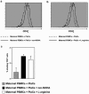
Down-regulation of CD3ζ chain and proliferation by PlaC is restored by inhibition of arginase or by addition of l-arginine. PlaC (2 × 10⁵) were stimulated with plate-bound anti-CD3 mAb and anti-CD28 mAb in a final volume of 100 μl DMEM (0.1 mM l-arginine). Then 10 μL nor-NOHA (5.6 μM) was added to some of the wells, and 30 min later maternal PBMC (1 × 10⁵, labeled with CFSE to differentiate maternal TCR+ from placental TCR+) were added in a final volume of 100 μL. l-Arginine (2 μL 100 mM) was added to some of the wells. (a, b) After 2 days, the cells were harvested and the expression of CD3ζ was determined in maternal TCR+ cells (CFSE+ cells). Data show the results of one representative experiment out of three independent experiments. (c) The frequency of cells having undergone at least one cell division was determined in maternal TCR+ CFSE+ cells. Data show the results ± SD of one representative experiment out of three independent experiments.

Similar articles

Cited by

References

    1. Medawar PB. Some immunological and endocrinological problems raised by the evolultion of viviparity in vertebrates. Symp. Soc. Exp. Biol. 1953;7:320–338.
    1. Hunt JS, Petroff MG, McIntire RH, Ober C. HLA-G and immune tolerance in pregnancy. FASEB J. 2005;19:681–693. - PubMed
    1. Petroff MG. Immune interactions at the maternal-fetal interface. J. Reprod. Immunol. 2005;68:1–13. - PubMed
    1. Tafuri A, Alferink J, Moller P, Hammerling GJ, Arnold B. T cell awareness of paternal alloantigens during pregnancy. Science. 1995;270:630–633. - PubMed
    1. Guleria I, Khosroshahi A, Ansari MJ, Habicht A, Azuma M, Yagita H, Noelle RJ, et al. A critical role for the programmed death ligand 1 in fetomaternal tolerance. J. Exp. Med. 2005;202:231–237. - PMC - PubMed

Publication types

LinkOut - more resources