. 2007 Jul 1;405(1):157-64.
doi: 10.1042/BJ20070025.

Is the mammalian serine palmitoyltransferase a high-molecular-mass complex?

Affiliations

Is the mammalian serine palmitoyltransferase a high-molecular-mass complex?

Thorsten Hornemann et al. Biochem J. .

Abstract

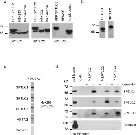
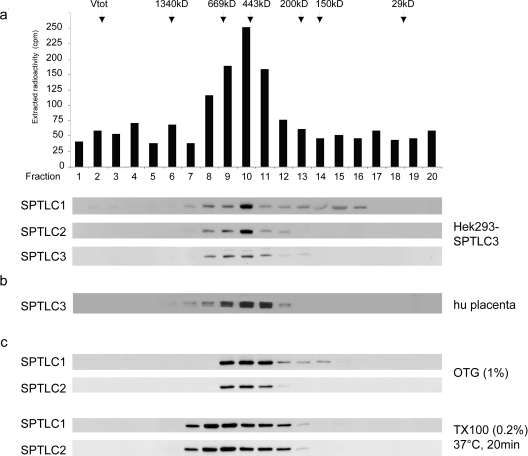
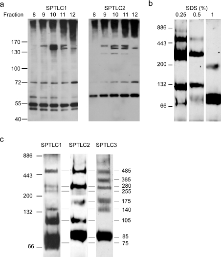
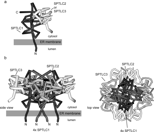
SPT (serine palmitoyltransferase) catalyses the rate-limiting step for the de novo synthesis of sphingolipids. Mammalian SPT is believed to be a heterodimer composed of two subunits, SPTLC1 and SPTLC2. We reported previously the identification of a new third SPT subunit, SPTLC3. In the present study, we have investigated the structure of the SPT complex in more detail. Pull-down assays with antibodies against SPTLC3 concomitantly co-precipitated SPTLC1 and SPTLC2 in human placenta extracts and SPTLC3 overexpressing human embryonic kidney-293 cells. By size exclusion chromatography, we determined the molecular mass of the functional SPT complex to be approx. 480 kDa. By Blue-native-PAGE experiments we demonstrated that all three SPT subunits (SPTLC1-3) are co-localized within a single SPT complex. On the basis of these results we conclude that the functional SPT is not a dimer, but a higher organized complex, composed of three distinct subunits (SPTLC1, SPTLC2 and SPTLC3) with a molecular mass of 480 kDa. The stoichiometry of SPTLC2 and SPTLC3 in this complex seems not to be fixed and is probably changed dynamically in dependence of the tissue specific SPTLC2 and SPTLC3 expression levels. Based on our own and earlier published data we propose a model of an octameric SPT structure. The observed dynamic composition of the SPT complex could provide a cellular mechanism to adjust SPT activity to tissue specific requirements in sphingolipid synthesis.


Figures

Figure 1
Figure 1. Expression and immunoprecipitation of the SPT complex
(a) Expression of the SPT subunits in HEK-293 cells and human placenta tissue. Total cell extract (50 μg/lane) was separated by SDS/PAGE (10% gels), blotted on to PVDF and probed for the SPT subunits using subunit specific antibodies against SPTLC1, SPTLC2 and SPTLC3. The first lanes show transfected HEK-293 cells overexpressing the respective subunit. All subunits were specifically detected by their respective antibodies. The heterologously expressed subunits migrate to a slightly larger size due to the mass addition of the added V5-tag. The polyclonal antibodies detect both the transfected (upper band) and the endogenous (lower band) subunits. (b) Extracts from SPTLC1 and SPTLC2 overexpressing HEK-293 cells were incubated with the anti-V5 antibody to specifically precipitate the V5-tagged subunits. The precipitate was then separated by SDS/PAGE, blotted on to PVDF and probed with the polyclonal antibody against the respective subunit. In both cases we could see the precipitation of not only the V5-tagged subunit, but also the endogenously encoded untagged subunit. This indicates that the tagged and untagged subunits are located within the same protein complex. This contradicts the concept of a heterodimeric SPT structure. (c) The V5-tagged SPTLC3 subunit from overexpressing HEK-293 cells was captured by an anti-V5 antibody and the precipitate probed for the co-precipitation of SPTLC1 and SPTLC2. Surprisingly, all three SPT subunits could be detected in the precipitate. Calnexin, as an abundant ER protein, was not found in the precipitate, showing the specificity of the assay. (d) In order to confirm these results we repeated this experiment with human placenta tissue. Placenta tissue was shown to natively express high levels of SPTLC3. The positive control (left-hand lanes) shows that all proteins, including calnexin, are expressed in human placenta. The extract was incubated with polyclonal antibodies against SPTLC1, SPTLC2 and SPTLC3, and precipitated by the addition of Protein G–agarose. In the negative control we added an excess (5 μg/ml) of target-specific blocking peptides (immunization peptides) as a competitor. After separation by SDS/PAGE (10% gels) and blotting on to PVDF, the precipitate was cross-probed for the co-precipitation of the other two subunits. For all of the samples we observed the co-precipitation of the other two subunits, which had not been targeted directly. No signal was seen in the presence of the blocking peptides or when the beads were used alone. To further exclude that the observed complex is possibly caused by an unspecific aggregation of ER proteins, we again probed for calnexin, which was not detected. This suggests that all three SPT subunits are located within a single complex. Molecular mass markers given in kDa are shown to the left-hand side of the Figures in (a), (b) and (d). Ab, antibody; hu placenta, human placenta.
Figure 2
Figure 2. Size exclusion chromatography of the functional SPT complex
(a) Size-exclusion chromatography of a 0.2% Triton X-100 cell extract obtained from SPTLC3 overexpressing HEK-293 cells. The fractions were assayed for SPT activity and probed for the presence of SPTLC1, SPTLC2 and SPTLC3 on a Western blot. The arrows mark the elution fractions of the marker proteins (masses shown in kDa). The maximal SPT activity eluted in fraction 10, which corresponds to a mass of 460–480 kDa. In all fractions that displayed SPT activity we observed the co-elution of the three SPT subunits. The intensity of the signal correlated with the measured SPT activity, and showed a maximum intensity in fraction 10. This result suggests that the mammalian SPT enzyme has a functional size of 460–480 kDa. (b) Fractionation of a 0.2% Triton X-100 extract from human placenta tissue. The experimental conditions are the same as (a). The elution profile for SPTLC1 and SPTLC2 was identical to (a) (results not shown). (c) To exclude that the observed SPT complex is due to Triton X-100 insoluble complexes, wild-type HEK-293 cells were lysed in either 1% (w/v) OTG or 0.2% Triton X-100 (TX100) with a subsequent incubation at 37°C for 20 min. Both conditions are reported to resolve the formation of detergent insoluble complexes [11]. As before, we observed a parallel elution of the subunits at a molecular mass of approx 480 kDa.
Figure 3
Figure 3. Crosslinking and electrophoretic analysis of the SPT complex
(a) HEK-293 cells were treated with limiting amounts of formaldehyde for 1 h at 4°C, lysed in 0.5% SDS and fractionated over a size-exclusion column. Fractions 8–12 were separated by SDS/PAGE (7% gels) and immunostained for SPTLC1 and SPTLC2. We observed the formation of a stably cross-linked complex of approx. 160 kDa, which was positive for SPTLC1 and SPTLC2. Due to the limited spatial resolution and the appearance of a smeary signal on top of the gel we could not detect higher molecular mass cross-linked complexes. (b) HEK-293 cells were lysed in BN buffer with increasing amounts of SDS (0.25–1%). The extract was separated by BN-PAGE, blotted on to PVDF and probed for the SPTLC2 subunit. The pattern shows the gradual breakdown of the full SPT complex into several smaller sub-complexes. The intensity of the breakdown correlates with the SDS concentration used in the lysis buffer. (c) BN-PAGE of human placenta extract. The homogenized tissue was lysed in BN-PAGE buffer containing 0.5% SDS and separated by BN-PAGE (7–15% gels) as described above. The gel was blotted and stained separately for SPTLC1, SPTLC2 and SPTLC3. As before, we observed a specific breakdown of the SPT complex into various sub-complexes. Most of those complexes showed a positive signal for two or all three SPT subunits. The masses of these complexes was deduced from the molecular mass of defined marker proteins. The largest complex was detected with a molecular mass of approx 480 kDa, which is in very good agreement with the deduced mass of 480 kDa from the size exclusion chromatography shown in Figure 2. Molecular mass markers are shown to the left-hand side of the Figures in kDa.
Figure 4
Figure 4. Theoretical model of the SPT complex structure
The model is based on the following assumptions. First, that the N-terminal end of SPTLC1 is bound to the ER membrane; secondly, that SPTLC2 and SPTLC3 are not interacting directly with the ER membrane; thirdly, that the active site of SPT is formed at the interface between two monomers; and, finally, that SPTLC2 can be dynamically replaced by SPTLC3 within the complex. (a) The SPTLC2 or SPTLC3 monomers form a dimeric base structure with SPTLC1, which is then assembled to its final octameric state. (b) Putative model of the octameric SPT complex. In the conformation shown, two SPTLC1–SPTLC2 and two SPTLC1–SPTLC3 dimers are assembled together to form an octameric circular structure. The stoichiometry of SPTLC2 and SPTLC3 within the complex depends on their individual cellular expression levels. The final complex contains four active sites, which are located at the monomer–monomer interface. The N-termini of the SPTLC1 subunits are bound to the ER membrane in a manner that the catalytic head group protrudes towards the cytosolic compartment of the cell.

References

    1. Hannun Y. A., Luberto C., Argraves K. M. Enzymes of sphingolipid metabolism: from modular to integrative signaling. Biochemistry. 2001;40:4893–4903. - PubMed
    1. Hannun Y. A., Obeid L. M. The ceramide-centric universe of lipid-mediated cell regulation: stress encounters of the lipid kind. J. Biol. Chem. 2002;277:25847–25850. - PubMed
    1. Merrill A. H., Jr, Nixon D. W., Williams R. D. Activities of serine palmitoyltransferase (3-ketosphinganine synthase) in microsomes from different rat tissues. J. Lipid Res. 1985;26:617–622. - PubMed
    1. Hojjati M. R., Li Z., Jiang X. C. Serine palmitoyl-CoA transferase (SPT) deficiency and sphingolipid levels in mice. Biochim. Biophys. Acta. 2005;1737:44–51. - PubMed
    1. Hanada K. Serine palmitoyltransferase, a key enzyme of sphingolipid metabolism. Biochim. Biophys. Acta. 2003;1632:16–30. - PubMed

Publication types