. 2007 Mar;210(3):318-27.
doi: 10.1111/j.1469-7580.2007.00699.x.

Long-term morphology of a healing bone-tendon interface: a histological observation in the sheep model

Affiliations

Long-term morphology of a healing bone-tendon interface: a histological observation in the sheep model

R Newsham-West et al. J Anat. 2007 Mar.

Abstract

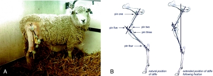
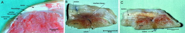
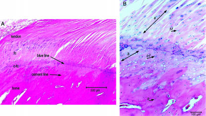
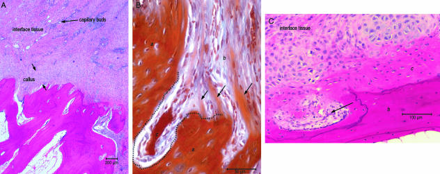
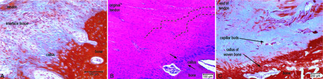
The purpose of this study was to examine and describe the sequence of events involved in long-term biological reconstruction of a tendon-bone interface following surgical reattachment. Patellar tendon re-attachment in the adult sheep was used to investigate and describe the biological components involved in healing and repair of a tendon enthesis. Light microscopy was used to describe the healing morphology at time intervals of 8, 12, 26, 52 and 104 weeks. By 8 weeks a collagen continuum was observed between the tendon and bone. Over time this fibrous bridge became anchored into the original tissues (tendon and bone), with the resultant enthesis resembling more a fibrous rather than the original fibrocartilagenous enthesis. The associated collagen fibrils between the two tissues gradually changed in morphology over time to reflect the fibres seen in the original tendon tissue. The fibrous tissue of the forming enthesis remained hypercellular when compared with the controls. The resultant long-term morphology may be a reflection of functional adaptation rather than anatomical replication.


Figures

Fig. 1
Fig. 1
The external fixation used to maintain the stifle in extension in initial stages of healing. (A) The triangular arrangement of the steel rods that formed the external fixator on the sheep; and (B) the location of the four 5.0-mm Schanz pins along the midline of the femur, and the tibia (pins 1, 2, 3 and 4) and the 3.0-mm Kirschner wire in the patella.
Fig. 2
Fig. 2
Macroscopic healing of the tendon–bone junction. (A) The macroscopic view of the insertion of the normal patellar tendon enthesis in the sheep. (B) At 8 weeks, macroscopically a large band of scar tissue was seen between the tendon and the bone in samples taken from around the suture anchors. (C) At 52 weeks, macroscopically the interface seen between the tendon and the bone showed an observable collagen continuity between the tendon and bone.
Fig. 3
Fig. 3
Histological appearance of the normal enthesis of the patella tendon. (A) The histological appearance of the zones of tissue within a normal patellar tendon enthesis of the sheep (H&E stain); (B) the change in cellular appearance from across the ‘blue line’ from the rounder cells within lacunae (f-f) of the zone of fibrocartilage, small cells within the zone of calcified fibrocartilage (c-f), to the small oseocytes of the bone (o), and the change in fibre orientation through fibrocartilage layer (a) and calcified fibrocartilage (b) in the normal enthesis (alcian blue/H&E stain).
Fig. 4
Fig. 4
Histological appearance of the healing junction between bone and tendon at 8 and 12 weeks. (A) The interfacing scar tissue between the tendon stump and the bone at 8 weeks and the formation of a callus tissue covering the bone surface (alcian blue/H&E stain). (B) The vertically orientated fibre within the callus (b) butted-up to, but integrate within, the original bone (a). What appeared to be new cancellous cavities (c) are seen at 12 weeks (MALT stain). (C) The cells within the callus of woven bone (c) seen against the original bone (b) and the cells of the interface tissue that resembled chondrocytes. What appeared to be small cancellous cavities formed (arrow) across the bone surface at 12 weeks (H&E stain).
Fig. 5
Fig. 5
Histological appearance of the healing junction between bone and tendon at 26, 52 and 104 weeks. (A) Microscopically the formation of a callus of what appeared woven bone integrated the fibrous component of the interface tissue over the bone surface at 26 weeks (MALT stain). (B) The interface between the tendon and bone at 52 weeks was filled by a layer of tissue that resembled woven bone (between arrows) (H&E stain). (C) The interface between the tendon and the bone was still disorganized and hypercellular especially around the vascular buds compared with controls at 102 weeks (MALT stain).

References

    1. Andrews EJ, Bennett BT, Clark JD, et al. Report of the AVMA panel on euthanasia. J Am Vet Med Assoc. 1993;202:229–249. - PubMed
    1. Aoki M, Isogai S, Okamura K, Fukushima S, Ishii S. Healing of the rotator cuff at tendon insertion to bone: a study using canine infraspinatus. Proceedings of the 44th Annual Meeting, Orthopaedic Res Soc New Orleans; 1998; Louisiana. p. 627.
    1. Benjamin M, Evans EJ, Copp L. The histology of tendon attachments to bone in man. J Anat. 1986;149:89–100. - PMC - PubMed
    1. Benjamin M, Evans EJ, Dontheneni RR, Findlay JA, Pemberton DJ. Quantitative differences in the histology of the attachment zones of the meniscal horns in the knee joint of man. J Anat. 1991;177:127–134. - PMC - PubMed
    1. Benjamin M, Ralphs JR. Tendons and ligaments – an overview. Histol Histopathol. 1997;12:1135–1144. - PubMed

LinkOut - more resources