. 2007 Feb;25(4):1097-111.
doi: 10.1111/j.1460-9568.2007.05344.x.

Alteration of the cell adhesion molecule L1 expression in a specific subset of primary afferent neurons contributes to neuropathic pain

Affiliations

Alteration of the cell adhesion molecule L1 expression in a specific subset of primary afferent neurons contributes to neuropathic pain

Hiroki Yamanaka et al. Eur J Neurosci. 2007 Feb.

Abstract

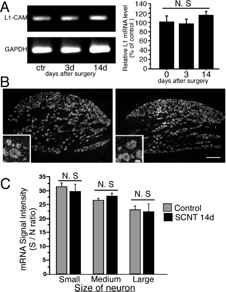
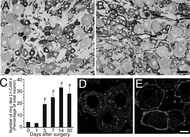
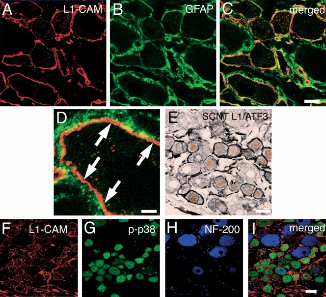
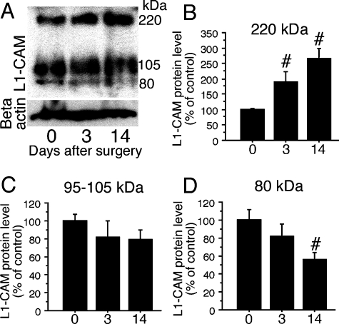
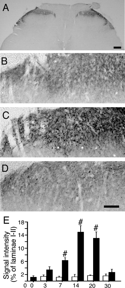
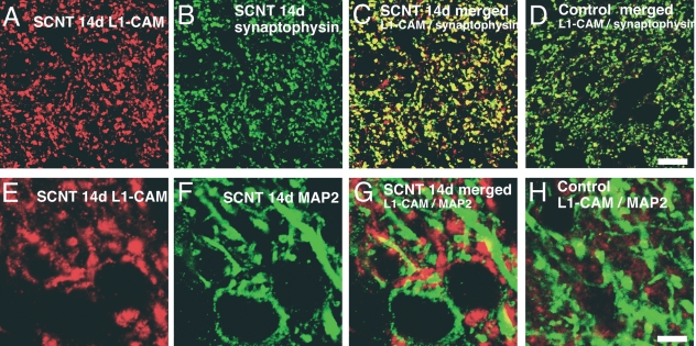
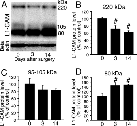
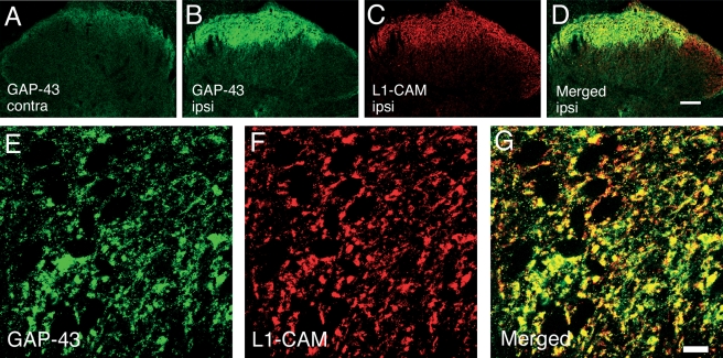
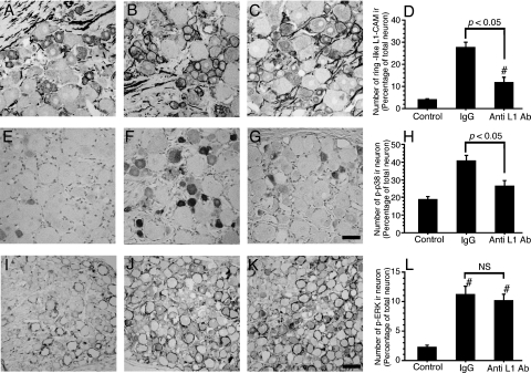
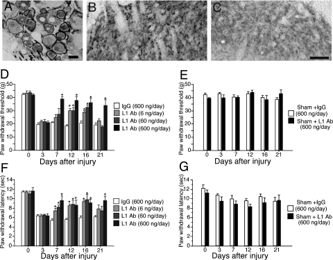
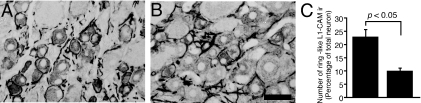
The cell adhesion molecule L1 (L1-CAM) plays important functional roles in the developing and adult nervous systems. Here we show that peripheral nerve injury induced dynamic post-transcriptional alteration of L1-CAM in the rat dorsal root ganglia (DRGs) and spinal cord. Sciatic nerve transection (SCNT) changed the expression of L1-CAM protein but not L1-CAM mRNA. In DRGs, SCNT induced accumulation of the L1-CAM into the surface of somata, which resulted in the formation of immunoreactive ring structures in a number of unmyelinated C-fiber neurons. These neurons with L1-CAM-immunoreactive ring structures were heavily colocalized with phosphorylated p38 MAPK. Western blot analysis revealed the increase of full-length L1-CAM and decrease of fragments of L1-CAM after SCNT in DRGs. Following SCNT, L1-CAM-immunoreactive profiles in the dorsal horn showed an increase mainly in pre-synaptic areas of laminae I-II with a delayed onset and colocalized with growth-associated protein 43. In contrast to DRGs, SCNT increased the proteolytic 80-kDa fragment of L1-CAM and decreased full-length L1-CAM in the spinal cord. The intrathecal injection of L1-CAM antibody for the extracellular domain of L1-CAM inhibited activation of p38 MAPK and emergence of ring structures of L1-CAM immunoreactivity in injured DRG neurons. Moreover, inhibition of extracellular L1-CAM binding by intrathecal administration of antibody suppressed the mechanical allodynia and thermal hyperalgesia induced by partial SCNT. Collectively, these data suggest that the modification of L1-CAM in nociceptive pathways might be an important pathomechanism of neuropathic pain.


Figures

Fig. 1
Fig. 1
Expression of cell adhesion molecule L1 (L1-CAM) mRNA in L4,5 dorsal root ganglia (DRGs) after sciatic nerve transection. (A) The levels of L1-CAM mRNA in the ipsilateral L4,5 DRGs were determined by the reverse transcription-polymerase chain reaction (PCR) technique. Gel panels show PCR products from the L4,5 DRGs taken at 3 and 14 days after surgery. Right graph shows the mRNA levels of L1-CAM expressed as percentages of the mRNA level in the normal control ganglia (mean ± SEM). N.S., not significant compared with the naive control (P > 0.05; anova). (B) Dark-field images of in-situ hybridization (ISH) show L1-CAM mRNA in L5 DRGs of a control rat (left) and 14 days after sciatic nerve transection (SCNT) (right). Insets show highly magnified pictures. (C) Quantification of mean signal density of ISH in each size population in DRG neurons of control rats (grey bars) and 14 days after SCNT (black bars) (mean ± SEM; n = 4, total 2099 neurons for SCNT, total 2122 neurons for control naive). N.S., not significant compared with control (P > 0.05; Student's t-test). Scale bars: 250 µm (low magnification), 25 µm (insets). GAPDH, glyceraldehyde 3-phosphate dehydrogenase.
Fig. 2
Fig. 2
Changes of cell adhesion molecule L1 (L1-CAM) protein expression and the formation of L1-CAM-immunoreactive (ir) ring structures in the dorsal root ganglia (DRGs) after sciatic nerve transection. Photomicrographs show L1-CAM immunoreactivity in the DRGs of normal control (A) and 14 days after sciatic nerve transection (SCNT) (B). (C) Time course of the formation of L1-CAM-ir ring structures after SCNT shown by the percentage of neurons with L1-CAM-ir ring structures in the total L5 DRGs (mean ± SEM; each time point n = 4, more than 500 total neurons from each rat). #P < 0.05 (anova) compared with naive control (day 0). (D and E) Highly magnified confocal two-dimensional images show disappearance of intracellular L1-CAM-ir profiles in DRG neuron and the accumulation around the cytoplasm after SCNT. Control L5 DRG neuron (D) and 14 days after SCNT (E). Scale bars: A and B, 50 µm; D and E, 10 µm.
Fig. 3
Fig. 3
Characterization of sciatic nerve transection (SCNT)-induced cell adhesion molecule L1 (L1-CAM)-immunoreactive (ir) ring structure in dorsal root ganglia (DRGs). (A–D) Double staining of L1-CAM-ir profiles (red) with glial fibrillary acidic protein (GFAP) (green), a marker of satellite cells and Schwann cells, in the L5 DRGs 7 days after SCNT. (C and D) Merged images of L1-CAM-ir profiles and GFAP-ir staining. (D) Higher magnification image of C. Arrows indicate the L1-CAM-ir profiles that are located in neuronal somata. (E) Double staining of L1-CAM (black) with activating transcription factor 3 (ATF3) (brown), a marker of injured neurons, shows the L1-CAM-ir ring structures formed around injured DRG neurons. (F–I) Triple staining demonstrates heavy colocalization of L1-CAM (red) with phospho-p38 MAPK (p-p38) (green) and not with NF-200 (purple), a marker for myelinated A fibers in injured DRG neurons. Scale bars: A–C, 10 µm; D, 2.5 µm; E, 25 µm; F and G, 25 µm.
Fig. 4
Fig. 4
Alteration of cell adhesion molecule L1 (L1-CAM) fragments in dorsal root ganglia (DRGs) after sciatic nerve transection (SCNT). (A) Western blot analysis indicates an increase in full-length L1-CAM levels and decrease in 80-kDa fragment in DRGs. (B–D) Quantification of the L1-CAM protein levels in DRGs after SCNT. The quantified band length is listed above. #P < 0.05 (anova) compared with naive control (day 0).
Fig. 5
Fig. 5
Increases in cell adhesion molecule L1 (L1-CAM) immunoreactivity in dorsal horn after sciatic nerve transection (SCNT). (A) Low-power magnification images of L1-CAM in spinal cord. Higher magnification images of the dorsal horn in a control naive rat (B), 14 days after SCNT (C) and 30 days after SCNT (D). (C) Increased L1-CAM-immunoreactive (ir) profiles in button-like structures. (E) Statistical quantification shows the effect of SCNT on the area of the L1-CAM-ir buttons per section (mean ± SEM; n = 4). #P < 0.05 (anova) compared with naive control (day 0). Solid bars: ipsilateral, open bars: contralateral. Scale bars: A, 200 µm; B–D, 50 µm.
Fig. 6
Fig. 6
Characterization of sciatic nerve transection (SCNT)-induced cell adhesion molecule L1 (L1-CAM)-immunoreactive (ir) button in dorsal horn. (A–C) Double staining of L1-CAM (red) with synaptophysin (green), a marker of pre-synaptic buttons, at day 14 after injury shows the increase in L1-CAM-ir profiles in synaptic terminals. (D) Merged image of L1-CAM and synaptophysin in the dorsal horn of control rat. (E–G) Double staining of L1-CAM (red) with microtubule-associated protein 2 (MAP2) (green), a marker of dendrite of central nervous system neuron, at day 14 after SCNT demonstrates L1-CAM accumulation in post-synaptic areas. (H) Merged images of the dorsal horn of control rat. Scale bars: A–D, 10 µm; E–H, 5 µm.
Fig. 7
Fig. 7
Proteolysis of cell adhesion molecule L1 (L1-CAM) in the spinal cord after sciatic nerve transection (SCNT). (A) Western blot analysis indicates a decrease in full-length L1-CAM levels and an increase in 80-kDa fragment in dorsal root ganglia (DRGs) after SCNT. (B–D) Quantification of the L1-CAM protein levels in DRGs after SCNT (mean ± SEM; n = 3 at each time point). The quantified band lengths are listed above. #P < 0.05 (anova) compared with naive control (day 0).
Fig. 8
Fig. 8
Double-labeling confocal two-dimensional images of growth-associated protein 43 (GAP-43) (green) and cell adhesion molecule L1 (L1-CAM) (red) in dorsal horn 14 days after sciatic nerve transection (SCNT). (A and B) GAP-43 increased in laminae I–II of the dorsal horn and colocalized with L1-CAM. (A) Contralateral and (B) ipsilateral to the SCNT. (C) L1-CAM immunoreactivity and (D) a merged image of B and C. (E–G) Higher magnification images of double labeling show colocalization with GAP-43- (E) and L1-CAM-immunoreactive buttons (F). (G) A merged image of E and F. Scale bars: A–D, 100 µm; E–G, 10 µm.
Fig. 9
Fig. 9
Effects of the anti-cell adhesion molecule L1 (L1-CAM) antibody on sciatic nerve transection (SCNT)-induced alteration of L1-CAM and phosphorylation of MAPKs in the dorsal root ganglia (DRGs). Photomicrographs show the expression of L1-CAM-immunoreactive (ir) ring structure (A–C), phosphorylated p38-ir profiles (E–G) and phosphorylated extracellular signal-regulated kinase (p-ERK)-ir profiles (I–K). Tissue sections are L5 DRGs of a control naive rat (A, E and I), 7 days after SCNT treatment with intrathecal control IgG (B, F and J) and 7 days after SCNT treatment with intrathecal chronic injection of anti-L1-CAM antibody (600 ng/day) (C, G and K). Quantification of the percentage of neurons with L1-CAM-ir ring structures (D), phospho-p38 MAPK (p-p38)-ir neurons (H) and p-ERK-ir neurons (L) at 7 days after SCNT (mean ± SEM; n = 4, more than 500 total neurons from each rat) (Ab, antibody). P < 0.05 compared with saline-treated group (anova); NS, not significant (P > 0.05); #P < 0.05 (anova) compared with naive control. Scale bars: A–C and E–G, 50 µm; I–K, 100 µm.
Fig. 10
Fig. 10
Effects of the p38 inhibitor 4-(4-fluorophenyl)-2-(4-methylsulfonylphenyl)-5-(4-pyridyl)-1H-imidazole (SB) on the formation of cell adhesion molecule L1 (L1-CAM)-immunoreactive (ir) ring structures in the dorsal root ganglia (DRGs) following sciatic nerve transection (SCNT). Tissue sections are L5 DRGs of 7 days after SCNT treatment with intrathecal control vehicle (A) and 7 days after SCNT treatment with intrathecal chronic injection of SB (4 µg/day) (B). (C) Quantification of the percentage of neurons with L1-CAM-ir ring structures (mean ± SEM; n = 4, more than 500 total neurons from each rat). NS, not significant (P > 0.05, Student's t-test). Scale bar, 50 µm.
Fig. 11
Fig. 11
Changes of cell adhesion molecule L1 (L1-CAM) protein expression and pain behaviors in neuropathic pain model. (A–C) Expression of L1-CAM in dorsal root ganglia (DRGs) and dorsal horn of partial sciatic nerve transection (PSNL) rats at 14 days after injury. (A) Double labeling of L1-CAM with activating transcription factor 3 in DRGs at 14 days after PSNL. (B and C) PSNL induced L1-CAM-immunoreactive buttons in dorsal horn. Dorsal horn ipsilateral (B) and contralateral (C) to the PSNL. Scale bar: A–C, 25 µm. (D–G) Effects of intrathecal chronic administration of anti-L1-CAM antibody on neuropathic pain behaviors. (D) The effects of chronic intrathecal administration of anti-L1-CAM antibody on mechanical allodynia in the PSNL model. The highest concentration of anti-L1-CAM antibody (600 ng/day) administration reversed the thermal hyperalgesia for up to 21 days after surgery. The lower concentrations of L1-CAM (60 and 6 ng/day) had a limited effect on the withdrawal threshold, except at day 16 after injury. (F) The withdrawal latency to the radiant heat stimuli was obtained from the same rats that received the mechanical stimuli. Injury-induced thermal hyperalgesia was suppressed by the administration of the L1-CAM antibody. (E and G) Intrathecal anti-L1-CAM antibody and control IgG have no effects on the basal mechanical and thermal sensitivity. *P < 0.05 compared with saline-treated group. In all graphs, values were represented by mean ± SEM (n = 6 in each group) (Ab, antibody).
Fig. 12
Fig. 12
Effects of the anti-cell adhesion molecule L1 (L1-CAM) antibody on partial sciatic nerve transection (PSNL)-induced alteration of L1-CAM in the dorsal root ganglia (DRGs). Tissue sections are L5 DRGs of 7 days after PSNL treatment with intrathecal control IgG (A) and 7 days after PSNL treatment with intrathecal chronic injection of anti-L1-CAM antibody (600 ng/day) (B). (C) Quantification of the percentage of neurons with L1-CAM-immunoreactive (ir) ring structures (mean ± SEM; n = 4, more than 500 total neurons from each rat). P < 0.05 compared with control IgG-treated group (Student's t-test). Left bar: IgG treatment, right bar: L1-CAM antibody treatment. Scale bar, 50 µm.

Similar articles

Cited by

References

    1. Aoki E. Localization of nitric oxide-related substances in the peripheral nervous tissues. Brain Res. 1993;620:142–145. - PubMed
    1. Baba H. Silent NMDA receptor-mediated synapses are developmentally regulated in the dorsal horn of the rat spinal cord. JNeurophysiol. 2000;83:955–962. - PubMed
    1. Benowitz LI. GAP-43 as a marker for structural plasticity in the mature CNS. ProgBrain Res. 1990;86:309–320. - PubMed
    1. Brock JH. Distribution and injury-induced plasticity of cadherins in relationship to identified synaptic circuitry in adult rat spinal cord. JNeurosci. 2004;24:8806–8817. - PMC - PubMed
    1. Brummendorf T. The axonal recognition molecule F11 is a multifunctional protein: specific domains mediate interactions with Ng-CAM and restrictin. Neuron. 1993;10:711–727. - PubMed

Publication types

Substances