Review
. 2007 Mar 1:2:11.
doi: 10.1186/1750-1172-2-11.

Cardiac tumours in children

Affiliations
Review

Cardiac tumours in children

Orhan Uzun et al. Orphanet J Rare Dis. .

Abstract

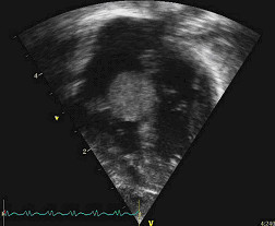
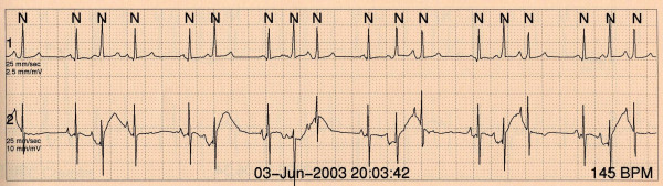
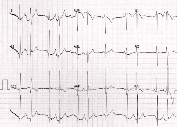
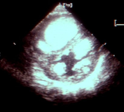
Cardiac tumours are benign or malignant neoplasms arising primarily in the inner lining, muscle layer, or the surrounding pericardium of the heart. They can be primary or metastatic. Primary cardiac tumours are rare in paediatric practice with a prevalence of 0.0017 to 0.28 in autopsy series. In contrast, the incidence of cardiac tumours during foetal life has been reported to be approximately 0.14%. The vast majority of primary cardiac tumours in children are benign, whilst approximately 10% are malignant. Secondary malignant tumours are 10-20 times more prevalent than primary malignant tumours. Rhabdomyoma is the most common cardiac tumour during foetal life and childhood. It accounts for more than 60% of all primary cardiac tumours. The frequency and type of cardiac tumours in adults differ from those in children with 75% being benign and 25% being malignant. Myxomas are the most common primary tumours in adults constituting 40% of benign tumours. Sarcomas make up 75% of malignant cardiac masses. Echocardiography, Computing Tomography (CT) and Magnetic Resonance Imaging (MRI) of the heart are the main non-invasive diagnostic tools. Cardiac catheterisation is seldom necessary. Tumour biopsy with histological assessment remains the gold standard for confirmation of the diagnosis. Surgical resection of primary cardiac tumours should be considered to relieve symptoms and mechanical obstruction to blood flow. The outcome of surgical resection in symptomatic, non-myxomatous benign cardiac tumours is favourable. Patients with primary cardiac malignancies may benefit from palliative surgery but this approach should not be recommended for patients with metastatic cardiac tumours. Surgery, chemotherapy and radiotherapy may prolong survival. The prognosis for malignant primary cardiac tumours is generally extremely poor.


Figures

Figure 1
Figure 1
A foetal tumour in the left ventricular (LV) cavity causing LV outflow tract obstruction.
Figure 2
Figure 2
Right ventricular (RV) tumour in a 2-year-old child resulting in RV outflow tract obstruction. The Doppler velocity is 2.77 m/sec.
Figure 3
Figure 3
Multiple rhabdomyomas are found on a foetal ultrasound at 20 weeks of gestation. Rhabdomyomas appears brighter than the surrounding myocardium.
Figure 4
Figure 4
A tumour in the RV cavity results in RV midcavity obstruction.
Figure 5
Figure 5
Rhabdomyoma on the tricuspid valve with some inflow obstruction in a different patient.
Figure 6
Figure 6
A 24-hour Holter on a child a month after birth shows pre-excitation and premature atrial beats.
Figure 7
Figure 7
In the same child aged 3 years the 12-lead ECG reveals loss of pre-excitation but ongoing premature atrial complexes.
Figure 8
Figure 8
Echocardiography on a newborn child shows multiple rhabdomyomas in both ventricles. In this child the diagnosis of tuberous sclerosis was confirmed.
Figure 9
Figure 9
Fibroma of the heart of a foetus. There is macerated myocardium.
Figure 10
Figure 10
The second image shows that the tumour composed of fibroblasts arranged in fascicles.
Figure 11
Figure 11
Cystic teratoma (*) attached to the aortic root (Ao).
Figure 12
Figure 12
Pericardial effusion (*) and cystic teratoma (♥) in a child.
Figure 13
Figure 13
Typical cystic and multi-lobular appearance of a teratoma excised from heart.
Figure 14
Figure 14
Histology of a teratoma showing multiple immature elements in it.
Figure 15
Figure 15
Microscopically histiocytoid nodules contain large oval cardiac myocytes with coarse granular pale cytoplasm.
Figure 16
Figure 16
High power microscopy showing enlarged, granular-appearing myocytes.

Similar articles

Cited by

References

    1. McAllister HA Jr. Primary tumours of the heart and pericardium. Pathol Annu. 1979;14:325–355. - PubMed
    1. Nadas AS, Ellison RC. Cardiac tumors in infancy. Am J Cardiol. 1968;21:363–366. doi: 10.1016/0002-9149(68)90140-9. - DOI - PubMed
    1. Holley DG, Martin GR, Brenner JI. Diagnosis and management of fetal cardiac tumors: a multicenter experience and review of published reports. J Am Coll Cardiol. 1995;26:516–520. doi: 10.1016/0735-1097(95)80031-B. - DOI - PubMed
    1. Lam KY, Dickens P, Chan AC. Tumors of the heart. A 20-year experience with a review of 12,485 consecutive autopsies. Arch Pathol Lab Med. 1993;117:1027–1031. - PubMed
    1. Bulkley BH, Hutchins GM. Atrial myxomas: a fifty year review. Am Heart J. 1979;97:639–643. doi: 10.1016/0002-8703(79)90193-5. - DOI - PubMed

MeSH terms

LinkOut - more resources