

In vivo assay of presynaptic microtubule cytoskeleton dynamics in Drosophila
- PMID: 17331586
- PMCID: PMC2713775
- DOI: 10.1016/j.jneumeth.2007.01.013
In vivo assay of presynaptic microtubule cytoskeleton dynamics in Drosophila
Abstract
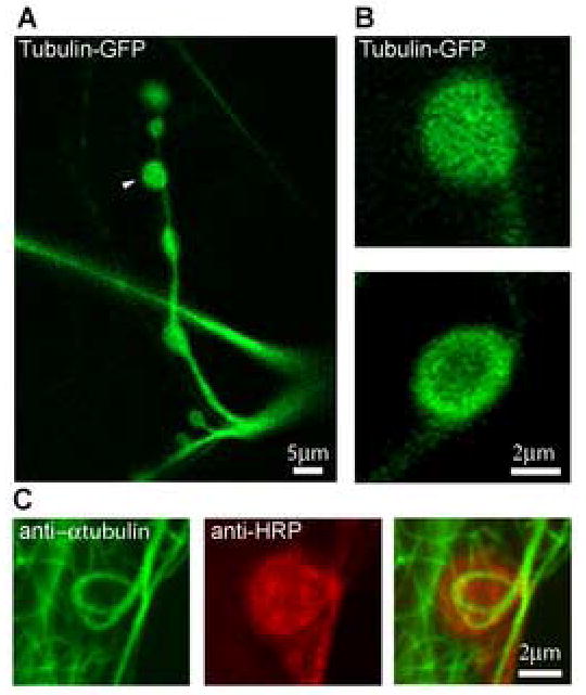
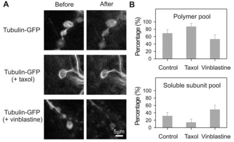
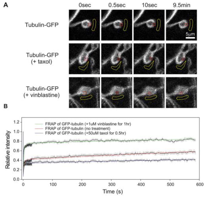
Disrupted microtubule dynamics in neuronal synapses has been suggested as an underlying cause for several devastating neurological diseases, including Hereditary Spastic Paraplegia (HSP) and Fragile X Syndrome (FXS). However, previous studies have been restricted to indirect assays of synaptic microtubules, i.e. immunocytochemistry of microtubule-associated proteins and post-translationally modified tubulins characteristic of microtubules with different stabilities. Very little is known about synaptic microtubule dynamics in vivo, or how microtubule dynamics may be disrupted in disease states. In this study, we develop methods to analyze microtubule dynamics directly in living synaptic boutons in situ. We use fluorescence recovery after photobleaching (FRAP) of transgenic green fluorescent protein (GFP) tagged tubulin at the well-characterized Drosophila neuromuscular junction (NMJ) synapse. FRAP measurements of tubulin-GFP demonstrate biphasic recovery kinetics. Treatment with taxol to stabilize microtubules and promote microtubule assembly reduces both recovery phases. Treatment with vinblastine to disassemble microtubules increases the fast recovery phase and decreases the slow recovery phase. These data indicate that the fast recovery phase is generated by rapid diffusion of tubulin subunits and the slow phase is generated by the relatively slow turnover of microtubules. This study demonstrates that tubulin-GFP fluorescence recovery after photobleaching can be used to assay microtubule dynamics directly in living synapses.
Figures
Similar articles
-
The hereditary spastic paraplegia gene, spastin, regulates microtubule stability to modulate synaptic structure and function.Curr Biol. 2004 Jul 13;14(13):1135-47. doi: 10.1016/j.cub.2004.06.058. Curr Biol. 2004. PMID: 15242610
-
Specific in vivo labeling of tyrosinated α-tubulin and measurement of microtubule dynamics using a GFP tagged, cytoplasmically expressed recombinant antibody.PLoS One. 2013;8(3):e59812. doi: 10.1371/journal.pone.0059812. Epub 2013 Mar 28. PLoS One. 2013. PMID: 23555790 Free PMC article.
-
Altered skeletal muscle microtubule-mitochondrial VDAC2 binding is related to bioenergetic impairments after paclitaxel but not vinblastine chemotherapies.Am J Physiol Cell Physiol. 2019 Mar 1;316(3):C449-C455. doi: 10.1152/ajpcell.00384.2018. Epub 2019 Jan 9. Am J Physiol Cell Physiol. 2019. PMID: 30624982 Free PMC article.
-
Morphological features of central synapses, with emphasis on the presynaptic terminal.Life Sci. 1977 Aug 15;21(4):477-92. doi: 10.1016/0024-3205(77)90086-8. Life Sci. 1977. PMID: 333215 Review. No abstract available.
-
Synaptic cytoskeleton at the neuromuscular junction.Int Rev Neurobiol. 2006;75:217-36. doi: 10.1016/S0074-7742(06)75010-3. Int Rev Neurobiol. 2006. PMID: 17137930 Review. No abstract available.
Cited by
-
Harvesting and preparing Drosophila embryos for electrophysiological recording and other procedures.J Vis Exp. 2009 May 20;(27):1347. doi: 10.3791/1347. J Vis Exp. 2009. PMID: 19488027 Free PMC article.
-
Bicaudal-D binds clathrin heavy chain to promote its transport and augments synaptic vesicle recycling.EMBO J. 2010 Mar 3;29(5):992-1006. doi: 10.1038/emboj.2009.410. Epub 2010 Jan 28. EMBO J. 2010. PMID: 20111007 Free PMC article.
-
Conformational flexibility of tubulin dimers regulates the transitions of microtubule dynamic instability.bioRxiv [Preprint]. 2025 Jul 2:2025.06.30.662375. doi: 10.1101/2025.06.30.662375. bioRxiv. 2025. PMID: 40631249 Free PMC article. Preprint.
-
Dynamic microtubules at the synapse.Curr Opin Neurobiol. 2020 Aug;63:9-14. doi: 10.1016/j.conb.2020.01.003. Epub 2020 Feb 12. Curr Opin Neurobiol. 2020. PMID: 32062144 Free PMC article. Review.
-
Fruit fly for anticancer drug discovery and repurposing.Ann Med Surg (Lond). 2023 Feb 7;85(2):337-342. doi: 10.1097/MS9.0000000000000222. eCollection 2023 Feb. Ann Med Surg (Lond). 2023. PMID: 36845805 Free PMC article. No abstract available.
References
-
- Ashburner M. Drosophila: A laboratory manual. Cold Spring Harbor Press. 1989
-
- Baas PW, Brown A. Slow axonal transport: the polymer transport model. Trends in Cell Bio. 1997;7:380–84. - PubMed
-
- Barth AI, Nathke IS, Nelson WJ. Cadherins, catenins and APC protein: interplay between cytoskeletal complexes and signaling pathways. Curr Opin Cell Biol. 1997;9:683–90. - PubMed
Publication types
MeSH terms
Substances
Grants and funding
LinkOut - more resources
Full Text Sources
Other Literature Sources
Molecular Biology Databases
Miscellaneous
