. 2007 Apr 22;274(1613):1035-41.
doi: 10.1098/rspb.2006.0347.

Feedback control in active sensing: rat exploratory whisking is modulated by environmental contact

Affiliations

Feedback control in active sensing: rat exploratory whisking is modulated by environmental contact

Ben Mitchinson et al. Proc Biol Sci. .

Abstract

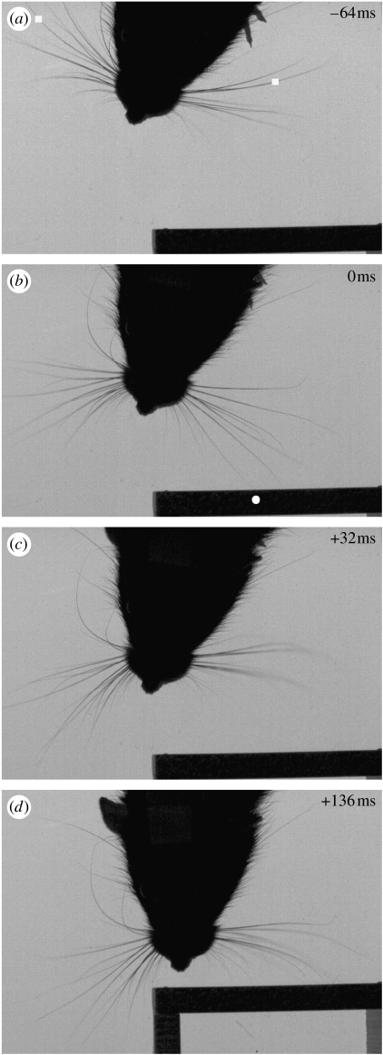
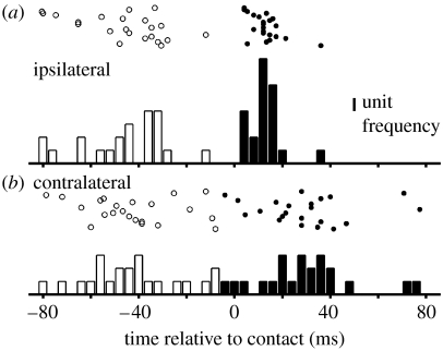
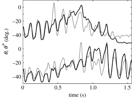
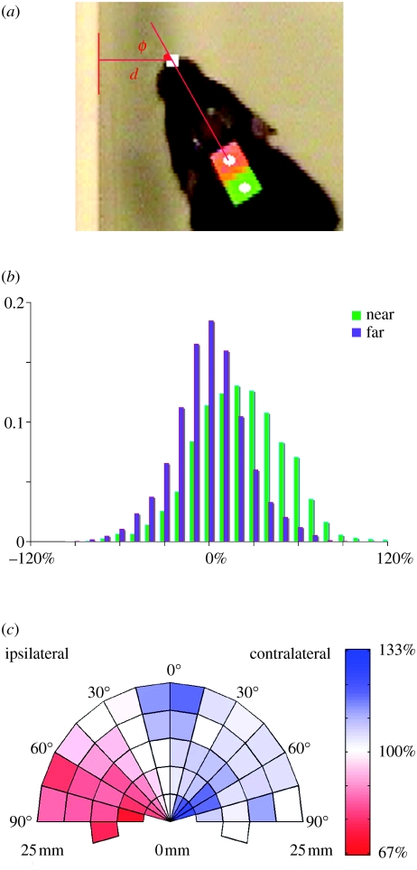
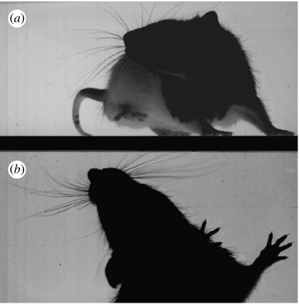
Rats sweep their facial whiskers back and forth to generate tactile sensory information through contact with environmental structure. The neural processes operating on the signals arising from these whisker contacts are widely studied as a model of sensing in general, even though detailed knowledge of the natural circumstances under which such signals are generated is lacking. We used digital video tracking and wireless recording of mystacial electromyogram signals to assess the effects of whisker-object contact on whisking in freely moving animals exploring simple environments. Our results show that contact leads to reduced protraction (forward whisker motion) on the side of the animal ipsilateral to an obstruction and increased protraction on the contralateral side. Reduced ipsilateral protraction occurs rapidly and in the same whisk cycle as the initial contact. We conclude that whisker movements are actively controlled so as to increase the likelihood of environmental contacts while constraining such interactions to involve a gentle touch. That whisking pattern generation is under strong feedback control has important implications for understanding the nature of the signals reaching upstream neural processes.


Figures

Figure 1
Figure 1
High-speed video frames showing the effect of unilateral object contact (at t=0) on bilateral whisker protraction. (a) (t=−64 ms) Protraction commences approximately synchronously on both sides of the snout; the filled white squares show the tracked rear column whiskers. (b) (t=0 ms) A deflection occurs on a forward whisker, the filled white circle indicates the point of contact with the vertical surface. (c) (t=+32 ms) Protraction ends on the side contralateral to the contact, note that whiskers on the ipsilateral side are already partially retracted having ceased protraction at t=+12 ms. (d) (t=+136 ms) Contralateral whiskers reach maximum protraction in the whisk cycle subsequent to the initial contact (note that the interposed retraction is not shown). Protraction amplitude in this whisk is notably increased contralaterally, compared with whisks preceding contact, and reduced ipsilaterally such that the whiskers on that side are only gently deflected by the surface contact. Video 1 of electronic supplementary material shows the full whisk cycle bracketing the initial deflection.
Figure 2
Figure 2
(a) Ipsilateral and (b) Contralateral whisker protraction onset (open bars) and cessation (filled bars) relative to time of contact (t=0) of a forward whisker with an obstacle. Protraction onset times are similar on both sides of the snout, but cessation occurs earlier on the ipsilateral side than on the contralateral side and appears more closely tied to the time of contact (note the pronounced peak ipsilaterally approximately 13 ms after contact). Data are from 22 high-speed video clips recorded with nine different animals.
Figure 3
Figure 3
Mystacial EMG as a proxy measure of whisking, example in one animal. Black trace is whisker angle (θ), determined by high-speed video tracking, of the left/right (top/bottom) whisker field (column 1); note separate y-axis origins for each trace. Grey trace (θ*) is an estimate of θ derived from left/right whisking EMG data. Increasing θ corresponds to whisker protraction for all traces.
Figure 4
Figure 4
Proximity to a wall induces a systematic bias in whisking asymmetry. (a) Distance (d) and bearing (ϕ) to a single nearby wall was determined by automated tracking of normal speed video, allowing the data to be partitioned into NEAR (d≤25 mm) and FAR (d≥100 mm) subsets, and for left and right whisking amplitudes to be classified as ipsilateral or contralateral. (b) Normalized frequency histograms of NEAR (green, n=3965) and FAR (purple, n=5989) whisking bias (contralateral–ipsilateral amplitude) as a percentage of average whisk strength. The distributions show a strong bias (mean 21.5%) in favour of contralateral whisking for NEAR only. Plots for individual animals are given in the electronic supplementary material. (c) Polar plot of ipsilateral (shown left of the midline) and contralateral (shown right of midline) mean whisking amplitude binned, for NEAR frames only, according to d and ϕ. For the purpose of this display, the nearby wall is always to the left with its bearing and distance mirrored across the midline (frames in which the wall was originally to the right have therefore been reflected in the midline). The colour scale (red–white–blue) shows increasing percentage of average whisk strength. Bins with a count of less than five frames are omitted (3955 frames displayed); included bins represent 5–541 frames each (median 92). The presence of a nearby wall leads to reduced ipsilateral and increased contralateral whisking. Ipsilateral reduction is most pronounced in the range 45–90°, where the rat is more ‘side-on’ to the obstruction. Where the wall lies directly in front (0–15°), but at some distance (d>15 mm), there is evidence of increased whisking bilaterally. Data for individual animals are given in the electronic supplementary material.
Figure 5
Figure 5
High-speed video frame showing asymmetric whisking against a wall. (a) The image of the animal in the vertical plane obtained using a front-silvered mirror, positioned behind a glass wall, and slanted at an oblique angle with respect to the camera. (b) The view in horizontal plane, with the viewpoint of the camera aligned with the vertical surface of the wall. (Note that the dark strip across the centre of the frame therefore corresponds to the floor of the arena for (a) and the wall for (b).) The snapshot shows the moment of maximum protraction for a single whisk. Protraction is strongly asymmetric in a manner that appears to reduce bending of the whiskers on the side of the snout closest to the wall, while increasing the likelihood of surface contact by whiskers on the opposite side. An extract from the video clip from which this frame was taken is provided in video 4 of electronic supplementary material.

References

    1. Ahissar E, Arieli A. Figuring space by time. Neuron. 2001;32:185–201. doi:10.1016/S0896-6273(01)00466-4 - DOI - PubMed
    1. Aloimonos J.Y, Weiss I, Bandopadhay A. Active vision. Int. J. Comput. Vis. 1988;1:333–356. doi:10.1007/BF00133571 - DOI
    1. Arabzadeh E, Zorzin E, Diamond M.E. Neuronal encoding of texture in the whisker sensory pathway. PLoS Biol. 2005;3:e17. doi:10.1371/journal.pbio.0030017 - DOI - PMC - PubMed
    1. Ballard D.H. Animate vision. Artif. Intell. 1991;48:57–96. doi:10.1016/0004-3702(91)90080-4 - DOI
    1. Berg R.W, Kleinfeld D. Rhythmic whisking by rat: retraction as well as protraction of the vibrissae is under active muscular control. J. Neurophysiol. 2003;89:104–117. doi:10.1152/jn.00600.2002 - DOI - PubMed

Publication types