. 2007;35(6):1885-96.
doi: 10.1093/nar/gkm085. Epub 2007 Mar 1.

Studies on the function of the riboregulator 6S RNA from E. coli: RNA polymerase binding, inhibition of in vitro transcription and synthesis of RNA-directed de novo transcripts

Affiliations

Studies on the function of the riboregulator 6S RNA from E. coli: RNA polymerase binding, inhibition of in vitro transcription and synthesis of RNA-directed de novo transcripts

Nina Gildehaus et al. Nucleic Acids Res. 2007.

Abstract

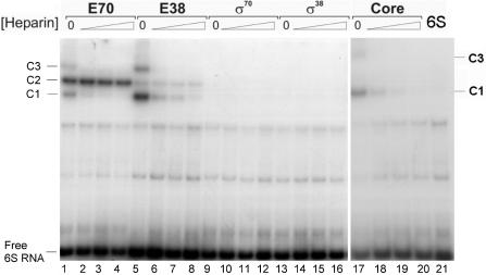
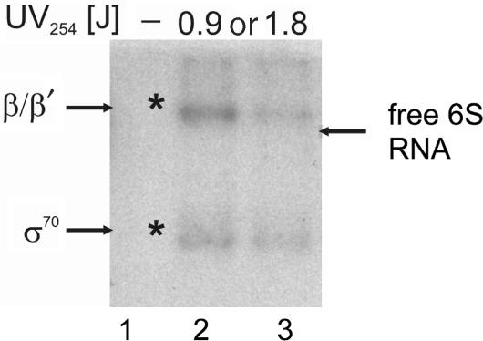
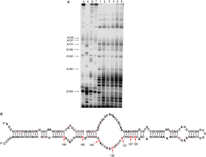
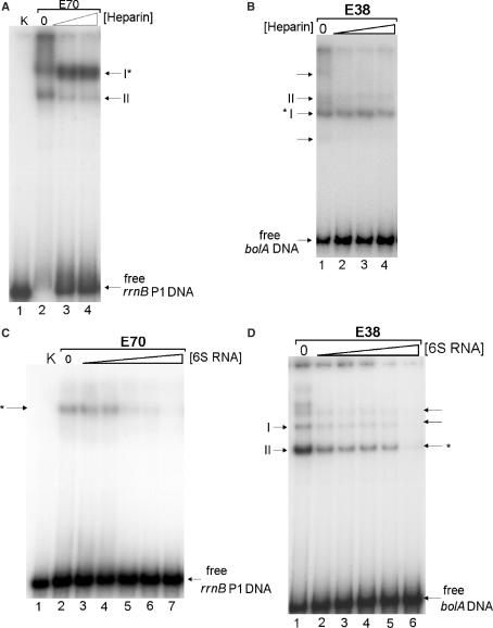
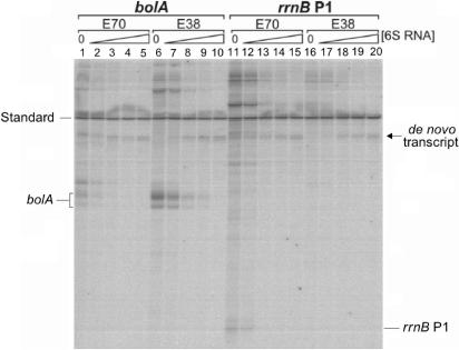
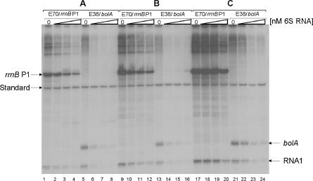
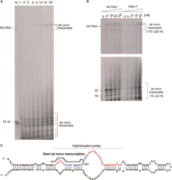
Escherichia coli 6S RNA represents a non-coding RNA (ncRNA), which, based on the conserved secondary structure and previous functional studies, had been suggested to interfere with transcription. Selective inhibition of sigma-70 holoenzymes, preferentially at extended -10 promoters, but not stationary-phase-specific transcription was described, suggesting a direct role of 6S RNA in the transition from exponential to stationary phase. To elucidate the underlying mechanism, we have analysed 6S RNA interactions with different forms of RNA polymerase by gel retardation and crosslinking. Preferred binding of 6S RNA to Esigma(70) was confirmed, however weaker binding to Esigma(38) was also observed. The crosslinking analysis revealed direct contact between a central 6S RNA sequence element and the beta/beta' and sigma subunits. Promoter complex formation and in vitro transcription analysis with exponential- and stationary-phase-specific promoters and the corresponding holoenzymes demonstrated that 6S RNA interferes with transcription initiation but does not generally distinguish between exponential- and stationary-phase-specific promoters. Moreover, we show for the first time that 6S RNA acts as a template for the transcription of defined RNA molecules in the absence of DNA. In conclusion, this study reveals new aspects of 6S RNA function.


Figures

Figure 1.
Figure 1.
Complex formation of 6S RNA with different forms of RNA polymerase. A gel retardation analysis of 6S RNA complexed to different RNA polymerase preparations is shown. Different forms of RNA polymerase were complexed with 9 nM [32P] 3′- end- labelled 6S RNA. Lanes 1–4: 3 nM reconstituted holoenzyme Eσ70; lanes 5–8: 3 nM reconstituted holoenzyme Eσ38; lanes 9–12: 60 nM free σ70; lanes 13–16: 60 nM free σ38; lanes 17–20: 6 nM core RNA polymerase. Complex formation for each enzyme preparation was challenged with increasing heparin concentrations, from left to right: 0, 50, 100 or 200 ng/μl. Lane 21 on the extreme right shows 6S RNA in the absence of protein. The positions of free 6S RNA and of the three complexes with different mobilities are indicated at the margin and labelled 6S RNA, C1, C2 and C3, respectively.
Figure 2.
Figure 2.
UV crosslinking analysis of E. coli 6S RNA complexes formed with Eσ70. Complexes between radiolabelled 6S RNA and Eσ70 holoenzyme were formed and irradiated at 254 nm at increasing UV dosages of 0.9 or 1.8 J. In the lane marked by a dash, irradiation was omitted. Samples were subjected to RNaseA digestion and separated by SDS gel electrophoresis. Radioactive bands were visualized by autoradiography. Asterisks indicate radioactive bands that correspond to marker positions of RNA polymerase subunits. Bands were assigned according to non-radioactive marker proteins separated alongside the digested samples after Coomassie staining. The positions for the β/β′ and σ70 subunits are indicated at the margin. The position of a free 6S RNA sample separated without digestion is indicated by an arrow on the right margin.
Figure 3.
Figure 3.
Regions of 6S RNA in contact with RNA polymerase. (A) The example of an autoradiogram of a primer extension analysis of isolated 6S RNA samples after crosslinking is shown. Samples have been separated on 10% denaturing polyacrylamide gels. A, C, G and T indicate primer extension sequencing reactions, which have been performed with a 6S RNA template. In lanes labelled 1–6, different 6S RNA-polymerase complexes were analysed after crosslinking; 1: 6S RNA complexed with Eσ70, 2: 6S RNA complexed with Eσ38, 3: 6S RNA complexed with core enzyme, 4: 6S RNA complexed with the isolated σ70 subunit, 5: 6S RNA complexed with the isolated σ38 subunit, 6: 6S RNA after UV irradiation in the absence of protein. Characteristic positions that deviate in the primer extension pattern between free 6S RNA and RNA polymerase complexes are indicated at the left margin. (B) Location of crosslink sites within the 6S RNA secondary structure. Positions of the 6S RNA nucleotides, which have been identified to be in contact with the Eσ70 holoenzyme, are indicated by arrows and sequence numbers.
Figure 4.
Figure 4.
Effect of 6S RNA on transcription initiation complex formation. (A) Formation of initiation complexes at the rrnB P1 promoter was analysed by gel retardation. Initiation complexes were formed with a radioactive DNA fragment, containing the rrnB P1 promoter (1.5 nM) and 3 nM active RNA polymerase Eσ70 in the presence of 65 μM ATP and CTP as starting nucleotides. Samples were treated with various heparin concentrations (0, 100 and 200 ng/μl) and separated on a native 5% polyacrylamide gel. The positions of two RNA polymerase complexes (holoenzyme: I and core: II) and the free DNA are indicated. Different concentrations of heparin (lane 3: 100 ng/μl, lane 4: 200 ng/μl) were included. The essentially heparin resistant rrnB P1 ternary initiation complex is indicated by an asterisk. The gel band II corresponds to a core enzyme–DNA complex. In K, only free P1 DNA was separated. (B) Formation of initiation complexes at the bolA promoter is shown for the Eσ38 holoenzyme. The ternary initiation complex at the bolA promoter (I) is indicated by an asterisk. Bands labelled II correspond to the RNA polymerase core complex. Minor complexes are indicated by arrows. Various heparin concentrations were applied: lane 1: 0, lane 2: 50 ng/μl, lane 3: 100 ng/μl, lane 4: 200 ng/μl.(C) Effects of 6S RNA on initiation complex formation at rrnB P1. Complexes were formed as in (A), but in the presence of increasing amounts of 6S RNA (lane 2: 0, lane 3: 5 nM, lane 4: 10 nM, lane 5: 50 nM, lane 6: 100 nM, lane 7: 1 μM). An asterisk denotes the positions of specific rrnB P1 initiation complexes. In K, only free P1 DNA was separated. (D) Complexes were formed as in (B) but in the presence of increasing amounts of 6S RNA (lane 1: 0, lane 2: 5 nM, lane 3: 10 nM, lane 4: 50 nM, lane 5: 100 nM, lane 6: 1 μM). Bands labelled II, and indicated by an asterisk, denote the position of specific bolA initiation complex. Bands labelled I correspond to the RNA polymerase core complex. Minor complexes are indicated by arrows.
Figure 5.
Figure 5.
Effect of 6S RNA on the in vitro transcription from σ70 and σ38-specific promoters on linear DNA fragments. Products from in vitro transcription reactions were separated on denaturing polyacrylamide gels and visualized by autoradiography. Reactions with the bolA or rrnB P1 promoters are shown on the left or right side, respectively. The different holoenzymes employed (E70, E38) are indicated above the lanes. For each system the amount of 6S RNA present in the reaction was varied (lanes 1, 6, 11, 16: 0 nM, lane 2, 7, 12, 17: 10 nM, lane 3, 8, 13, 18: 50 nM, lane 4, 9, 14, 19: 100 nM, lane 5, 10, 15, 20: 250 nM). A 260 bp radiolabelled DNA fragment, indicated at the margin, was included as internal standard for quantification. The positions of the run-off transcripts for the bolA (∼124 nt) and rrnB P1 (64 nt) promoters are marked. An arrow denotes a de novo product that consistently arises when 6S RNA is incubated with E. coli RNA polymerase, even in the absence of any template DNA.
Figure 6.
Figure 6.
Effect of 6S RNA addition on the in vitro transcription with supercoiled templates. Three different reactions, corresponding to different steps of the transcription cycle, were directed by the controlled addition of 6S RNA. In A (lanes 1–8), 6S RNA was added to the RNA polymerase reconstitution reaction. In B (lanes 9–16), 6S RNA was applied to the transcription reaction, which was initiated by the addition of template DNA as the last component. In C (lanes 17–24), 6S RNA was present in the reaction mixture before transcription was started by the addition of the enzyme. The amount of 6S RNA was varied in each series (lanes: 1, 5, 9, 13, 17, 21: 0 nM, lanes: 2, 6, 10, 14, 18, 22: 50 nM, lanes: 3, 7, 11, 15, 19, 23: 100 nM, lanes: 4, 8, 12, 16, 20, 24: 250 nM, respectively). The different holoenzymes (Eσ70, Eσ38) and supercoiled template DNAs (rrnB P1, bolA) are indicated above the lanes. Terminated transcripts started from either rrnB P1 or bolA and the σ70-dependent plasmid-encoded RNA1 promoter are marked at the margin. A 260 bp radiolabelled reference DNA fragment (standard) was included in each lane for quantification.
Figure 7.
Figure 7.
Formation of 6S RNA-dependent de novo transcripts. (A) The time-dependent formation of 6S RNA-directed formation of de novo transcripts is shown. Reaction mixtures contained 20 nM Eσ70 RNA polymerase holoenzyme and 100 nM 6S RNA, free of any noticeable DNA. Reaction was performed in standard in vitro transcription buffer (see Materials and methods section) for the times in minutes indicated on top of the lanes. Products were separated on 15% denaturing polyacrylamide gels. The positions of a 18-mer DNA oligonucleotide and a 6S RNA marker are indicated. (B) Effect of 6S RNA and RNA polymerase concentrations on the formation of de novo transcripts. The upper and lower part of the figure represent different exposures of the upper and lower part of the same gel in order to better visualize the longer products. Reactions have been performed as in (A) except that 6S RNA or RNA polymerase was varied as indicated on top of the lanes (6S RNA: 0–100 nM; RNA polymerase: 0–40 nM). Reactions with increasing 6S RNA contained constant 20 nM RNA polymerase and reactions with increasing RNA polymerase constant amounts of 6S RNA (50 nM). The lengths of the different de novo transcripts are indicated at the margin. (C) Scheme depicting the de novo transcription start site within the secondary structure of 6S RNA, according to the analysis presented in Supplementary Data (Figure S1A–D). The start site (U44), direction of synthesis and approximate length of the short de novo transcripts are indicated by a grey arrow. The conserved CRI sequence is shown in blue, and the hybridization sequence region is shown in red and indicated by a bracket.

Similar articles

Cited by

References

    1. Barrick JE, Sudarsan N, Weinberg Z, Ruzzo WL, Breaker RR. 6S RNA is a widespread regulator of eubacterial RNA polymerase that resembles an open promoter. RNA. 2005;11:774–784. - PMC - PubMed
    1. Suzuma S, Asari S, Bunai K, Yoshino K, Ando Y, Kakeshita H, Fujita M, Nakamura K, Yamane K. Identification and characterization of novel small RNAs in the aspS-yrvM intergenic region of the Bacillus subtilis genome. Microbiology. 2002;148:2591–2598. - PubMed
    1. Ando Y, Asari S, Suzuma S, Yamane K, Nakamura K. Expression of a small RNA, BS203 RNA, from the yocI-yocJ intergenic region of Bacillus subtilis genome. FEMS Microbiol. Lett. 2002;207:29–33. - PubMed
    1. Brown JW, Ellis JC. Comparative Analysis of RNA Secondary Structures: 6S RNA. Weinheim: Wiley-VCH GmbH & Co; 2005. pp. 491–512.
    1. Wassarman KM, Storz G. 6S RNA regulates E. coli RNA polymerase activity. Cell. 2000;101:613–623. - PubMed

MeSH terms