Comparative Study
. 2007;35(6):1868-84.
doi: 10.1093/nar/gkm066. Epub 2007 Mar 1.

An improved definition of the RNA-binding specificity of SECIS-binding protein 2, an essential component of the selenocysteine incorporation machinery

Affiliations
Comparative Study

An improved definition of the RNA-binding specificity of SECIS-binding protein 2, an essential component of the selenocysteine incorporation machinery

A Cléry et al. Nucleic Acids Res. 2007.

Abstract

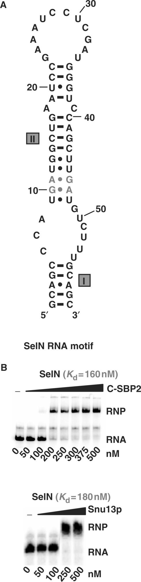
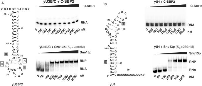
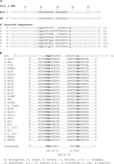
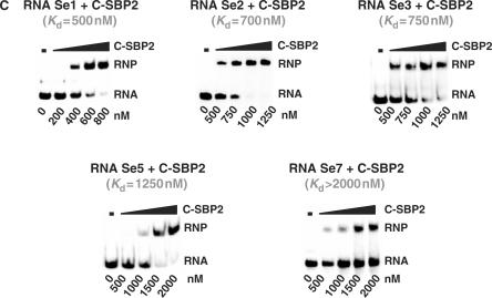
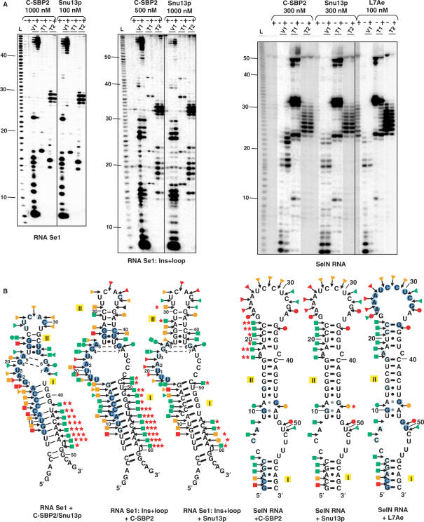
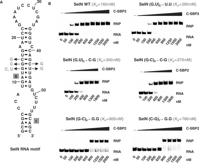
By binding to SECIS elements located in the 3'-UTR of selenoprotein mRNAs, the protein SBP2 plays a key role in the assembly of the selenocysteine incorporation machinery. SBP2 contains an L7Ae/L30 RNA-binding domain similar to that of protein 15.5K/Snu13p, which binds K-turn motifs with a 3-nt bulge loop closed by a tandem of G.A and A.G pairs. Here, by SELEX experiments, we demonstrate the capacity of SBP2 to bind such K-turn motifs with a protruding U residue. However, we show that conversion of the bulge loop into an internal loop reinforces SBP2 affinity and to a greater extent RNP stability. Opposite variations were found for Snu13p. Accordingly, footprinting assays revealed strong contacts of SBP2 with helices I and II and the 5'-strand of the internal loop, as opposed to the loose interaction of Snu13p. Our data also identifies new determinants for SBP2 binding which are located in helix II. Among the L7Ae/L30 family members, these determinants are unique to SBP2. Finally, in accordance with functional data on SECIS elements, the identity of residues at positions 2 and 3 in the loop influences SBP2 affinity. Altogether, the data provide a very precise definition of the SBP2 RNA specificity.


Figures

Figure 1.
Figure 1.
C-SBP2 and Snu13p interact with SelN RNA. (A) The secondary structure of the SelN RNA motif is according to Fagegaltier et al. (52). The G.A sheared base pairs are shown in gray and helices I and II are indicated. (B) The affinity of C-SBP2 and Snu13p for SelN RNA was tested by gel-shift assay using 5 fmol of labeled SelN RNA and protein concentrations ranging from 0 to 500 nM, as indicated below the lanes. Incubation conditions are described in the Materials and Methods section. RNP formation was revealed by electrophoresis on 6% non-denaturing polyacrylamide gels. The apparent Kd values (indicated above the autoradiograms) were calculated with the SigmaPlot Software (SPSS Science Software), by measuring the radioactivity signals corresponding to the free and bound RNAs.
Figure 2.
Figure 2.
C-SBP2 does not interact with yU3B/C and yU4 RNAs. The binding of C-SBP2 and Snu13p on yU3B/C or yU4 RNAs was tested by gel-shift assays. The secondary structures of yU3B/C (Panel A) and yU4 (Panel B) are according to Marmier-Gourrier et al. (26) and Mougin et al. (43), respectively. The G.A sheared pairs are in gray and helices I and II are indicated. Nucleotides involved in the K-turn folding are numbered from 1 to 7. Complexes were formed between 5 fmol of uniformly labeled yU3B/C (Panel A) or yU4 (Panel B) RNAs, and C-SBP2 or Snu13p at concentrations ranging from 0 to 2500 nM, as indicated below the lanes. Incubation conditions were as described in the Materials and Methods section. The autoradiograms obtained after electrophoresis on 6% polyacrylamide gels are shown. For the Snu13p–RNP complexes shown as controls, the apparent Kd values were calculated as described in the Materials and Methods section.
Figure 3.
Figure 3.
Sequences of the RNAs recovered from the SELEX experiment and test of their affinities for C-SBP2. (A) Alignment of the WT yU3B/C RNA sequence with the degenerated N18 RNA and the selected Se1-Se7 RNAs sequences. Nucleotides in Se1-Se7 RNA, are numbered according to the positions of the homolog nucleotides in the WT yU3B/C RNA. The number of sequenced plasmids encoding each selected RNA is indicated in brackets on the right of the sequences. The nucleotides corresponding to the constant sequence are shown in gray, nucleotides in the degenerated sequence and nucleotides mutated during the RT-PCR cycles are shown in black. The GA dinucleotides are underlined. (B) The nucleotide sequences of a series of SECIS motifs from various genes and species (30,52) were aligned with the Se1 RNA sequence taking as references the UGA and GA conserved nucleotides of the K-turn structure (bold characters). A consensus sequence of the SECIS K-turn motifs is deduced from the alignment and indicated below. The positions of the conserved nucleotides in the two strands of helix II are indicated (C) Estimation of the affinity of C-SBP2 for the Se1, Se2, Se3, Se5 and Se7 RNAs by gel-shift assays. RNA–protein complexes formed with 5 fmol of labeled RNA and increasing concentrations of C-SBP2 (as indicated below the lanes) were fractionated by gel electrophoresis as in Figure 1. The apparent Kd values are indicated above the autoradiograms.
Figure 3.
Figure 3.
Sequences of the RNAs recovered from the SELEX experiment and test of their affinities for C-SBP2. (A) Alignment of the WT yU3B/C RNA sequence with the degenerated N18 RNA and the selected Se1-Se7 RNAs sequences. Nucleotides in Se1-Se7 RNA, are numbered according to the positions of the homolog nucleotides in the WT yU3B/C RNA. The number of sequenced plasmids encoding each selected RNA is indicated in brackets on the right of the sequences. The nucleotides corresponding to the constant sequence are shown in gray, nucleotides in the degenerated sequence and nucleotides mutated during the RT-PCR cycles are shown in black. The GA dinucleotides are underlined. (B) The nucleotide sequences of a series of SECIS motifs from various genes and species (30,52) were aligned with the Se1 RNA sequence taking as references the UGA and GA conserved nucleotides of the K-turn structure (bold characters). A consensus sequence of the SECIS K-turn motifs is deduced from the alignment and indicated below. The positions of the conserved nucleotides in the two strands of helix II are indicated (C) Estimation of the affinity of C-SBP2 for the Se1, Se2, Se3, Se5 and Se7 RNAs by gel-shift assays. RNA–protein complexes formed with 5 fmol of labeled RNA and increasing concentrations of C-SBP2 (as indicated below the lanes) were fractionated by gel electrophoresis as in Figure 1. The apparent Kd values are indicated above the autoradiograms.
Figure 4.
Figure 4.
All the selected RNAs that recognize C-SBP2 can form a K-turn structure. (A) Secondary structure analysis of RNAs Se1, Se3, Se5, Se6 or Se7 by enzymatic probing. The RNAs were 5′-end labeled with 32P, renatured and digested with V1 (0.001 U, lane 2), T1 (0.8 U, lane 3) or T2 (2.4 U, lane 4) RNases, under conditions described in the Materials and Methods section. As a control, undigested RNA was fractionated in parallel (lane 1). Lane L corresponds to the alkaline hydrolysis of the RNA used for localization of the RNase cleavage sites. Electrophoresis was performed on a 10% 8M urea–polyacrylamide gel. Nucleotide positions are indicated on the left. (B) Secondary structure models proposed for the selected RNAs. Models were proposed based on thermodynamic considerations and the results of the enzymatic digestions are shown in A. Regions corresponding to the degenerated sequences are shown by gray characters. For RNAs Se1, 3, 5, 6 and 7, V1, T1 and T2 RNase cleavages are represented by arrows surmounted of squares, dots and triangles, respectively. The color of symbols reflects the intensity of cleavages (gray, dark gray and black for low, medium and strong, respectively). Nucleotide numbering is as in Figure 3A. The apparent Kd values established for each RNA by gel retardation are indicated. The free energies of the proposed secondary structures, expressed in kcal/mol, were calculated by using the M-Fold software.
Figure 5.
Figure 5.
Mutations in helix II of RNA Se1 are more deleterious for C-SBP2 than for Snu13p binding. (A) Positions of base substitutions in the Se1 RNA are represented in gray on the proposed secondary structure. The nature of the mutations in the variant Se1 RNAs is shown on the right of helix II. (B) The affinities of C-SBP2 and Snu13p for Se1 RNA and its variants were estimated by gel-shift assays using 5′-end labeled RNAs and protein concentrations ranging from 0 to 4000 nM. The apparent Kd values obtained for each of the RNA–protein complexes are indicated.
Figure 6.
Figure 6.
A K-turn motif with an extended internal loop increases C-SBP2 affinity. The variant Se1:Ins (A) and Se1:Ins + loop RNAs (B and C) are shown. The additional residues in these variant RNAs compared to Se1 RNA are shown in gray. The affinities of C-SBP2 and Snu13p for Se1:Ins (A) and Se1:Ins + loop (B) were tested by gel-shift assays. Complex formation was performed as described in Figure 1, using 5 fmol of 5′-end labeled RNA and increasing concentrations of C-SBP2 or Snu13p proteins. In Panels A and B, the apparent Kds are indicated above the autoradiograms. (C) The base substitutions generated at positions 2 and 3 in the internal loop of the Se1:Ins + loop RNA are indicated in gray. The table gives the apparent Kd values established by gel-shift assays for complexes formed between C-SBP2 and the variant Se1:Ins + loop RNAs.
Figure 7.
Figure 7.
C-SBP2 forms highly stable complexes with RNAs containing an extended internal loop. The stabilities of the complexes formed between C-SBP2 and Snu13p and the Se1:Ins + loop (A) and SelN (B) RNAs were tested by competition experiments. RNA–protein complexes were formed by using 5 fmol of 5′-end labeled Se1:Ins + loop or SelN RNAs and C-SBP2 (300 nM) or Snu13p (1000 or 300 nM). The RNA–protein complexes were challenged with increasing concentrations of cold Se1:Ins + loop or SelN RNAs (10–100 000- and 10–40 000-fold molar excess, respectively, as indicated below the lanes). The remaining complexes were fractionated by gel electrophoresis. (C) Comparison of the relative stabilities of the Snu13p–SelN and C-SBP2–SelN complexes. RNP complexes formed with C-SBP2 at 300 nM were challenged by addition of an excess of Snu13p protein and vice versa. Complexes formed with Snu13p at 300 nM were challenged by addition of an excess of C-SBP2. The remaining complexes were fractionated by gel electrophoresis. The identities and concentrations of the protein competitors used in the assays are indicated below the lanes.
Figure 8.
Figure 8.
C-SBP2 protects a larger region of the Se1:Ins + loop and SelN RNAs than Snu13p. (A) The in vitro transcribed 5′-end labeled Se1, Se1:Ins + loop and SelN RNAs (25 fmol) were incubated in the absence (−) or presence (+) of C-SBP2, Snu13p or L7Ae. The protein concentrations used in the assays are indicated above each panel, 2 µg of tRNA were added in each assay and the digestion was carried out for 6 min at 20°C, in buffer D, in the presence of 0.8 U RNase T1, 2.4 U RNase T2 or 0.001 U RNase V1, as described in the Materials and Methods section. The cleavage products were fractionated on a 10% polyacrylamide–8M urea gel. L: alkaline hydrolysis. Nucleotide positions are indicated on the left. (B) Schematic representation of the results shown in panel A on the secondary structures proposed for the three studied RNAs. Helices I and II are indicated. V1, T1 and T2 RNase cleavages are represented by arrows surmounted of squares, dots and triangles, respectively. The color of symbols reflects the intensity of cleavages (green, orange and red for low, medium and strong, respectively). Nucleotides with decreased sensitivity to RNase in the presence of the proteins are circled in blue (pale and dark for low and strong protection, respectively). Nucleotides with increased sensitivity to RNase in the presence of the proteins are indicated by a red star. The number of stars reflects the increased sensitivity to cleavage (one, two and three representing low, medium and strong, respectively).
Figure 9.
Figure 9.
The sequence and stability of helix II are important for C-SBP2 binding onto SelN RNA. (A) The base-pair substitutions generated at positions 5, 6 and 7 in helix II of the SelN RNA are shown. (B) Complexes were formed with 5 fmol of radiolabeled WT or mutated SelN RNA and increasing concentrations of the C-SBP2 protein (from 50 to 2000 nM). The RNP complexes were fractionated on 6% polyacrylamide 8–M urea gel and apparent dissociation constants were determined by measuring the radioactivity in the bands of gel corresponding to free RNA and the RNP. The determined Kds are indicated above each autoradiogram.

References

    1. Klein DJ, Schmeing TM, Moore PB, Steitz TA. The kink-turn: a new RNA secondary structure motif. EMBO J. 2001;20:4214–4221. - PMC - PubMed
    1. Wimberly BT, Brodersen DE, Clemons WM, Jr, Morgan-Warren RJ, Carter AP, Vonrhein C, Hartsch T, Ramakrishnan V. Structure of the 30S ribosomal subunit. Nature. 2000;407:327–339. - PubMed
    1. Schluenzen F, Tocilj A, Zarivach R, Harms J, Gluehmann M, Janell D, Bashan A, Bartels H, Agmon I, et al. Structure of functionally activated small ribosomal subunit at 3.3 angstroms resolution. Cell. 2000;102:615–623. - PubMed
    1. Ban N, Nissen P, Hansen J, Moore PB, Steitz TA. The complete atomic structure of the large ribosomal subunit at 2.4 Å resolution. Science. 2000;289:905–920. - PubMed
    1. Vidovic I, Nottrott S, Hartmuth K, Luhrmann R, Ficner R. Crystal structure of the spliceosomal 15.5kD protein bound to a U4 snRNA fragment. Mol. Cell. 2000;6:1331–1342. - PubMed

Publication types

MeSH terms