. 2007 Jun 15;109(12):5260-9.
doi: 10.1182/blood-2006-10-054015. Epub 2007 Mar 1.

Ligand density dramatically affects integrin alpha IIb beta 3-mediated platelet signaling and spreading

Affiliations

Ligand density dramatically affects integrin alpha IIb beta 3-mediated platelet signaling and spreading

Markéta Jirousková et al. Blood. .

Abstract

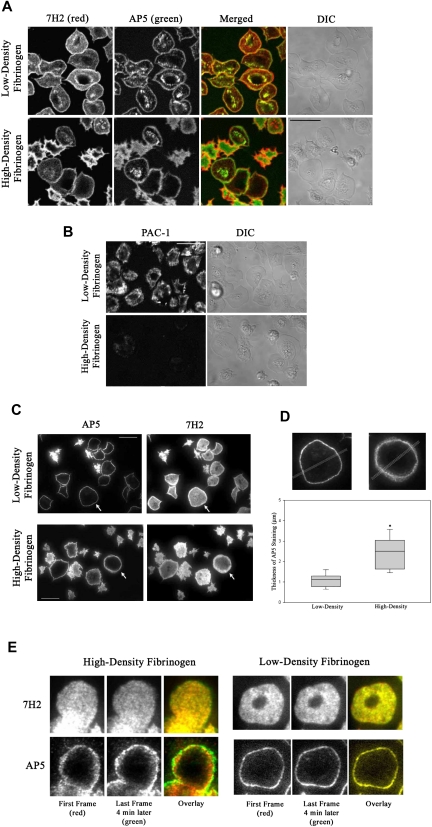
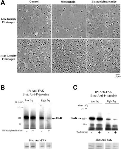
The impact of ligand density on integrin-mediated cell adhesion and outside-in signaling is not well understood. Using total internal reflection fluorescent microscopy, conformation-specific antibodies, and Ca(2+) flux measurements, we found that the surface density of fibrinogen affects alpha II b beta 3-mediated platelet signaling, adhesion, and spreading. Adhesion to fibrinogen immobilized at low density leads to rapid increases in cytosolic Ca(2+) and sequential formation of filopodia and lamellipodia. In contrast, adhesion to high-density fibrinogen results in transient or no increases in Ca(2+) and simultaneous formation of filopodia and lamellipodia. alpha II b beta 3 receptors at the basal surface of platelets engage fibrinogen in a ringlike pattern at the cell edges under both conditions. This engagement is, however, more dynamic and easily reversed on high-density fibrinogen. Src and Rac activity and actin polymerization are important for adhesion to low-density fibrinogen, whereas PKC/PI3 kinases contribute to platelet spreading on high-density fibrinogen. We conclude that 2 fundamentally different signaling mechanisms can be initiated by a single integrin receptor interacting with the same ligand when it is immobilized at different densities.


Figures

Figure 1
Figure 1
Filopodia and lamellipodia formation in platelets adhering to low- and high-density fibrinogen. Time-lapse TIR-FM imaging of platelets adhering to low- or high-density fibrinogen in the presence of Alexa 488-7H2Fab was recorded for 30 minutes after the addition of platelets into the fibrinogen-coated wells (Video S2). Individual platelets were analyzed for the appearance of new filopodia and onset of lamellipodia formation, counting the time they first appeared in the evanescent field (ie, within 200 nm of the substrate) as t = 0 seconds. The boxes in the figure represent the median and the 25th and 75th percentiles of the time to the onset and end of filopodia formation and the onset of lamellipodia formation. A total of 79 platelets were analyzed on low-density and 62 platelets on high-density fibrinogen from 2 independent experiments; *P = .03, **P = .04 low-density versus high-density fibrinogen.
Figure 2
Figure 2
Patterns of total and activated αIIbβ3 in platelets adherent to low- and high-density fibrinogen. Platelets were added to wells coated with fibrinogen and incubated for 1 hour. Wells were then washed and adherent platelets incubated with fluorescently labeled antibodies as described in “Materials and methods.” (A) By confocal microscopy, 7H2 and AP5 staining produced similar patterns on platelets adherent to both low- and high-density fibrinogen, with AP5 staining being more pronounced in the granulomere region in platelets on low-density fibrinogen. (B) PAC-1, mAb specific for activated αIIbβ3, intensely stained the surface of spread platelets on low-density fibrinogen but, under the same conditions, stained only weakly the surface of platelets spread on high-density fibrinogen. Differential interference contrast images (DIC) are shown on the right side for comparison of the platelet morphology. (C) TIR-FM of platelets double stained with AP5 and 7H2 revealed that AP5 staining on the basal surface of platelets spread on low-density fibrinogen appears in a very thin rim at the edge, whereas AP5 staining of spread platelets on high-density fibrinogen is much thicker and diffuse. Cells marked with arrows are magnified in panel D. Bars represent 10 μm. Images shown are representative of at least 2 independent experiments. (D) TIR-FM images were analyzed for the width of AP5 staining by line scan analysis. The box plot shows the median and the 25th and 75th percentiles for 21 and 16 platelets on low- and high-density fibrinogen, respectively, from 3 independent experiments, *P < .001. (E) 7H2 staining of receptors on a platelet spread on high-density fibrinogen is static, whereas AP5 staining shows radial movement. Alexa 546-7H2–labeled platelets were allowed to adhere for 1 hour and then Alexa 488-AP5 was added and both antibodies were imaged using TIR-FM for 5 minutes. The image of a platelet spread on high-density fibrinogen (Video S3) was taken at the beginning of the acquisition period (red) and then 4 minutes later (green). On both low- and high-density fibrinogen, 7H2 staining did not change between the first and last frame as demonstrated by overlay (yellow). AP5 staining of platelet on high-density fibrinogen showed radial extension as judged by the appearance of strong green ring outside the red/yellow staining on the overlaid image. AP5 staining on low-density fibrinogen was without a change between the first and last frame (yellow overlay).
Figure 3
Figure 3
Platelet adhesion to low-density fibrinogen leads to more rapid and sustained calcium response. Platelets loaded with calcium-sensitive dyes were added to wells coated with low- or high-density fibrinogen, and the cytosolic calcium fluxes during the first 30 minutes of the adhesion process were recorded using confocal microscopy as described in “Materials and methods” (Video S3). The adherent platelets were then analyzed according to their morphology and the characteristics of their calcium responses. (A) Population analysis demonstrated that most platelets adherent to high-density fibrinogen did not show any elevation in intracellular Ca2+ (*P < .001 high vs low-density fibrinogen, n = 3), whereas on low-density fibrinogen, approximately 40% of adherent platelets showed sustained Ca2+ oscillation (**P = .01). (B) Selected single platelet recordings of intracellular Ca2+ fluxes typical of nonresponsive platelet on high-density fibrinogen, a transient Ca2+ elevation in a platelet on high-density fibrinogen, and sustained oscillatory Ca2+ response in a platelet on low-density fibrinogen. Results are presented as mean ± SD.
Figure 4
Figure 4
Platelet adhesion to low-density fibrinogen induces greater protein tyrosine phosphorylation than adhesion to high-density fibrinogen. Platelets were allowed to adhere to fibrinogen (fbg)–coated or collagen (coll)–coated wells for 1 hour. After washing, adherent platelets were lysed in a buffer containing phosphatase inhibitors as described in “Materials and methods.” (A) Equal amounts of protein were subjected to electrophoresis and immunoblotting with mAbs specific for phosphotyrosine. Phosphotyrosine staining of proteins was less intense in platelets adherent to high-density compared with low-density fibrinogen; a protein of Mr approximately 100 kDa demonstrated approximately 50% less intense staining in platelets adherent to high-density than to low-density fibrinogen. For comparison, phosphotyrosine staining of proteins from platelets in suspension prior to adhesion is shown. (B) Equal amounts of protein lysates were used to immunoprecipitate FAK. Immunoprecipitated proteins were analyzed by immunoblotting for phosphotyrosine. Thereafter, the membranes were stripped and reanalyzed with antibody to FAK, to verify that the amounts of immunoprecipitated proteins were equal in all lanes. Results shown are representative of 3 independent experiments.
Figure 5
Figure 5
PKC and PI3K inhibition decreases FAK tyrosine phosphorylation and platelet spreading on high-density fibrinogen. Platelets were incubated with bisindolylmaleimide (10 μM; A-B), wortmannin (20 nM; A,C), or vehicle (control) and then allowed to adhere to wells precoated with fibrinogen. (A) Morphology of platelets adherent to low-density fibrinogen did not change with treatment with bisindolylmaleimide or wortmannin. Platelets adherent to high-density fibrinogen in the presence of these inhibitors showed less spreading than the control platelets. (B-C) Presence of PKC or PI3K inhibitors led to a decrease in FAK tyrosine phosphorylation in platelets adherent to high-density fibrinogen only. Noncontiguous lanes from a single blot are shown in both panels B and C. Results shown are representative of 3 independent experiments.
Figure 6
Figure 6
Apyrase and inhibitors of Src, Rac-1, or Rho kinase decrease platelet spreading on fibrinogen. Platelets were allowed to adhere to fibrinogen in the presence of a Src-kinase inhibitor (20 μM PP2), a Rac-1 inhibitor (100 μM NSC23766), or a Rho kinase inhibitor (5 μM H1152) for 1 hour. (A-B) Adherent platelets were fixed, permeabilized, and stained with fluorescently labeled phalloidin for F-actin. The Src (A) and Rac-1 (B) inhibitor and to a lesser extent the Rho kinase inhibitor (B) led to impaired spreading of platelets adhering to both low- and high-density fibrinogen. (C) Analysis of FAK immunoprecipitated from platelets adherent in the presence of Rac-1 and Rho kinase inhibitors revealed decreased phosphorylation of FAK in platelets adherent to high- but not to low-density fibrinogen. (D-E) Presence of apyrase led to less extensive spreading in platelets on both high- and low-density fibrinogen, but to decrease in FAK phosphorylation in platelets on high-density fibrinogen only. Noncontiguous lanes from a single blot are shown. Results shown are representative of 3 independent experiments.
Figure 7
Figure 7
Summary of differences in platelets signaling and spreading during αIIbβ3-mediated interaction with low- versus high-density fibrinogen. Attachment of platelets to low-density fibrinogen requires activity of Src, PKC, and PI3 kinases and actin polymerization. Full platelet spreading depends on Rac-1 and Rho kinase. Platelet adhesion to low-density fibrinogen further leads to inside-out signaling, resulting in αIIbβ3 activation and recruitment of additional platelets on top of the adherent platelets. In contrast, platelet attachment to high-density fibrinogen is possible even in the presence of inhibitors of Src, PKC, PI3-kinase, and actin polymerization. Platelet spreading depends on Src, PI3K, PKC, Rac-1, and Rho kinase activation. Few platelets are recruited to the adherent platelets indicating inadequate inside-out signaling or inhibition of luminal αIIbβ3 activation.

Similar articles

Cited by

References

    1. Hynes RO. Integrins: bidirectional, allosteric signaling machines. Cell. 2002;110:673–687. - PubMed
    1. Shattil SJ, Newman PJ. Integrins: dynamic scaffolds for adhesion and signaling in platelets. Blood. 2004;104:1606–1615. - PubMed
    1. Xiao T, Takagi J, Coller BS, Wang JH, Springer TA. Structural basis for allostery in integrins and binding to fibrinogen-mimetic therapeutics. Nature. 2004;432:59–67. - PMC - PubMed
    1. Xiong JP, Stehle T, Zhang R, et al. Crystal structure of the extracellular segment of integrin αVβ3 in complex with an Arg-Gly-Asp ligand. Science. 2002;296:151–155. - PubMed
    1. Hatton MW, Moar SL, Richardson M. Deendothelialization in vivo initiates a thrombogenic reaction at the rabbit aorta surface: correlation of uptake of fibrinogen and antithrombin III with thrombin generation by the exposed subendothelium. Am J Pathol. 1989;135:499–508. - PMC - PubMed

Publication types