Review
. 2007 Mar 2;100(4):489-501.
doi: 10.1161/01.RES.0000258451.44949.d7.

Regulation of phosphodiesterase 3 and inducible cAMP early repressor in the heart

Affiliations
Review

Regulation of phosphodiesterase 3 and inducible cAMP early repressor in the heart

Chen Yan et al. Circ Res. .

Abstract

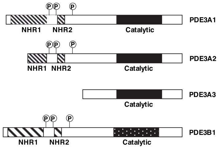
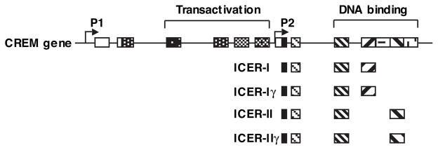
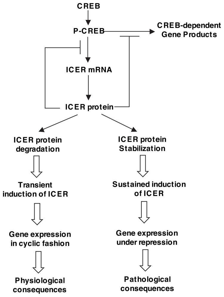
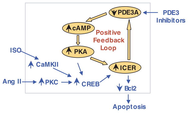
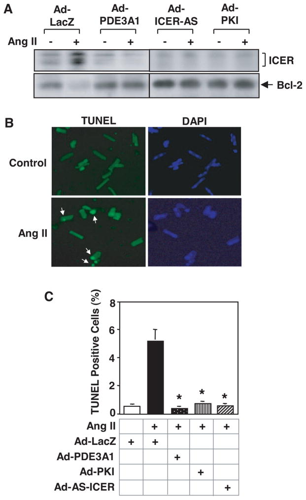
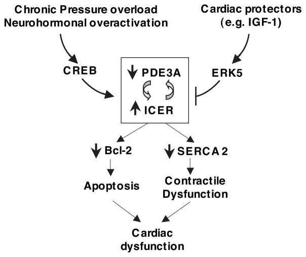
Growing evidence suggests that multiple spatially, temporally, and functionally distinct pools of cyclic nucleotides exist and regulate cardiac performance, from acute myocardial contractility to chronic gene expression and cardiac structural remodeling. Cyclic nucleotide phosphodiesterases (PDEs), by hydrolyzing cAMP and cyclic GMP, regulate the amplitude, duration, and compartmentation of cyclic nucleotide-mediated signaling. In particular, PDE3 enzymes play a major role in regulating cAMP metabolism in the cardiovascular system. PDE3 inhibitors, by raising cAMP content, have acute inotropic and vasodilatory effects in treating congestive heart failure but have increased mortality in long-term therapy. PDE3A expression is downregulated in human and animal failing hearts. In vitro, inhibition of PDE3A function is associated with myocyte apoptosis through sustained induction of a transcriptional repressor ICER (inducible cAMP early repressor) and thereby inhibition of antiapoptotic molecule Bcl-2 expression. Sustained induction of ICER may also cause the change of other protein expression implicated in human and animal failing hearts. These data suggest that the downregulation of PDE3A observed in failing hearts may play a causative role in the progression of heart failure, in part, by inducing ICER and promoting cardiac myocyte dysfunction. Hence, strategies that maintain PDE3A function may represent an attractive approach to circumvent myocyte apoptosis and cardiac dysfunction.


Figures

Figure 1
Figure 1
Structural organization of PDE3 isozymes. Three PDE3A gene products of PDE3A (PDE3A1, PDE3A2, and PDE3A3) and 1 PDE3B (PDE3B1) have been reported. The various PDE3A products differ considerably in the N-terminal portion via alternative splicing, transcription promoters, or translation start sites, while having an identical C-terminal portion that contains the PDE catalytic domain. The longest PDE3A1 contains a large N-terminal hydrophobic domain (NHR1, ≈200 aa) with 6 predicted transmembrane helices, followed by a small hydrophobic domain (NHR2, ≈50 aa) and 3 sites for phosphorylation by PKA, PKB, and PKA, respectively, between NHR1 and NHR2.
Figure 2
Figure 2
Schematic diagram showing the organization of CREM gene and ICER proteins. Line indicates intron; box, exon; open box, noncoding region; filled box, coding region; P1 and P2, promoters.
Figure 3
Figure 3
The role of transient versus sustained induction of ICER. Transcription activator CREB is activated on phosphorylation at serine 133. Phosphorylated CREB (p-CREB) binds to CRE in the promoter region and activates transcription. ICER is also transcriptionally induced by p-CREB via a CRE-binding sequence in the ICER promoter. Induction of ICER, in turn, blocks CREB-mediated gene transcription, including ICER itself, by competing with CREB in binding CRE sequences. ICER proteins are subjected to proteasome-dependent degradation. Reduction of ICER allows cAMP-inducible gene expression to be activated repeatedly. Therefore, ICER induction is a transient phenomenon. This transient expression of ICER is physiologically important for negative-feedback regulation of CREB-dependent gene expression and allows CREB-dependent genes to be activated in a cyclic fashion. In contrast, sustained induction of ICER may cause persistent suppression of CREB-regulated gene expression and thus lead to pathological consequences.
Figure 4
Figure 4
Schematic diagram showing the PDE3A-ICER positive-feedback loop stimulated by Ang II, ISO, and PDE3 inhibitors, leading to myocyte apoptosis. With respect to Ang II, activation of PKC and CREB by Ang II via AT1R initiates the PDE3A-ICER feedback loop probably by CREB-dependent ICER gene transcription (shown in blue). Induction of ICER leads to a reduction of PDE3A expression, and reduction of PDE3A leads to ICER elevation because of PKA-dependent ICER stabilization that prevents ICER degradation; this constitutes the positive PDE3A-ICER feedback loop (shown in orange) and allows a persistent induction of ICER, which plays a key role in cardiac myocyte apoptosis. CaMKII indicates Ca2+/calmodulin kinase II. Adapted from Ding et al.
Figure 5
Figure 5
Role of PDE3-ICER feedback loop in the regulation of rat adult cardiomyocyte apoptosis. Rat adult cardiac myocytes were transduced with 100 multiplicities of infection of Ad-LacZ, Ad-PDE3A1, Ad-PKI, or Ad-AS-ICER, followed by the treatment with vehicle or Ang II (200 nmol/L) for 48 hours. A, Western blots showing the expression levels of ICER and Bcl2. B, Microscopic images of cardiomyocytes stained with TUNEL (left panels) and DAPI (4′,6′-diamido-2-phenylindole) (right panels). Arrows point to apoptotic cells showing positive nuclear TUNEL staining. C, Quantitative results of TUNEL experiments. Data are means±SEM (n=3). *P<0.vs Ang II+Ad-LacZ. Adapted from Ding et al.
Figure 6
Figure 6
Proposed model showing the regulation and function the PDE3A-ICER feedback loop. Pathological stimuli (such as chronic pressure overload and neurohormonal overactivation) promote the PDE3A-ICER feedback loop via initial activation of CREB, whereas cardiac protectors (such as IGF-1) attenuate it through activation of ERK5. The persistent ICER induction resulting from the PDE3A-ICER feedback loop dysregulates the expression of a number of proteins (such as downregulation of Bcl-2 and SERCA2) and may, in turn, lead to cardiac myocyte death and contractile dysfunction, which is associated with impaired cardiac function.

References

    1. Lohse MJ, Engelhardt S, Eschenhagen T. What is the role of beta-adrenergic signaling in heart failure? Circ Res. 2003;93:896–906. - PubMed
    1. Lohmann SM, Fischmeister R, Walter U. Signal transduction by cGMP in heart. Basic Res Cardiol. 1991;86:503–514. - PubMed
    1. Soderling SH, Beavo JA. Regulation of cAMP and cGMP signaling: new phosphodiesterases and new functions. Curr Opin Cell Biol. 2000;12:174–179. - PubMed
    1. Jurevicius J, Fischmeister R. cAMP compartmentation is responsible for a local activation of cardiac Ca2+ channels by beta-adrenergic agonists. Proc Natl Acad Sci U S A. 1996;93:295–299. - PMC - PubMed
    1. Zaccolo M, Pozzan T. Discrete microdomains with high concentration of cAMP in stimulated rat neonatal cardiac myocytes. Science. 2002;295:1711–1715. - PubMed

Publication types

MeSH terms

Substances