. 2007 May;18(5):1701-9.
doi: 10.1091/mbc.e06-08-0747. Epub 2007 Mar 1.

Dynamic regulation of p53 subnuclear localization and senescence by MORC3

Affiliations

Dynamic regulation of p53 subnuclear localization and senescence by MORC3

Keiko Takahashi et al. Mol Biol Cell. 2007 May.

Abstract

The tumor suppressor p53 is a key transcriptional factor regulating the induction of cellular senescence by oncogenic signals. The activity of p53 is regulated by recruitment into promyelocytic leukemia (PML)-nuclear bodies (NBs) as well as by stabilization through posttranslational modifications such as phosphorylation and acetylation. Here we found that MORC3 (microrchidia3)-ATPase activated p53 and induced cellular senescence in normal human and mouse fibroblasts but not p53-/- fibroblasts. Conversely, genotoxic stress-induced phosphorylation and stabilization of p53 but barely increased its transcriptional activity in Morc3-/- fibroblasts. MORC3 localized on PML-NBs in presence of PML and mediated recruitment of p53 and CREB-binding protein (CBP) into PML-NBs. In contrast, expression of ATPase activity-deficient mutant MORC3-E35A or siRNA repression of MORC3 impaired the localization of p53 and Sp100 but not CBP on PML-NBs. These results suggest that MORC3 regulates p53 activity and localization into PML-NBs. We identified a new molecular mechanism that regulates the activity of nuclear proteins by localization to a nuclear subdomain.


Figures

Figure 1.
Figure 1.
MORC3 induces p53-dependent premature senescence. Data in A, C, and E represent mean ± SD of triplicate experiments. (A) Human normal fibroblasts WI38 infected with a retroviral vector carrying EGFP (Vector, •), EGFP-MORC3 (MORC3, □) or EGFP-PML IV (PML IV, ▴) were selected with puromycin for 4 d, and the number of the cells was counted at days 3, 6, 9, and 12. Day 0 indicates the first day after the selection. (B) Representative senescence-associated β-galactosidase (SA-β-Gal) staining in WI38 cells infected with a vector carrying EGFP (Vector, left), EGFP-MORC3 (MORC3, middle) or EGFP-PML IV (PML IV, right). SA-β-Gal–positive cells were blue (top images). Nuclei were stained with 1 μg/ml 4′,6-diamidino-2-phenylindole (DAPI, bottom images). (C) Quantitation of SA-β-Gal–positive cells as shown in B. More than 249 cells were counted in each experiment. (D) Induction of p16 in WI38 cells infected with vector (lane 1), vector carrying MORC3 (lane 2), or PML IV (lane 3). Lysates were collected at day 6 and immunoblotted with anti-p16 or anti-β-tubulin antibody. (E) The retrovirus control vector (circles) or the vector carrying MORC3 (squares) was infected into p53+/+ (filled) or p53−/− (open) mouse embryonic fibroblasts (MEFs), and the population doublings of these cells were measured. p53+/+ and p53−/− MEFs were generated from littermate embryos.
Figure 2.
Figure 2.
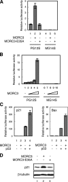
MORC3 transactivates p53. (A) U-2 OS cells were cotransfected with the control vector, pMEPy-FLAG-MORC3, or pMEPy-FLAG-MORC3-E35A, and firefly luciferase reporter constructs (PG12S-Luc containing 12 p53-binding elements or MG14S-Luc containing 14 mutated p53-binding elements). Relative activities were calculated from arbitrary light units of firefly luciferase activity normalized for transfection efficiency (see Materials and Methods). Data represent mean ± SD of triplicate transfections. (B) U-2 OS cells were cotransfected with firefly luciferase constructs (PG12S-Luc or MG14S-Luc, 35 ng) and increasing amounts of the MORC3 expression vector (0, 8.75, 17.5, 35, and 70 ng). Total amount of plasmid DNA was kept constant by the addition of empty vector. Luciferase activities were measured as described in A. (C) MORC3 induces p53-transactivation of the p21 (left) and Bax (right) promoters. H1299 cells were transfected with p21-CBG99luc containing the p21 promoter or Bax-CBG99luc containing the Bax promoter together with pMEPy-FLAG-MORC3 and pMEPy-HA-p53. The CBG99 luciferase reporter construct, pMEPy-FLAG-MORC3, pMEPy-HA-p53, and CBR luciferase control vector were transfected at amount ratios of 40:80:1:320 in the p21 promoter assay and 20:12.5:1:10 in the Bax promoter assay, respectively. (D) U-2 OS cells were transfected with the pMEPy vector (lane 1), pMEPy-FLAG-MORC3 (lane 2), or its E35A mutant (lane 3). Three days after transfection, total lysates of transfectants were immunoblotted for p21 and β-tubulin.
Figure 3.
Figure 3.
Impairment of p53 activation by adriamycin (ADR) treatment in MEFs derived from Morc3−/− mice. (A) Organization of mouse Morc3 gene, targeting vector, and knockout allele. Gray boxes denote exons (2–9). Black, gray, and white arrows indicate hrGFP (Stratagene), neomycin resistance gene (neo) and Diphtheria toxin fragment A (DT-A) expression cassette, respectively. White arrowheads indicate loxP sites. P, PvuII; N, NcoI. (B) Southern blot analysis of genomic tail DNA digested with PvuII using the 5′ probe shown in A. An 8-kbp band in wild-type mice (lanes 1 and 2), a 6-kbp band in homozygous mice (lanes 5 and 6), and both bands in heterozygous mice (lanes 3 and 4) were detected. (C) Western blot analysis of lysates of MEFs from wild-type (lane 1), heterozygous (lane 2), and homozygous (lane 3) mice. Morc3 protein was detected with a rabbit polyclonal antibody. (D) Morc3+/+ and Morc3−/− MEFs were treated with 1 μg/ml adriamycin (ADR) for 0–12 h. Total cell lysates at 0, 4, 8, and 12 h were immunoblotted for p21, p53, p53 with acetylated lysine 373 and lysine 382 (Acetyl-p53; mouse K370 and K379), Morc3, and β-tubulin. This experiment was performed at least three times, with similar results. (E) Morc3+/+ and Morc3−/− MEFs were treated with ADR for 12 h. Total cell lysates from treated or untreated MEFs were separated by SDS-PAGE and immunoblotted with antibodies for p21, p53, p53 with phosphorylated serine 15 (p53 Ser15; mouse S18) and serine 20 (p53 Ser20; mouse S23), Acetyl-p53, Morc3, and β-tubulin.
Figure 4.
Figure 4.
MORC3 localization to PML-NBs depends on its GHL-ATPase activity. Overlapping images are shown in the right side (Merge) of B–F and H. (A) Morc3+/+ and −/− MEFs were stained by indirect immunofluorescence with anti-MORC3 monoclonal antibody (17A9; green). This antibody specifically detected nuclei of Morc3+/+ (right image) but not Morc3−/− (left image) MEFs. (B) Representative images of Saos-2 cells stained with anti-MORC3 antibody (17A9; green), anti-PML antibody (red, top images) and anti-Sp100 antibody (red, bottom images). MORC3 colocalized with PML and Sp100 on PML-NBs. (C) HeLa cells were transiently transfected with the FLAG-MORC3 expression vector and stained for FLAG (green), and PML (red, top image), or Sp100 (red, bottom image). Overexpressed MORC3 colocalized with PML and with Sp100. (D) Acute promyelocytic leukemia NB4 cells were incubated in the presence of 1 μM of all-trans-retinoic acid (ATRA, bottom images) or vehicle (DMSO, top images) and stained for MORC3 (green) and PML (red). In the absence of ATRA, MORC3 was diffusely localized to entire nuclei except for nucleoli. Treatment with ATRA-mediated localization of MORC3 into PML-NBs in cells with reorganized PML-NBs. (E) PML-knockdown vector-transfected Saos-2 cells were stained for MORC3 (green) and PML (red). MORC3 was completely dispersed or localized to two or fewer obvious nuclear foci in 73.3% of cells in which PML was repressed, as indicated by two or fewer obvious PML-nuclear foci. In contrast, we detected cells containing two or fewer MORC3 nuclear foci in 38.5% of cells transfected with a control vector (data not shown). (F) WI38 cells were infected with a retrovirus vector carrying FLAG-PML IV, selected with puromycin, and stained for MORC3 (green) and for FLAG (red) at day 1. PML-NBs localization of endogenous MORC3 was enhanced in FLAG-PML IV expressing cells. (G) ATPase-activity–deficient mutant (FLAG-MORC3-E35A)-expressing HeLa cells were stained for FLAG (E35A). MORC3-E35A did not accumulate in PML-NBs and diffusely localized to entire nuclei except for nucleoli. (H) FLAG-MORC3-expressing HeLa cells were fixed after ATP depletion for 30 min and stained for FLAG (green) and PML (red). Left image shows low-power view; other images show magnified view of boxed region. ATP depletion caused nuclear dispersion of MORC3 but not PML from PML-NBs. (I) ATP in HeLa cells transfected with pME-EGFP-MORC3-I-P and pME-FLAG-MORC3-I-P at a 1:8 ratio was depleted by the addition of sodium azide and 2-deoxyglucose to the culture medium. Cells in the same field were observed by phase contrast (left) or epifluorescence microscopy after the indicated times. EGFP-MORC3 gradually dispersed from nuclear foci with ATP depletion.
Figure 5.
Figure 5.
ATPase-dependent recruitment of Sp100, CBP, and p53 into PML-NBs by MORC3. (A) and (B) FLAG-MORC3–expressing U-2 OS cells (A) and WI38 cells (B) were stained for FLAG, and endogenous p53 (top images) or CBP (bottom images). WI38 cells infected with pLNCX2-FLAG-MORC3 retrovirus vector were selected with puromycin for 4 d and stained at day 1. Both p53 and CBP were completely colocalized with FLAG-MORC3 in all FLAG-MORC3–expressing U-2 OS cells. In WI38 cells, CBP accumulated in PML-NBs of all FLAG-MORC3–expressing cells, but p53 accumulated in only a portion of FLAG-MORC3-expressing cells (14.3% of cells at day 1 and 47.4% of cells at day 3). (C) HeLa cells transfected with control vector pH1′-DsRed2-I-P (Vector, lanes 1 and 2), mismatched shRNA vector pH1′-shRNA695-DsRed2-I-P containing three mismatched nucleotides (Mismatched shRNA, lanes 3 and 4) or knockdown shRNA vector pH1′-shRNA732-DsRed2-I-P (Knockdown shRNA, lane 5 and 6) were selected with 1 μg/ml puromycin for 2 d. Lysates of the transfected cells were immunoblotted with anti-MORC3 rabbit polyclonal antibody or anti-β-tubulin antibody. MORC3 expression was assayed in two independent shRNA transfections. (D) HeLa cells were transfected with knockdown shRNA vector shRNA732. After selection with puromycin, the transfected cells were stained for Sp100 (green). Sp100 was dispersed in 41.3% of DsRed2-positive cells (DsRed) in which expression of MORC3 should be suppressed by shRNA. (E) Saos-2 cells were transfected with the MORC3-knockdown shRNA vector shRNA732 (shRNA, middle and bottom images) or the control vector pH1′-DsRed2-I-P (Vector, top images) (1 μg), and the HA-p53 (1 μg) and FLAG-PML IV (3 μg) expression vectors, selected with puromycin for 2 d and treated with 1 μg/ml ADR for 6 h. These cells were permeabilized before fixation and stained for HA (green, top and middle images) or CBP (green, bottom images), and FLAG (red). After ADR treatment, HA-p53 accumulated in nuclear bodies containing FLAG-PML IV in vector-only–transfected cells (Vector, top images) but not in knockdown shRNA vector-transfected cells (middle images). Endogenous CBP colocalized with FLAG-PML IV in both vector-only and MORC3-knockdown vector-transfected cells (bottom images). (F) Localization of endogenous Sp100 (red, top image) and endogenous PML (red, bottom image) in HeLa cells expressing FLAG-MORC3-E35A (green). Expression of MORC3-E35A caused complete dispersion of Sp100 in 91% of cells, whereas PML was still localized in NBs.

References

    1. Ban C., Junop M., Yang W. Transformation of MutL by ATP binding and hydrolysis: a switch in DNA mismatch repair. Cell. 1999;97:85–97. - PubMed
    1. Bischof O., Kirsh O., Pearson M., Itahana K., Pelicci P. G., Dejean A. Deconstructing PML-induced premature senescence. EMBO J. 2002;21:3358–3369. - PMC - PubMed
    1. Bode A. M., Dong Z. Post-translational modification of p53 in tumorigenesis. Nat. Rev. Cancer. 2004;4:793–805. - PubMed
    1. Borden K. L. Pondering the promyelocytic leukemia protein (PML) puzzle: possible functions for PML nuclear bodies. Mol. Cell Biol. 2002;22:5259–5269. - PMC - PubMed
    1. Braig M., Lee S., Loddenkemper C., Rudolph C., Peters A. H., Schlegelberger B., Stein H., Dorken B., Jenuwein T., Schmitt C. A. Oncogene-induced senescence as an initial barrier in lymphoma development. Nature. 2005;436:660–665. - PubMed

Publication types

MeSH terms