Comparative Study
. 2007 Mar 21;26(6):1737-48.
doi: 10.1038/sj.emboj.7601631. Epub 2007 Mar 1.

Composition and three-dimensional EM structure of double affinity-purified, human prespliceosomal A complexes

Affiliations
Comparative Study

Composition and three-dimensional EM structure of double affinity-purified, human prespliceosomal A complexes

Nastaran Behzadnia et al. EMBO J. .

Abstract

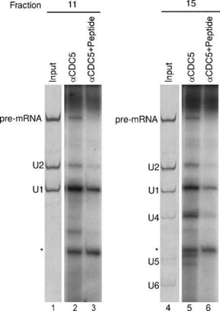
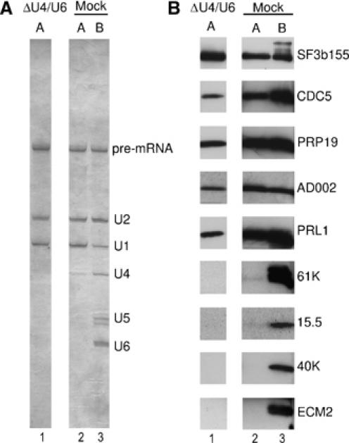
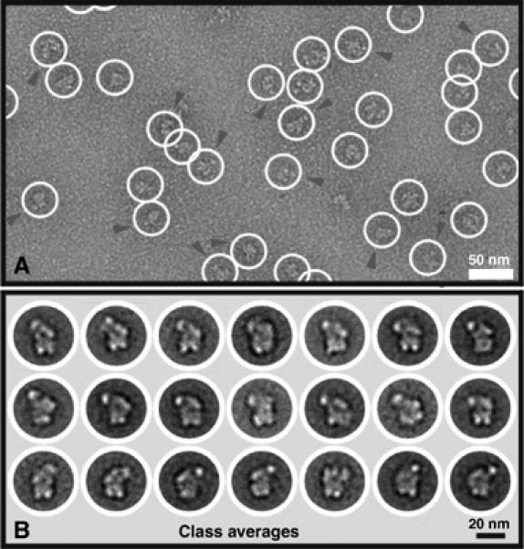
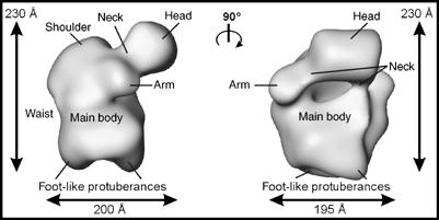
Little is known about the higher-order structure of prespliceosomal A complexes, in which pairing of the pre-mRNA's splice sites occurs. Here, human A complexes were isolated under physiological conditions by double-affinity selection. Purified complexes contained stoichiometric amounts of U1, U2 and pre-mRNA, and crosslinking studies indicated that these form concomitant base pairing interactions with one another. A complexes contained nearly all U1 and U2 proteins plus approximately 50 non-snRNP proteins. Unexpectedly, proteins of the hPrp19/CDC5 complex were also detected, even when A complexes were formed in the absence of U4/U6 snRNPs, demonstrating that they associate independent of the tri-snRNP. Double-affinity purification yielded structurally homogeneous A complexes as evidenced by electron microscopy, and allowed for the first time the generation of a three-dimensional structure. A complexes possess an asymmetric shape (approximately 260 x 200 x 195 angstroms) and contain a main body with various protruding elements, including a head-like domain and foot-like protrusions. Complexes isolated here are well suited for in vitro assembly studies to determine factor requirements for the A to B complex transition.


Figures

Figure 1
Figure 1
Characterization of double affinity-purified spliceosomal A complexes. Complexes were purified by a combination of tobramycin and anti-SF3a66 affinity selection, and fractionated on a glycerol gradient. RNA was isolated from the indicated fractions, analyzed by denaturing PAGE and visualized by silver staining (upper panel) or autoradiography (lower panel). RNA identities are indicated on the right. Contaminating high-molecular-weight RNA (most likely rRNA from the 40S subunit) is indicated by an asterisk. 30S and 50S correspond to migration positions of Escherichia coli ribosomal subunits in parallel gradients.
Figure 2
Figure 2
Identification of RNA–RNA interactions in double affinity-purified spliceosomal complexes. Gradient fractions 7–15 containing double affinity-purified complexes or 0.5% of the unfractionated tobramycin eluate (E) were UV irradiated in the presence (+; lanes 1 and 3–12) or absence (−; lane 2) of psoralen (AMT); note that the input of the gradient fractionated material, that is, the anti-SF3a66 eluate, is not shown. Recovered RNA was separated by denaturing PAGE and transferred to a nylon membrane that was hybridized sequentially with 32P-labeled probes against the U2 snRNA (C), U1 (B) or pre-mRNA (A). A low level of 32P-labeled pre-mRNA (present in the purified complexes) is detected in panels B and C, but crosslinked pre-mRNA species were not observed before Northern blotting. The identity of the crosslinked species is indicated on the right, where ‘PRE' indicates pre-mRNA and ‘intPRE' internally crosslinked pre-mRNA. Asterisks: the identity of this double band is unknown.
Figure 3
Figure 3
Characterization of crosslinks by RNase H digestion. (A–C) Pooled gradient fractions 10–12 were UV irradiated in the presence of psoralen. Recovered RNA was incubated with RNase H in the absence (lane 1) or presence of DNA oligonucleotides against the pre-mRNA (lane 2), U1 (lane 3) or U2 snRNA (lane 4) and then analyzed by Northern blotting as described in Figure 2. A longer exposure of the upper portion of the gel is shown below in panel C. Additional minor crosslinks are discussed in the text.
Figure 4
Figure 4
A complexes are precipitated by anti-CDC5 antibodies. Immunoprecipitations were performed with glycerol gradient fractions 11 and 15 (as indicated) containing double affinity-purified complexes, and anti-CDC5 antibodies (lanes 2 and 5) or anti-CDC5 antibodies preblocked with cognate CDC5 peptide (lanes 3 and 6). Coprecipitated RNAs were labeled with 32P-pCp, fractionated by 10% denaturing PAGE and detected by autoradiography (lanes 2, 3, 5 and 6). Lanes 1 and 4: RNA recovered directly from gradient fraction 11 or 15 and visualized by silver staining. The identity of the various RNAs is indicated on the left. The asterisk indicates a band arising due to the addition of tRNA before pCp labeling.
Figure 5
Figure 5
hPrp19/CDC5 complex proteins bind to the A complex independent of the tri-snRNP. (A, B) Spliceosomal complexes, MS2 affinity-selected from U4/U6-depleted (lane 1) and mock-depleted (lanes 2 and 3) nuclear extract, were fractionated on a glycerol gradient. A complex (lanes 1 and 2) and B complex fractions (lane 3) were separately pooled. (A) RNA was isolated, analyzed by denaturing PAGE and visualized by silver staining. (B) Proteins were recovered, separated by SDS–PAGE and immunoblotting was performed with the indicated antibodies.
Figure 6
Figure 6
EM of the A complex and 2D analysis. (A) Typical EM image of the A complex taken with a 4k × 4k CCD camera in tile mode. The image reveals a homogenous population of globular particles (circled) with maximum dimensions of ∼260 Å (diagonal). In some views, the head-like domain has been marked by an arrowhead. (B) 2D analysis of the A complex. Multiple class averages are shown and reveal particles with a globular main body with two bottom protuberances and an upper side protuberance (pointing to the left in rows 1 and 2, and pointing to the right in row 3).
Figure 7
Figure 7
3D structure of the A complex. A 3D structure was computed using the RCT approach and two views of its surface representation are separated by a 90° rotation. The A complex has a maximum size of ∼260 Å (diagonal). Characteristic landmarks are labeled for orientation and are described in the main text.

References

    1. Ajuh P, Kuster B, Panov K, Zomerdijk JC, Mann M, Lamond AI (2000) Functional analysis of the human CDC5L complex and identification of its components by mass spectrometry. EMBO J 19: 6569–6581 - PMC - PubMed
    1. Ajuh P, Sleeman J, Chusainow J, Lamond AI (2001) A direct interaction between the carboxyl-terminal region of CDC5L and the WD40 domain of PLRG1 is essential for pre-mRNA splicing. J Biol Chem 276: 42370–42381 - PubMed
    1. Azubel M, Wolf SG, Sperling J, Sperling R (2004) Three-dimensional structure of the native spliceosome by cryo-electron microscopy. Mol Cell 15: 833–839 - PubMed
    1. Behzadnia N, Hartmuth K, Will CL, Lührmann R (2006) Functional spliceosomal A complexes can be assembled in vitro in the absence of a penta-snRNP. RNA 12: 1738–1745 - PMC - PubMed
    1. Boehringer D, Makarov EM, Sander B, Makarova OV, Kastner B, Lührmann R, Stark H (2004) Three-dimensional structure of a pre-catalytic human spliceosomal complex B. Nat Struct Mol Biol 11: 463–468 - PubMed

Publication types

LinkOut - more resources