. 2007 Mar 21;26(6):1681-90.
doi: 10.1038/sj.emboj.7601624. Epub 2007 Mar 1.

Analysis of interactions in a tapasin/class I complex provides a mechanism for peptide selection

Affiliations

Analysis of interactions in a tapasin/class I complex provides a mechanism for peptide selection

Mingnan Chen et al. EMBO J. .

Abstract

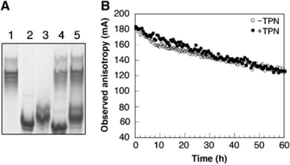
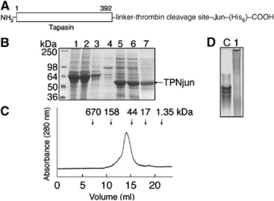
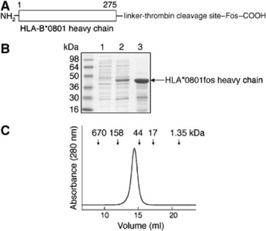
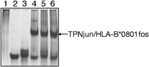
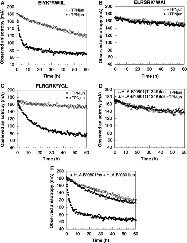
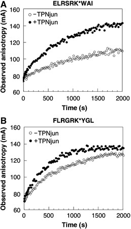
We examined interactions in a soluble tapasin (TPN)/HLA-B*0801 complex to gain mechanistic insights into the functions of TPN. Results show that TPN acts as a chaperone by increasing the ratio of active-to-inactive peptide-deficient HLA-B*0801 molecules in solution. TPN causes peptides to associate and dissociate faster owing to its effect on widening the binding groove of HLA-B*0801 molecules. Our data indicate that a TPN-assisted mechanism of peptide selection relies on disruption of conserved hydrogen bonds at the C-terminal end of the groove. Peptide sequence-dependent interactions along the entire length of the groove also play a role in this mechanism. We suggest that TPN influences presentation of antigenic peptides according to a mechanistically complicated process in which bound candidate peptides that are unable to conformationally disengage TPN from class I molecules are excluded from the repertoire. Overall, these studies unify our understanding of the functions of TPN.


Figures

Figure 1
Figure 1
Interaction between recombinant, soluble TPN and HLA-B*0801. (A) TPN (10 μg) and HLA-B*0801 (10 μg) (1:1 molar ratio) were incubated on ice in 20 mM Tris, 150 mM NaCl, and 10% glycerol (pH 7.5) for 30 min. Samples were analyzed on a native gel (8%): lane 1, TPN; lane 2, peptide-filled HLA-B*0801; lane 3, peptide-deficient HLA-B*0801; lane 4, mixture of TPN and peptide-filled HLA-B*0801; and lane 5, mixture of TPN and peptide-deficient HLA-B*0801. The gel was run at 4°C in 25 mM Tris and 200 mM glycine (pH 8.3). (B) At time=0, HLA-B*0801 (40 nM) loaded with EIYK*RWIIL (○) was added under stirring to a 1-cm cuvette containing 1000-fold molar excess of the nontagged peptide and 100-fold molar excess of β2m in 20 mM Hepes and 150 mM NaCl (pH 7.5). Peptide dissociation was monitored at 20°C (excitation and emission wavelengths were 495 and 524 nm, respectively). The experiment was repeated in the presence of TPN (400 nM) (•), which was pre-mixed in the buffer before adding HLA-B*0801. Data points were collected for 60 h.
Figure 2
Figure 2
Characterization of TPNjun. (A) The ER-lumenal domain of TPN was extended at its C-terminus with a linker (GGSGG), a thrombin site, a Jun leucine peptide, and a (His)6 sequence. (B) SDS–PAGE (12%) analysis of TPNjun. Lane 1, supernatant from High Five insect cells at 60 h after infection; lane 2, flow-through after washing column with 10 mM imidazole; lane 4, flow-through after washing column with 25 mM imidazole; lane 5, Ni-NTA matrix beads showing bound TPNjun; lane 6, flow-through after washing column with 250 mM imidazole to elute TPNjun; and lane 7, TPNjun after purification on a Superdex 200 HR 10/300 column at 10°C in 20 mM Tris and 150 mM NaCl (pH 7.5). (C) Gel filtration chromatogram of purified TPNjun (see (B) for conditions). The column was calibrated using protein standards that elute at positions corresponding to arrows at the top of the chromatogram. (D) Native PAGE (8%) analysis of purified TPNjun (lane 1) showing a protein smear while soluble TPN (lane C) migrates as a series of closely spaced bands.
Figure 3
Figure 3
Characterization of the ER-lumenal domain of HLA-B*0801fos heavy chain. (A) HLA-B*0801 heavy chain was extended at its C-terminus with a linker (GGSGG), a thrombin site, and a Fos leucine peptide. (B) SDS–PAGE (12%) analysis of HLA-B*0801fos heavy chain from bacterial cells. Lane 1, before induction; lane 2, after induction with IPTG; and lane 3, purified inclusion bodies. (C) Gel filtration chromatogram of purified peptide-filled HLA-B*0801fos (see Figure 2B for conditions).
Figure 4
Figure 4
Formation of the TPNjun/HLA-B*0801fos complex. TPNjun (10 μg) and HLA-B*0801fos (10 μg) (1:1 molar ratio) were incubated as described in Figure 1A. Samples were analyzed on native gel (8%): lane 1, TPNjun; lane 2, peptide-filled HLA-B*0801fos; lane 3, peptide-deficient HLA-B*0801fos; lane 4, mixture of TPNjun and peptide-filled HLA-B*0801fos; lane 5, mixture of TPNjun and peptide-deficient HLA-B*0801fos; and lane 6, TPNjun/peptide-deficient HLA-B*0801fos complex incubated with an excess of FLRGRAYGL. The gel was run as described in Figure 1A.
Figure 5
Figure 5
Effect of TPNjun on the kinetics of peptide dissociation. (A) Dissociation of EIYK*RWIIL was monitored as described in Figure 1B: HLA-B*0801fos alone (40 nM) (○) and in the presence of TPNjun (400 nM) (•). Data points were collected at various time points over a period of 166 h (−TPN) or continuously for 60 h (+TPN). (B) As described in Figure 1B for ELRSRK*WAI: HLA-B*0801fos alone (40 nM) (○) and in the presence of TPNjun (400 nM) (•). Data points were collected at various time points over a period of 576 h. (C) As described in Figure 1B for FLRGRK*YGL: HLA-B*0801fos alone (40 nM) (○) and in the presence of TPNjun (400 nM) (•). Data points were collected at various time points over a period of 546 h (−TPN) or continuously for 89 h (+TPN). (D) Dissociation of FLRGRK*YGL from HLA-B*0801(T134K)fos (40 nM) in the absence (▵) or presence (▴) of TPNjun (400 nM) was monitored as described in Figure 1B. Data points were collected continuously for 60 h. (E) Dissociation of EIYK*RWIIL from HLA-B*0801fos (40 nM) in the presence of HLA-B*0801jun/ELRSRKWAI (400 nM) (▴) was monitored as described in Figure 1B. Data points were collected continuously for 60 h. The decay curves shown in (A) are included for comparison purposes.
Figure 6
Figure 6
Effect of TPNjun on the kinetics of peptide association. (A) ELRSRK*WAI (160 nM) was mixed automatically with an equal volume of peptide-deficient HLA-B*0801fos (800 nM) (○) together with a 100-fold molar excess of β2m in 20 mM Hepes and 150 mM NaCl (pH 7.5). The time course of the reaction was monitored at 20°C. The binding of ELRSRK*WAI to peptide-deficient molecules in the presence of TPNjun (600 nM) (•) is shown. (B) As described in (A) for FLRGRK*YGL: peptide-deficient HLA-B*0801fos alone (800 nM) (○) and in the presence of TPNjun (600 nM) (•). For (A) and (B), data points were collected for 2000 s.

References

    1. Barber LD, Howarth M, Bowness P, Elliott T (2001) The quantity of naturally processed peptides stably bound by HLA-A*0201 is significantly reduced in the absence of tapasin. Tissue Antigens 58: 363–468 - PubMed
    1. Barnden MJ, Purcell AW, Gorman JJ, McCluskey J (2000) Tapasin-mediated retention and optimization of peptide ligands during the assembly of class I molecules. J Immunol 165: 322–330 - PubMed
    1. Belmares MP, Busch R, Wucherpfennig KW, McConnell HM, Mellins ED (2002) Structural factors contributing to DM susceptibility of MHC class II/peptide complexes. J Immunol 169: 5109–5117 - PubMed
    1. Binz AK, Rodriguez RC, Biddison WE, Baker BM (2003) Thermodynamic and kinetic analysis of a peptide-class I MHC interaction highlights the noncovalent nature and conformational dynamics of the class I heterotrimer. Biochemistry 42: 4954–4961 - PubMed
    1. Bouvier M, Wiley DC (1994) Importance of peptide amino and carboxyl termini to the stability of MHC class I molecules. Science 265: 398–402 - PubMed

Publication types