Comparative Study
. 2007 Mar 21;26(6):1713-25.
doi: 10.1038/sj.emboj.7601618. Epub 2007 Mar 1.

Structures and physiological roles of 13 integral lipids of bovine heart cytochrome c oxidase

Affiliations
Comparative Study

Structures and physiological roles of 13 integral lipids of bovine heart cytochrome c oxidase

Kyoko Shinzawa-Itoh et al. EMBO J. .

Abstract

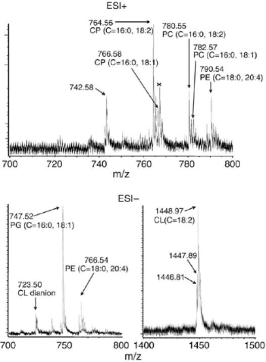
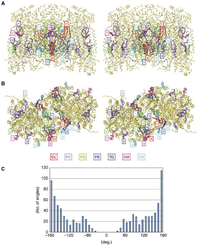
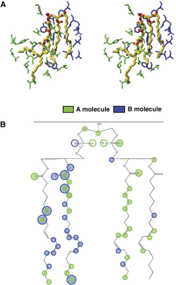
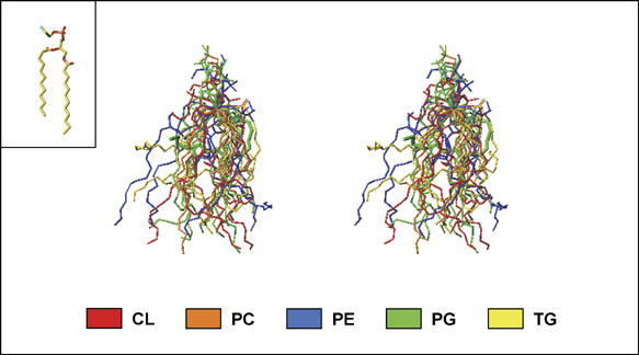
All 13 lipids, including two cardiolipins, one phosphatidylcholine, three phosphatidylethanolamines, four phosphatidylglycerols and three triglycerides, were identified in a crystalline bovine heart cytochrome c oxidase (CcO) preparation. The chain lengths and unsaturated bond positions of the fatty acid moieties determined by mass spectrometry suggest that each lipid head group identifies its specific binding site within CcOs. The X-ray structure demonstrates that the flexibility of the fatty acid tails facilitates their effective space-filling functions and that the four phospholipids stabilize the CcO dimer. Binding of dicyclohexylcarbodiimide to the O(2) transfer pathway of CcO causes two palmitate tails of phosphatidylglycerols to block the pathway, suggesting that the palmitates control the O(2) transfer process.The phosphatidylglycerol with vaccenate (cis-Delta(11)-octadecenoate) was found in CcOs of bovine and Paracoccus denitrificans, the ancestor of mitochondrion, indicating that the vaccenate is conserved in bovine CcO in spite of the abundance of oleate (cis-Delta(9)-octadecenoate). The X-ray structure indicates that the protein moiety selects cis-vaccenate near the O(2) transfer pathway against trans-vaccenate. These results suggest that vaccenate plays a critical role in the O(2) transfer mechanism.


Figures

Figure 1
Figure 1
Mass spectra of lipid fraction extracted from the crystalline bovine heart CcO preparation, using an Accu TOF (JEOL, JMS-T100LC) mass spectrometer with methanol. The upper and lower spectra are those in the positive and negative ion modes, respectively. Only the m/z regions where phospholipid signals as shown by arrows are observable are given.
Figure 2
Figure 2
Chemical structures of phospholipids detected in crystalline bovine heart CcO. The major configuration (cis) for vaccenate and oleate is shown. The cis-configuration is provisionally assigned for the other unsaturated fatty acids.
Figure 3
Figure 3
Mass spectra of lipid extracts from the bovine heart mitochondrial inner membrane (A) and the purified CcO (B) of P. denitrificans. These spectra were obtained under the same conditions described for Figure 1. No signal assignable to phospholipid was detected in the other m/z regions.
Figure 4
Figure 4
The locations of lipids and detergents in the X-ray structure of bovine heart CcO at 1.8 Å resolution in the oxidized state in side (A) and top (B) stereo-views. CH and DM denote cholate and decylmaltoside, respectively. Association of lipids and detergents with individual monomers is indicated by rectangles. The numbering system for each lipid and detergent species is arbitrary. The Cα-backbone traces are only given for the protein moiety. A histogram indicating the populations of torsion angles of single bonds of the hydrocarbon tails of all lipids in the X-ray structure is given in (C). Two plateau regions near ±120° are detectable in the population. There are no torsion angles less than ±30°.
Figure 5
Figure 5
Phospholipids in subunit III. (A) Stereo-view of atomic models of the three phospholipids within subunit III showing amino-acid residues close enough for hydrogen bonding or hydrophobic interactions. Temperature factors of each atom of the phospholipids are also given. The amino acids highlighted in yellow, green and purple denote those that belong to subunits I, III and VIa, respectively. The red structure is a portion of CL2 that interacts with PG2. (B) The (FOFC) difference electron density in which PG1 are omitted from the FC calculation in stereo-view.
Figure 6
Figure 6
CL1 bridging the two monomers. (A) The atomic model of CL1 in stereo-view. The amino acids of the same monomer (A molecule) as the one to which CL1 belongs and those of the other monomer (B molecule) are shown in green and dark blue, respectively. (B) A schematic representation of the hydrophobic interactions and hydrogen bonding interactions of CL1. The shadowed circles represent sites participating in hydrophobic interactions and the open circles indicate sites participating in hydrogen bonds. The green and dark blue colors denote the different monomers as defined in (A).
Figure 7
Figure 7
The conformational diversity of the hydrophobic tails of lipids in bovine heart CcO. The stick models of the lipids are superimposed by fixing the C-2 carbon of the glycerol frame and by fitting each glycerol moiety to the X-ray structure of 1,2-dilaurroyl-DL-phosphatidylethanolamine (inset) by the least squares method.
Figure 8
Figure 8
X-ray structures of the DCU derivative of bovine heart CcO. (A) The MR/DM electron density maps and the atomic models of the X-ray structure of the native and DCU derivative at Glu90 in stereo-view. The dotted lines denote hydrogen bonds. The red structures represent sections of fatty acid tails. (B) The effect of DCU derivative formation on the conformation of the phospholipids of subunit III. Blue and red structures denote the DCU derivative and native structures, respectively. The red dotted surfaces represent the molecular surfaces of the native structure determined by using a probe radius of 1.1 Å, which determines O2 accessible surface. (C) Effect of DCU derivative formation on the structure of the potential O2 pathway. The definition of the blue dotted surface is described in (B). Cross-sections of CcO parallel to the membrane surface at the level of heme a3 iron and CuB denoted by cyan balls are shown in the native and DCU derivative forms. Cα-backbone structures of subunits I (yellow) and III (green) are depicted as thin sticks. A possible O2 movement in the pathway is shown by a block dotted line. The connection of the two pathways in subunits I and III is clearly seen in the native form. PG1 and PG2 in subunit III are shown in pink and magenta, respectively.
Figure 9
Figure 9
Effect of chain elongation of palmitates of PG1 on the O2 transfer pathway. The blue dots denote the molecular surface calculated using a probe radius of 1.1 Å. (A) A cross-section of the native CcO at the level of heme a3 iron and CuB. (B) A close-up of the O2 transfer pathway near the subunit I/III interface. (C) Molecular surface calculated for the structure with a stearate substituted for the palmitate of PG1. Blockage of the O2 pathway is obvious. The conformation of the stearate was simulated by X-PLOR.

References

    1. Belrhali H, Nollert P, Royant A, Menzel C, Rosenbusch JP, Landau EM, Pebay-Peyroula E (1999) Protein, lipid and water organization in bacteriorhodopsin crystals: a molecular view of the purple membrane at 1.9 Å resolution. Structure 7: 909–917 - PubMed
    1. Buchanan SK, Walker JE (1996) Large-scale chromatographic purification of F1Fo-ATPase and complex I from bovine heart mitochondria. Biochem J 318: 343–349 - PMC - PubMed
    1. Camara-Artigas A, Brune D, Allen JP (2002) Interactions between lipids and bacterial reaction centers determined by protein crystallography. Proc Natl Acad Sci USA 99: 11055–11060 - PMC - PubMed
    1. Casey RA, Thelen M, Azzi A (1980) Dicyclohexylcarbodiimide binds specifically and covalently to cytochrome c oxidase while inhibiting its H+-translocating activity. J Biol Chem 255: 3994–4000 - PubMed
    1. Corcelli A, Colella M, Mascolo G, Fanizzi FP, Kates M (2000) A novel glycolipid and phospholipid in the purple membrane. Biochemistry 39: 3318–3326 - PubMed

Publication types