Review
. 2007 Mar;117(3):568-75.
doi: 10.1172/JCI31044.

ECM remodeling in hypertensive heart disease

Affiliations
Review

ECM remodeling in hypertensive heart disease

Bradford C Berk et al. J Clin Invest. 2007 Mar.

Abstract

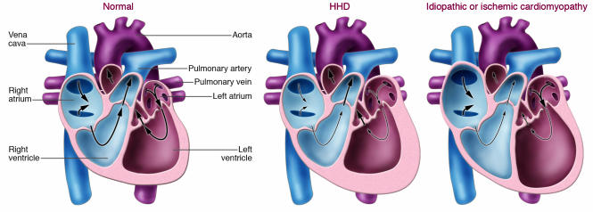
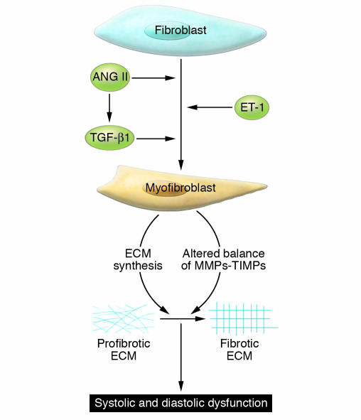
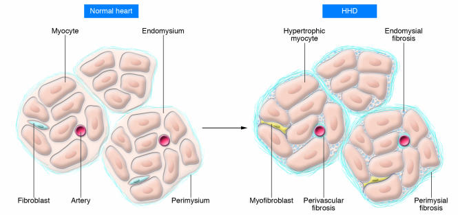
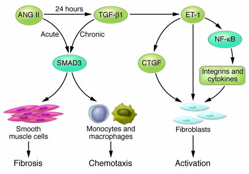
Hypertensive heart disease (HHD) occurs in patients that clinically have both diastolic and systolic heart failure and will soon become the most common cause of heart failure. Two key aspects of heart failure secondary to HHD are the relatively highly prevalent LV hypertrophy and cardiac fibrosis, caused by changes in the local and systemic neurohormonal environment. The fibrotic state is marked by changes in the balance between MMPs and their inhibitors, which alter the composition of the ECM. Importantly, the fibrotic ECM impairs cardiomyocyte function. Recent research suggests that therapies targeting the expression, synthesis, or activation of the enzymes responsible for ECM homeostasis might represent novel opportunities to modify the natural progression of HHD.


Figures

Figure 1
Figure 1. Schematic representation of changes in the cardiac chambers of an individual with HHD compared with idiopathic or ischemic cardiomyopathy.
The main difference between HHD and the other two main causes of heart failure (ischemic heart disease associated with prior myocardial infarction[s] and idiopathic dilated cardiomyopathy) is the nature of the geometric remodeling of the LV chamber. Patients with HHD usually present with LVH but with a normal-sized LV chamber and preserved systolic function. By contrast, patients with heart failure secondary to ischemia or idiopathic cardiomyopathy usually have an enlarged, dilated LV chamber and more frequently also have RV enlargement.
Figure 2
Figure 2. Mechanisms for transition of fibroblasts to myofibroblasts.
The transition of fibroblasts to myofibroblasts is an early event in HHD, regulated in part by increased expression of the hormones of the RAAS system (renin, ANG II, and aldosterone), ET-1, and TGF-β1. Myofibroblasts express a gene program that contributes to a progressive profibrotic state. Changes in the ECM occur in part due to an altered balance of MMPs and their inhibitors (TIMPs). These changes lead to a stiffening of the ECM and functional alterations that cause changes in signaling to myocytes. The altered physical and functional environment of the myocytes leads to progressive cardiac dysfunction.
Figure 3
Figure 3. Schematic representation of changes to the collagen network in HHD.
In the normal heart, thin layers of perimysium and endomysium surround myocardial bundles and myocytes, respectively. The walls of the blood vessels also contain adventitial fibroblasts that create an endomysial network. In HHD, there is hypertrophy of cardiomyocytes and transition of fibroblasts to myofibroblasts. These changes are associated in early disease with increases in ECM manifest by perivascular fibrosis and fibrosis of the endomysium and perimysium.
Figure 4
Figure 4. A laminin-dystroglycan-dystrophin signaling cascade.
Dystroglycan (DG) is a key component of the dystrophin-associated glycoprotein complex that provides mechanical support to the sarcolemma. β-Dystroglycan is the transmembrane subunit, and its extracellular domain noncovalently binds α-dystroglycan. Laminin is a major component of the basal laminae and a prominent protein in the endomysium. The binding of laminin to dystroglycan activates a growth factor receptor–bound protein 2–RAC1–PAK1–JNK (GRB2-RAC1-PAK1-JNK) pathway that promotes hypertrophy, an initial adaptive response to increased pressure. Decreased expression of laminin, as might occur during the transition to heart failure, might impair survival signaling to cardiomyocytes and predispose them to apoptosis, similar to the pathology in skeletal muscle dystrophies. Direct association between dystroglycan and MEK and between dystroglycan and ERK was recently demonstrated (82). MEK-dystroglycan association was localized to membrane ruffles, while ERK-dystroglycan association was found in focal adhesions. It is not known how these interactions are regulated.
Figure 5
Figure 5. Common signaling pathways activated by ANG II, ET-1, and TGF-β1 contribute to fibrosis.
SMAD3 mediates acute and chronic changes in gene expression that lead to inflammation and fibrosis. Chronic exposure to ANG II increases expression of TGF-β1. This cytokine promotes a profibrotic environment by multiple mechanisms, including stimulation of SMAD3-dependent gene expression in smooth muscle cells and monocytes and increased expression of ET-1 by endothelial cells. ET-1 promotes fibroblast activation by stimulating connective tissue growth factor (CTGF) expression and activating an NF-κB–dependent gene program that is profibrotic.

References

    1. Zile M.R., Brutsaert D.L. New concepts in diastolic dysfunction and diastolic heart failure: part II: causal mechanisms and treatment. Circulation. 2002;105:1503–1508. - PubMed
    1. Zile M.R., Brutsaert D.L. New concepts in diastolic dysfunction and diastolic heart failure: part I: diagnosis, prognosis, and measurements of diastolic function. Circulation. 2002;105:1387–1393. - PubMed
    1. Shirwany A., Weber K.T. Extracellular matrix remodeling in hypertensive heart disease. . J. Am. Coll. Cardiol. 2006;48:97–98. - PubMed
    1. Gandhi S.K., et al. The pathogenesis of acute pulmonary edema associated with hypertension. . N. Engl. J. Med. 2001;344:17–22. - PubMed
    1. Benjamin E.J., Levy D. Why is left ventricular hypertrophy so predictive of morbidity and mortality? Am. J. Med. Sci. 1999;317:168–175. - PubMed

Publication types

MeSH terms