

A nonrigid registration of MR breast images using complex-valued wavelet transform
- PMID: 17333413
- PMCID: PMC3043828
- DOI: 10.1007/s10278-007-9021-z
A nonrigid registration of MR breast images using complex-valued wavelet transform
Abstract
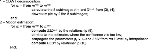
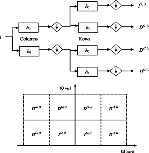
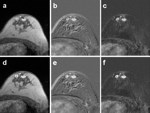
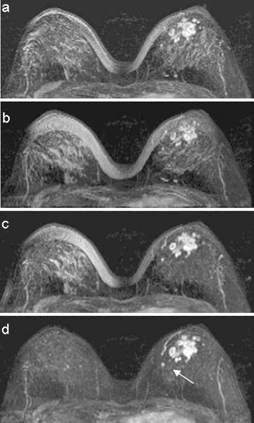
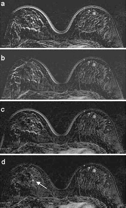
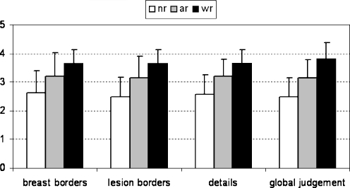
In this paper, a fast, slice-by-slice, nonrigid registration algorithm of dynamic magnetic resonance breast images is presented. The method is based on a multiresolution motion estimation of the breast using complex discrete wavelet transform (CDWT): the pyramid of oriented complex subimages is used to implement a hierarchical phase-matching-based motion estimation algorithm. The resulting motion estimate is nonrigid and pixel-independent. To assess the method performance, we computed the correlation coefficient and the normalized mutual information between pre- and postcontrast images with and without realignment. The indices increased after using our approach and the improvement was superior to rigid or affine registration. A set of clinical scores was also evaluated. The clinical validation demonstrated an increased readability in the subtraction images. In particular, CDWT registration allowed a best definition of breast and lesion borders and greater detail detectability.
Figures
Similar articles
-
Quantitative evaluation of free-form deformation registration for dynamic contrast-enhanced MR mammography.Med Phys. 2007 Apr;34(4):1221-33. doi: 10.1118/1.2712040. Med Phys. 2007. PMID: 17500454
-
Validation of an elastic matching algorithm based on complex wavelets for the realignment of dynamic MR breast images.Conf Proc IEEE Eng Med Biol Soc. 2004;2004:1745-6. doi: 10.1109/IEMBS.2004.1403523. Conf Proc IEEE Eng Med Biol Soc. 2004. PMID: 17272043
-
Volume-preserving nonrigid registration of MR breast images using free-form deformation with an incompressibility constraint.IEEE Trans Med Imaging. 2003 Jun;22(6):730-41. doi: 10.1109/TMI.2003.814791. IEEE Trans Med Imaging. 2003. PMID: 12872948 Clinical Trial.
-
Nonrigid registration using free-form deformations: application to breast MR images.IEEE Trans Med Imaging. 1999 Aug;18(8):712-21. doi: 10.1109/42.796284. IEEE Trans Med Imaging. 1999. PMID: 10534053
-
Integrating Morphological Edge Detection and Mutual Information for Nonrigid Registration of Medical Images.Curr Med Imaging Rev. 2019;15(3):292-300. doi: 10.2174/1573405614666180103163430. Curr Med Imaging Rev. 2019. PMID: 31989880 Review.
Cited by
-
A nonrigid registration algorithm for longitudinal breast MR images and the analysis of breast tumor response.Magn Reson Imaging. 2009 Nov;27(9):1258-70. doi: 10.1016/j.mri.2009.05.007. Epub 2009 Jun 13. Magn Reson Imaging. 2009. PMID: 19525078 Free PMC article.
-
Thick slices from tomosynthesis data sets: phantom study for the evaluation of different algorithms.J Digit Imaging. 2009 Oct;22(5):519-26. doi: 10.1007/s10278-007-9075-y. Epub 2007 Oct 23. J Digit Imaging. 2009. PMID: 17955296 Free PMC article.
References
-
- Tardivon A, Khoury C, Thibault F, Meunier M. New developments in breast imaging. Cancer Radiother. 2004;8(1):2–8. - PubMed
-
- Hulka CA, Smith BL, Sgroi DC, Tan L, Edmister WB, Semple JP, Campbell T, Kopans DB, Brady TJ, Weisskoff RM. Benign and malignant breast lesions: differentiation with echo-planar MR imaging. Radiology. 1995;197:33–38. - PubMed
-
- Stack JP, Redmond OM, Codd MB, Dervan PA, Ennis JT. Breast disease: tissue characterization with Gd-DTPA enhancement profiles. Radiology. 1990;174:491–494. - PubMed
Publication types
MeSH terms
Substances
LinkOut - more resources
Full Text Sources
Medical
