

Case Reports
Veterinary diagnostic imaging. Lobar hepatic infarction with hemorrhage, secondary to a condition of chronic lymphocytic portal hepatitis with dissecting fibrosis and abundant copper accumulation
Affiliations
- PMID: 17334039
- PMCID: PMC1780245
Item in Clipboard
Case Reports
Veterinary diagnostic imaging. Lobar hepatic infarction with hemorrhage, secondary to a condition of chronic lymphocytic portal hepatitis with dissecting fibrosis and abundant copper accumulation
Can Vet J.
2007 Feb.
No abstract available
Figures

A lateral ultrasonographic image demonstrating the presence of a fluid (asterix) filled density (arrow heads) that seems to be originating from the liver (L) parenchyma.

A series of axial computed tomography (CT) images demonstrating the appearance of the radiolucent density (white asterix) that originates from a single lobe of the liver (L). This is clearly outlined by a black arrow that identifies the fissure found between liver lobes on the left side of the scan (your right hand side). Both the spleen (SP) and stomach (ST) are visualized on these images.
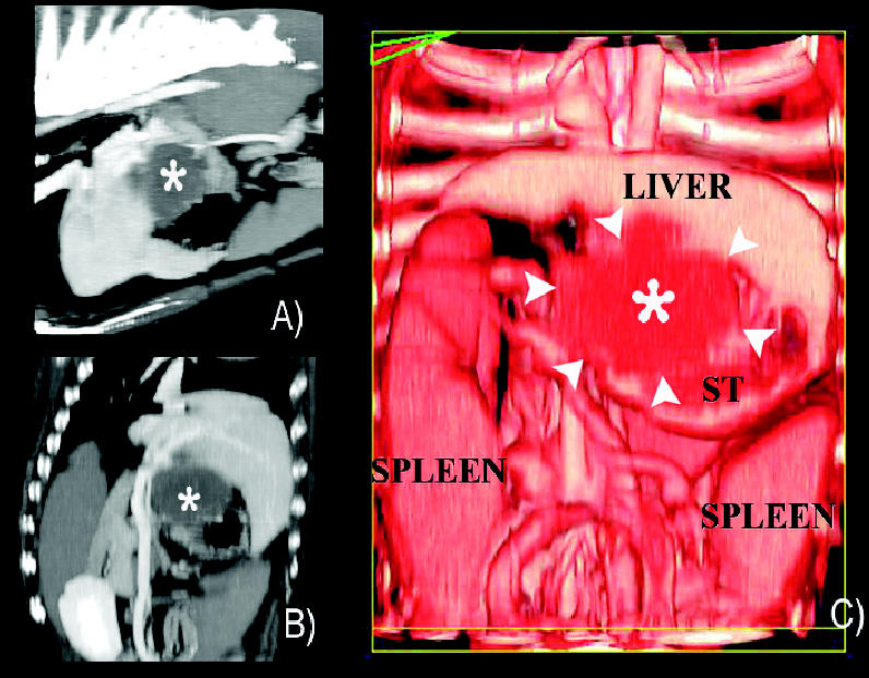
A) A lateral computed tomography (CT) reconstruction demonstrating the mass (asterix) in the dorsal part of the cranial quadrant of the abdomen and originating from the liver. B) A coronal oblique CT reconstruction again outlining the mass between the left lobes of the liver and the stomach. C) Color CT reconstruction of the mass within the liver outlined by arrowheads.

A 2.5-mm thick axial computed tomography (CT) image demonstrating the enhanced periphery of the mass (asterix) suggesting that it is enclosed within hepatic tissues (white arrows). The density within the lesion does not enhance with the iodine-based contrast agent and thereby is most probably a fluid-filled center that represents a previous intrahepatic hemorrhage. In this case, it was the result of the detrimental combination of a hepatic infarct secondary to hepatic fibrosis and copper storage disease.
Similar articles
-
What is your diagnosis? Cirrhosis.J Small Anim Pract. 2001 Oct;42(10):477, 517. J Small Anim Pract. 2001. PMID: 11688521 No abstract available.
-
Budd-Chiari-like syndrome in a dog due to liver lobe entrapment within the falciform ligament.J Am Anim Hosp Assoc. 2009 Sep-Oct;45(5):253-6. doi: 10.5326/0450253. J Am Anim Hosp Assoc. 2009. PMID: 19723850
-
[Chronic active or aggressive hepatitis and liver cirrhosis with copper accumulation in a Dobermann. Case report].Tierarztl Prax. 1991 Dec;19(6):675-81. Tierarztl Prax. 1991. PMID: 1796472 German.
-
Hepatic Fibrosis in Dogs.J Vet Intern Med. 2018 Jan;32(1):26-41. doi: 10.1111/jvim.14891. Epub 2017 Nov 30. J Vet Intern Med. 2018. PMID: 29194760 Free PMC article. Review.
-
Chronic hepatitis in Doberman pinschers. A review.Vet Q. 2004 Sep;26(3):98-106. doi: 10.1080/01652176.2004.9695173. Vet Q. 2004. PMID: 15559390 Review.
References
-
- Mandigers PJ, van den Ingh TS, Spee B, Penning LC, Bode P, Rothuizen J. Chronic hepatitis in Doberman pinschers. A review. Vet Q. 2004;26:98–106. - PubMed
-
- Müller V, Brummer D, Erhardt W, et al. Arterialisation of the portal vein as a model for the induction of hepatic fibrosis: Description of microsurgical models in the rat. Transpl Int. 2005;17:822–833. - PubMed
-
- Bressler C, Himes LC, Moreau RE. Portal vein and aortic thromboses in a Siberian husky with ehrlichiosis and hypothyroidism. J Small Anim Pract. 2003;44:408–410. - PubMed
Publication types
MeSH terms
Substances
LinkOut - more resources
Full Text Sources
Medical