. 2007 May;87(5):473-87.
doi: 10.1038/labinvest.3700533. Epub 2007 Mar 5.

H3 histamine receptor agonist inhibits biliary growth of BDL rats by downregulation of the cAMP-dependent PKA/ERK1/2/ELK-1 pathway

Affiliations

H3 histamine receptor agonist inhibits biliary growth of BDL rats by downregulation of the cAMP-dependent PKA/ERK1/2/ELK-1 pathway

Heather Francis et al. Lab Invest. 2007 May.

Abstract

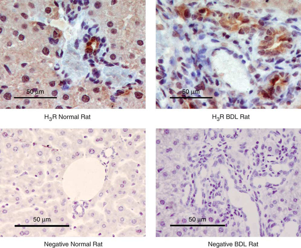
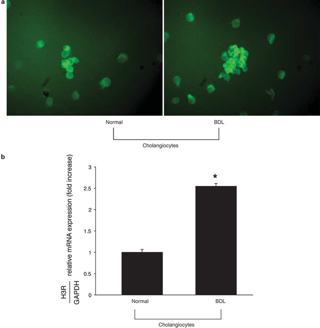
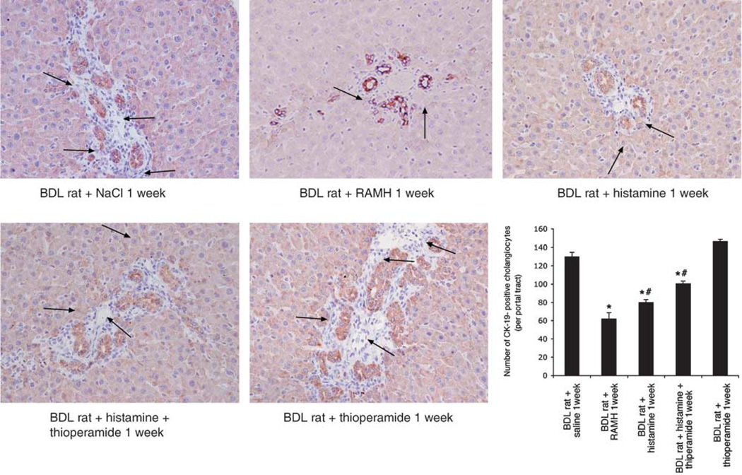
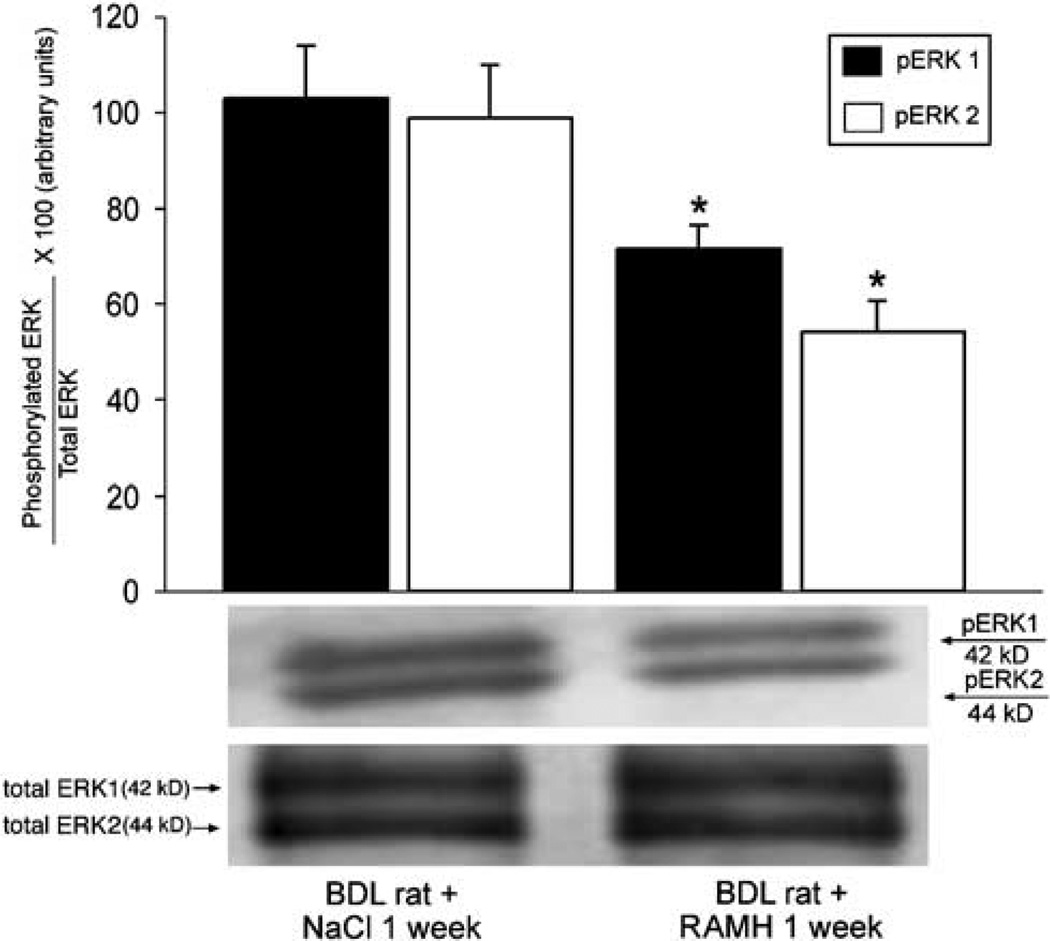
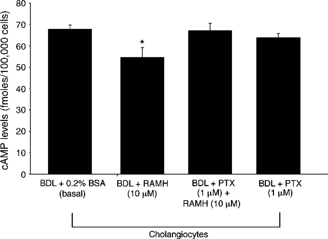
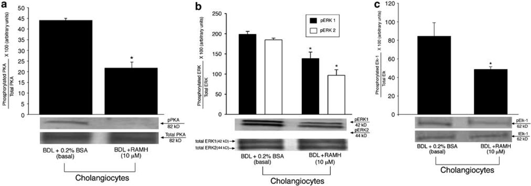
Histamine regulates many functions by binding to four histamine G-coupled receptor proteins (H1R, H2R, H3R and H4R). As H3R exerts their effects by coupling to Galpha(i/o) proteins reducing adenosine 3', 5'-monophosphate (cAMP) levels (a key player in the modulation of cholangiocyte hyperplasia/damage), we evaluated the role of H3R in the regulation of biliary growth. We posed the following questions: (1) Do cholangiocytes express H3R? (2) Does in vivo administration of (R)-(alpha)-(-)-methylhistamine dihydrobromide (RAMH) (H3R agonist), thioperamide maleate (H3R antagonist) or histamine, in the absence/presence of thioperamide maleate, to bile duct ligated (BDL) rats regulate cholangiocyte proliferation? and (3) Does RAMH inhibit cholangiocyte proliferation by downregulation of cAMP-dependent phosphorylation of protein kinase A (PKA)/extracellular signal-regulated kinase 1/2 (ERK1/2)/ets-like gene-1 (Elk-1)? The expression of H3R was evaluated in liver sections by immunohistochemistry and immunofluorescence, and by real-time PCR in cholangiocyte RNA from normal and BDL rats. BDL rats (immediately after BDL) were treated daily with RAMH, thioperamide maleate or histamine in the absence/presence of thioperamide maleate for 1 week. Following in vivo treatment of BDL rats with RAMH for 1 week, and in vitro stimulation of BDL cholangiocytes with RAMH, we evaluated cholangiocyte proliferation, cAMP levels and PKA, ERK1/2 and Elk-1 phosphorylation. Cholangiocytes from normal and BDL rats express H3R. The expression of H3R mRNA increased in BDL compared to normal cholangiocytes. Histamine decreased cholangiocyte growth of BDL rats to a lower extent than that observed in BDL RAMH-treated rats; histamine-induced inhibition of cholangiocyte growth was partly blocked by thioperamide maleate. In BDL rats treated with thioperamide maleate, cholangiocyte hyperplasia was slightly higher than that of BDL rats. In vitro, RAMH inhibited the proliferation of BDL cholangiocytes. RAMH inhibition of cholangiocyte growth was associated with decreased cAMP levels and PKA/ERK1/2/Elk-1 phosphorylation. Downregulation of cAMP-dependent PKA/ERK1/2/Elk-1 phosphorylation (by activation of H3R) is important in the inhibition of cholangiocyte growth in liver diseases.


Figures

Figure 1
Figure 1
Immunohistochemistry for H3R in paraffin-embedded liver sections from normal and 1-week BDL rats. By immunohistochemistry (scale bar = 50 µm), there was cytoplasmic and membranous staining for H3R in cholangiocytes from normal and BDL rats. H3R were also expressed by hepatocytes from normal and BDL rats. No immunohistochemical reaction was observed when normal or BDL liver sections were incubated with non-immune serum.
Figure 2
Figure 2
Immunofluorescence for H3R in frozen liver sections from normal and 1 week BDL rats. By immunofluorescence in liver sections, there was cytoplasmic and membranous staining (arrows) for H3R in cholangiocytes from normal and BDL rats. By immunofluorescence, H3R immunoreactivity (red) was colocalized with CK-19 immunoreactivity (green; indicated by arrows) demonstrating cholangiocyte expression (scale bar =20 µm); sections were counterstained with DAPI. H3R were also expressed by hepatocytes from normal and BDL rats. No immunofluorescent reaction was observed when normal or BDL liver sections were incubated with non-immune serum.
Figure 3
Figure 3
Immunofluorescence (a, top panel) for H3R in cytospin smears of isolated cholangiocytes, and real-time PCR (b, bottom panel) for H3R mRNA in cholangiocyte RNA (0.75 µg) from normal and 1 week BDL rats. By immunofluorescence (a, top panel), purified cholangiocytes from both normal and BDL rats express H3R. Original magnification, × 40. By real-time PCR (b, bottom panel), we found that (i) normal cholangiocytes express H3R mRNA (expressed as ratio to GAPDH mRNA); and (ii) the relative expression of H3R mRNA (expressed as ratio to GAPDH mRNA) significantly increased in cholangiocytes from BDL rats. Data are mean ± s.e.m. of three experiments. *P < 0.05 vs relative expression of H3R mRNA of normal cholangiocytes.
Figure 4
Figure 4
Measurement of the number of PCNA-positive cholangiocytes in liver sections from rats that (immediately after BDL) were treated with NaCl, RAMH, thioperamide maleate (H3R antagonist) or histamine in the absence or presence of thioperamide maleate for 1 week. In BDL rats, chronic RAMH administration induced a decrease in the number of PCNA-positive (see arrows) cholangiocytes compared to NaCl-treated rats. Administration of histamine to BDL rats significantly decreased the number of PCNA-positive cholangiocytes compared to BDL rats treated with NaCl; however, the decrease in the number of PCNA-positive cholangiocytes (by histamine administration alone) was less than that observed in BDL rats treated with RAMH (arrows). Histamine inhibition of the number of PCNA-positive cholangiocytes was partly blocked by thioperamide maleate; however, in the BDL rats treated with histamine + thioperamide maleate, the number of PCNA-positive cholangiocytes was still significantly lower than that of BDL rats treated with NaCl. In BDL rats treated with thioperamide maleate, the number of PCNA-positive cholangiocytes was slightly higher (although not significant) than that of BDL rats. Data are mean ± s.e.m. of five cumulative values from 10 randomly selected portal areas. *P <0.05 vs corresponding value from BDL rats treated with NaCl. #P <0.05 vs corresponding value from BDL rats treated with RAMH. Original magnification, × 20.
Figure 5
Figure 5
Measurement of the number of CK-19-positive cholangiocytes in liver sections from rats (that immediately after BDL) were treated with NaCl, RAMH, thioperamide maleate (H3R antagonist) or histamine in the absence or presence of thioperamide maleate for 1 week. In BDL rats, chronic RAMH administration induced a decrease in the number of CK-19-positive (see arrows) cholangiocytes compared to NaCl-treated rats. Administration of histamine to BDL rats significantly decreased the number of CK-19-positive cholangiocytes compared to BDL rats treated with NaCl; however, the decrease in the number of CK-19-positive cholangiocytes (by histamine administration alone) was less than that observed in BDL rats treated with RAMH (arrows). Histamine inhibition of the number of CK-19-positive cholangiocytes was partly blocked by thioperamide maleate; however, in the BDL rats treated with histamine + thioperamide maleate, the number of CK-19-positive cholangiocytes was still significantly lower than that of BDL rats treated with NaCl. In BDL rats treated with thioperamide maleate, the number of CK-19-positive cholangiocytes was slightly higher (although not significant) than that of BDL rats. Data are mean ± s.e.m. of five cumulative values from 10 randomly selected portal areas. *P <0.05 vs corresponding value from BDL rats treated with NaCl. #P < 0.05 vs corresponding value from BDL rats treated with RAMH. Original magnification, × 20.
Figure 6
Figure 6
Measurement of the number of γ-GT-positive ducts in liver sections from rats that (immediately after BDL) were treated with NaCl, RAMH, thioperamide maleate (H3R antagonist) or histamine in the absence or presence of thioperamide maleate for 1 week. In BDL rats, chronic RAMH administration induced a decrease in the number of γ-GT-positive ducts compared to NaCl-treated rats. Administration of histamine to BDL rats significantly decreased the number of γ-GT-positive ducts compared to BDL rats treated with NaCl; however, the decrease in the number of γ-GT-positive ducts (by histamine administration alone) was less than that observed in BDL rats treated with RAMH (arrows). Histamine inhibition of the number of γ-GT-positive ducts was partly blocked by thioperamide maleate; however, in the BDL rats treated with histamine + thioperamide maleate, the number of γ-GT-positive ducts was still significantly lower than that of BDL rats treated with NaCl. In BDL rats treated with thioperamide maleate, the number of γ-GT-positive ducts was slightly higher (although not significant) than that of BDL rats. Data are mean±s.e.m. of 16 cumulative values from 10 randomly selected portal areas. *P < 0.05 vs corresponding value from BDL rats treated with NaCl. #P < 0.05 vs corresponding value from BDL rats treated with RAMH.
Figure 7
Figure 7
(a, left panel) Measurement of PCNA protein expression in purified cholangiocytes from rats that (immediately after BDL) were treated with NaCl or RAMH for 1 week. PCNA protein expression was decreased in cholangiocytes from BDL rats treated with RAMH compared to cholangiocytes from NaCl-treated rats. Data are mean±s.e.m. of five experiments. *P < 0.05 vs corresponding value from BDL rats treated with NaCl. (b, right panel) Measurement of the phosphorylation of PKA in cholangiocytes from rats that (immediately after BDL) were treated with NaCl or RAMH for 1 week. Chronic in vivo administration of RAMH to BDL rats induced a decrease in the phosphorylation of PKA (expressed as ratio to total PKA protein expression) in purified cholangiocytes compared to cholangiocytes from BDL rats treated with NaCl. Data are mean±s.e.m. of three experiments. *P < 0.05 vs corresponding value from BDL rats treated with NaCl.
Figure 8
Figure 8
ERK1/2 phosphorylation (expressed as ratio to total ERK1/2 protein expression) was significantly decreased in cholangiocytes from BDL+ RAMH-treated animals compared to cholangiocytes from BDL rats treated with NaCl. Data are mean±s.e.m. of three experiments. *P < 0.05 vs corresponding value from BDL rats treated with NaCl.
Figure 9
Figure 9
(a) Chronic administration of RAMH to BDL rats decreased the phosphorylation of Elk-1 (expressed as ratio to total Elk-1 protein expression) in cholangiocytes compared to cholangiocytes from NaCl treated rats. Data are mean±s.e.m. of three experiments. *P<0.05 vs corresponding value from BDL rats treated with NaCl. (b) Measurement of PCNA protein expression in purified BDL cholangiocytes treated in vitro with 0.2% BSA (basal) or RAMH (10 µM) with 0.2% BSA for 3 h at 37°C. In vitro stimulation of purified BDL cholangiocytes with RAMH induced a significant decrease in PCNA protein expression compared to cholangiocytes treated with 0.2% BSA. Data are mean±s.e.m. of eight experiments. *P < 0.05 vs corresponding basal value.
Figure 10
Figure 10
Measurement of cAMP levels in purified BDL cholangiocytes stimulated at room temperature for 5 min with 0.2% BSA (basal) or RAMH (10 µM) in the absence or presence of PTX (1 µM). RAMH-induced inhibition of cAMP levels was blocked, in vitro, by pretreatment of BDL cholangiocytes with PTX. PTX alone did not change cAMP levels of BDL cholangiocytes. Data are mean±s.e.m. of six experiments. *P < 0.05 vs corresponding basal value.
Figure 11
Figure 11
Measurement of the phosphorylation of (a) PKA, (b) ERK1/2 and (c) Elk-1 in purified BDL cholangiocytes treated in vitro with 0.2% BSA (basal) or PAMH (10 µM). (a) In vitro, RAMH induced a decrease in PKA phosphorylation in purified BDL cholangiocytes compared to BDL cholangiocytes treated with BSA. Data are mean ± s.e.m. of three experiments. *P < 0.05 vs corresponding basal value. (b) RAMH decreased the phosphorylation of ERK1/2 (b) and Elk-1 (c) compared to cholangiocytes treated with BSA. Data are mean ± s.e.m. of three experiments. *P < 0.05 vs corresponding basal value.

Similar articles

Cited by

References

    1. Alvaro D, Mancino MG, Glaser S, et al. Proliferating cholangiocytes: a neuroendocrine compartment in the diseased liver. Gastroenterology. 2006;132:415–431. - PubMed
    1. LeSage G, Glaser S, Alpini G. Regulation of cholangiocyte proliferation. Liver. 2001;21:73–80. - PubMed
    1. Alpini G, Prall RT, LaRusso NF. The pathobiology of biliary epithelia. In: Arias IM, Boyer JL, Chisari FV, Fausto N, Jakoby W, Schachter D, Shafritz DA, editors. The Liver, Biology and Pathobiology. 4th edn. Philadelphia, PA: Lippincott Williams & Wilkins; 2001. pp. 421–435.
    1. Polimeno L, Azzarone A, Zeng QH, et al. Cell proliferation and oncogene expression after bile duct ligation in the rat: evidence of a specific growth effect on bile duct cells. Hepatology. 1995;21:1070–1078. - PMC - PubMed
    1. Francis H, Glaser S, Ueno Y, et al. cAMP stimulates the secretory and proliferative capacity of the rat intrahepatic biliary epithelium through changes in the PKA/Src/MEK/ERK1/2 pathway. J Hepatol. 2004;41:528–537. - PubMed

Publication types

MeSH terms