

Patients with type 2 diabetes have normal mitochondrial function in skeletal muscle
- PMID: 17334651
- PMCID: PMC1820754
- DOI: 10.1007/s00125-007-0594-3
Patients with type 2 diabetes have normal mitochondrial function in skeletal muscle
Abstract
Aims/hypothesis: Insulin resistance and type 2 diabetes are associated with mitochondrial dysfunction. The aim of the present study was to test the hypothesis that oxidative phosphorylation and electron transport capacity are diminished in the skeletal muscle of type 2 diabetic subjects, as a result of a reduction in the mitochondrial content.
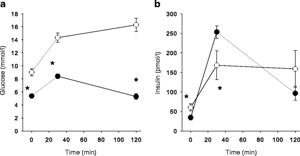
Materials and methods: The O(2) flux capacity of permeabilised muscle fibres from biopsies of the quadriceps in healthy subjects (n = 8; age 58 +/- 2 years [mean+/-SEM]; BMI 28 +/- 1 kg/m(2); fasting plasma glucose 5.4 +/- 0.2 mmol/l) and patients with type 2 diabetes (n = 11; age 62 +/- 2 years; BMI 32 +/- 2 kg/m(2); fasting plasma glucose 9.0 +/- 0.8 mmol/l) was measured by high-resolution respirometry.
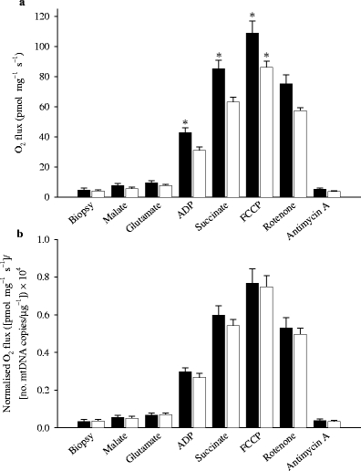
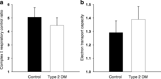
Results: O(2) flux expressed per mg of muscle (fresh weight) during ADP-stimulated state 3 respiration was lower (p < 0.05) in patients with type 2 diabetes in the presence of complex I substrate (glutamate) (31 +/- 2 vs 43 +/- 3 pmol O(2) s(-1) mg(-1)) and in response to glutamate + succinate (parallel electron input from complexes I and II) (63 +/- 3 vs 85 +/- 6 pmol s(-1) mg(-1)). Further increases in O(2) flux capacity were observed in response to uncoupling by FCCP, but were again lower (p < 0.05) in type 2 diabetic patients than in healthy control subjects (86 +/- 4 vs 109 +/- 8 pmol s(-1) mg(-1)). However, when O(2) flux was normalised for mitochondrial DNA content or citrate synthase activity, there were no differences in oxidative phosphorylation or electron transport capacity between patients with type 2 diabetes and healthy control subjects.
Conclusions/interpretation: Mitochondrial function is normal in type 2 diabetes. Blunting of coupled and uncoupled respiration in type 2 diabetic patients can be attributed to lower mitochondrial content.
Figures
References
-
- {'text': '', 'ref_index': 1, 'ids': [{'type': 'PubMed', 'value': '12351431', 'is_inner': True, 'url': 'https://pubmed.ncbi.nlm.nih.gov/12351431/'}]}
- Kelley DE, He J, Menshikova EV, Ritov VB (2002) Dysfunction of mitochondria in human skeletal muscle in type 2 diabetes. Diabetes 51:2944–2950 - PubMed
-
- {'text': '', 'ref_index': 1, 'ids': [{'type': 'DOI', 'value': '10.1007/BF01234508', 'is_inner': False, 'url': 'https://doi.org/10.1007/bf01234508'}, {'type': 'PubMed', 'value': '908476', 'is_inner': True, 'url': 'https://pubmed.ncbi.nlm.nih.gov/908476/'}]}
- Vondra K, Rath R, Bass A, Slabochová Z, Teisinger T, Vítek V (1977) Enzyme activities in quadriceps femoris muscle of obese diabetic male patients. Diabetologia 13:527–529 - PubMed
-
- {'text': '', 'ref_index': 1, 'ids': [{'type': 'PubMed', 'value': '11289047', 'is_inner': True, 'url': 'https://pubmed.ncbi.nlm.nih.gov/11289047/'}]}
- He J, Watkins S, Kelley DE (2001) Skeletal muscle lipid content and oxidative enzyme activity in relation to muscle fiber type in type 2 diabetes and obesity. Diabetes 50:817–823 - PubMed
-
- {'text': '', 'ref_index': 1, 'ids': [{'type': 'PubMed', 'value': '15894466', 'is_inner': True, 'url': 'https://pubmed.ncbi.nlm.nih.gov/15894466/'}]}
- Ørtenblad N, Mogensen M, Petersen I et al (2005) Reduced insulin-mediated citrate synthase activity in cultured skeletal muscle cells from patients with type 2 diabetes: evidence for an intrinsic oxidative enzyme defect. Biochim Biophys Acta 1741:206–214 - PubMed
-
- {'text': '', 'ref_index': 1, 'ids': [{'type': 'PubMed', 'value': '9216960', 'is_inner': True, 'url': 'https://pubmed.ncbi.nlm.nih.gov/9216960/'}]}
- Simoneau JA, Kelley DE (1997) Altered glycolytic and oxidative capacities of skeletal muscle contribute to insulin resistance in NIDDM. J Appl Physiol 83:166–171 - PubMed
Publication types
MeSH terms
Substances
LinkOut - more resources
Full Text Sources
Other Literature Sources
Medical
