

Molecular mechanisms of optic vesicle development: complexities, ambiguities and controversies
- PMID: 17335797
- PMCID: PMC1927083
- DOI: 10.1016/j.ydbio.2007.01.045
Molecular mechanisms of optic vesicle development: complexities, ambiguities and controversies
Abstract
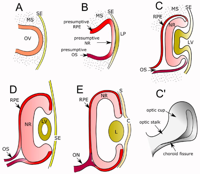
Optic vesicle formation, transformation into an optic cup and integration with neighboring tissues are essential for normal eye formation, and involve the coordinated occurrence of complex cellular and molecular events. Perhaps not surprisingly, these complex phenomena have provided fertile ground for controversial and even contradictory results and conclusions. After presenting an overview of current knowledge of optic vesicle development, we will address conceptual and methodological issues that complicate research in this field. This will be done through a review of the pertinent literature, as well as by drawing on our own experience, gathered through recent studies of both intra- and extra-cellular regulation of optic vesicle development and patterning. Finally, and without attempting to be exhaustive, we will point out some important aspects of optic vesicle development that have not yet received enough attention.
Figures

References
-
- Abrahante JE, Daul AL, Li M, Volk ML, Tennessen JM, Miller EA, Rougvie AE. The Caenorhabditis elegans hunchback-like gene lin-57/hbl-1 controls developmental time and is regulated by microRNAs. Dev Cell. 2003;4:625–37. - PubMed
-
- Adler R, Belecky-Adams TL. The role of bone morphogenetic proteins in the differentiation of the ventral optic cup. Development. 2002;129:3161–71. - PubMed
-
- Alfano G, Vitiello C, Caccioppoli C, Caramico T, Carola A, Szego MJ, McInnes RR, Auricchio A, Banfi S. Natural antisense transcripts associated with genes involved in eye development. Hum Mol Genet. 2005;14:913–23. - PubMed
-
- Ashery-Padan R, Gruss P. Pax6 lights-up the way for eye development. Curr Opin Cell Biol. 2001;13:706–14. - PubMed
Publication types
MeSH terms
Substances
Grants and funding
- R01 EY004859-19/EY/NEI NIH HHS/United States
- EY 1765/EY/NEI NIH HHS/United States
- EY 04859/EY/NEI NIH HHS/United States
- R01 EY004859/EY/NEI NIH HHS/United States
- R01 EY004859-22/EY/NEI NIH HHS/United States
- P30 EY001765/EY/NEI NIH HHS/United States
- R01 EY004859-17/EY/NEI NIH HHS/United States
- R01 EY004859-23/EY/NEI NIH HHS/United States
- R01 EY004859-18/EY/NEI NIH HHS/United States
- R01 EY004859-24/EY/NEI NIH HHS/United States
- R01 EY004859-21/EY/NEI NIH HHS/United States
- R01 EY004859-20/EY/NEI NIH HHS/United States
LinkOut - more resources
Full Text Sources
Other Literature Sources
