. 2007 Apr 15;304(2):811-24.
doi: 10.1016/j.ydbio.2007.01.033. Epub 2007 Jan 27.

Transactivation from Gal4-VP16 transgenic insertions for tissue-specific cell labeling and ablation in zebrafish

Affiliations

Transactivation from Gal4-VP16 transgenic insertions for tissue-specific cell labeling and ablation in zebrafish

Jon M Davison et al. Dev Biol. .

Abstract

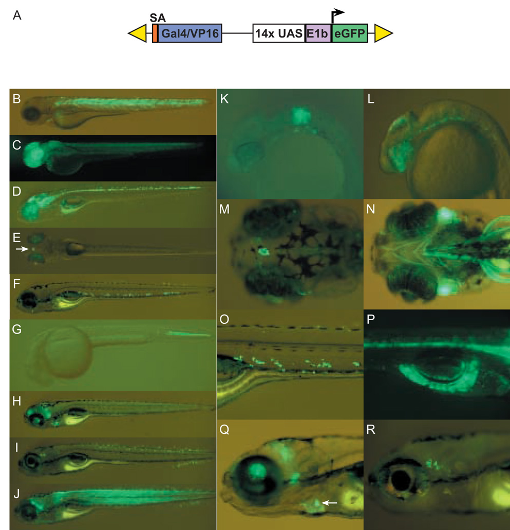
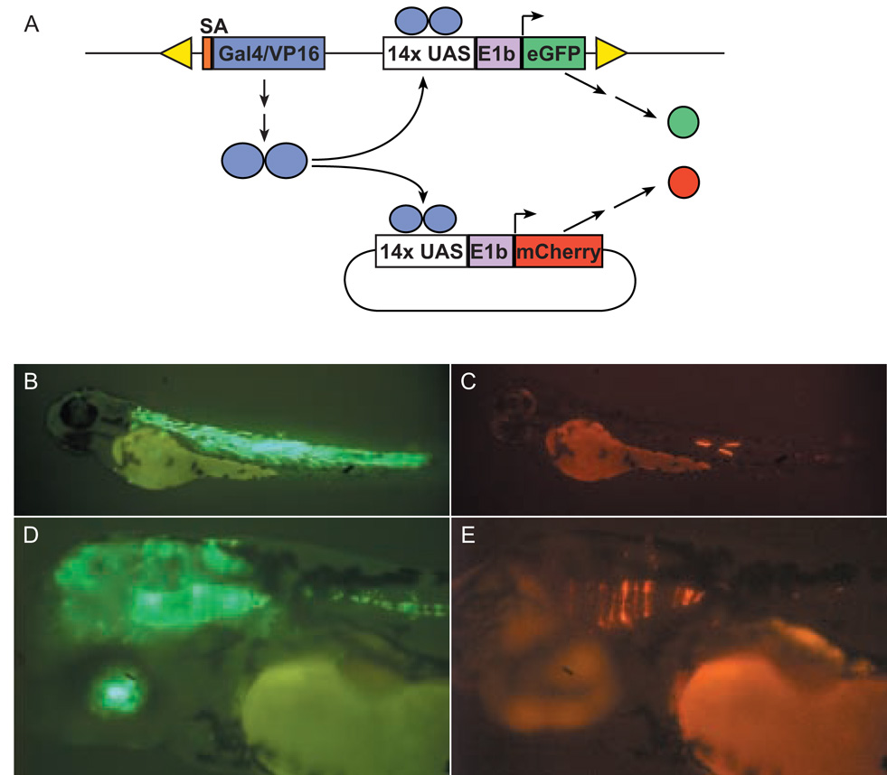
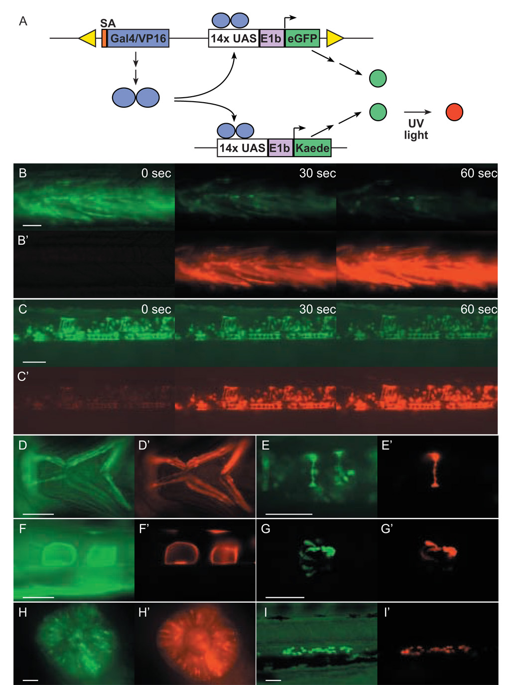
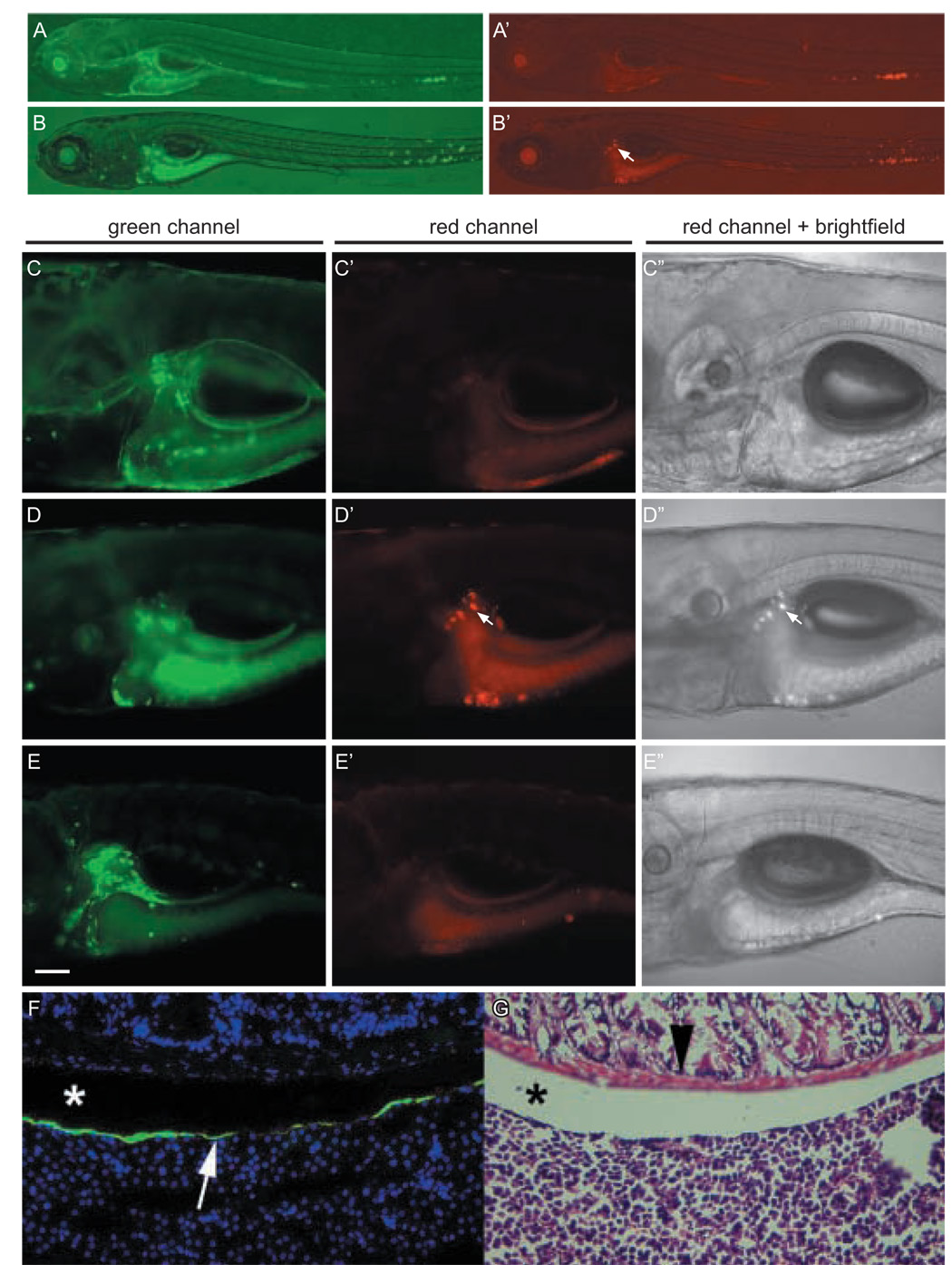
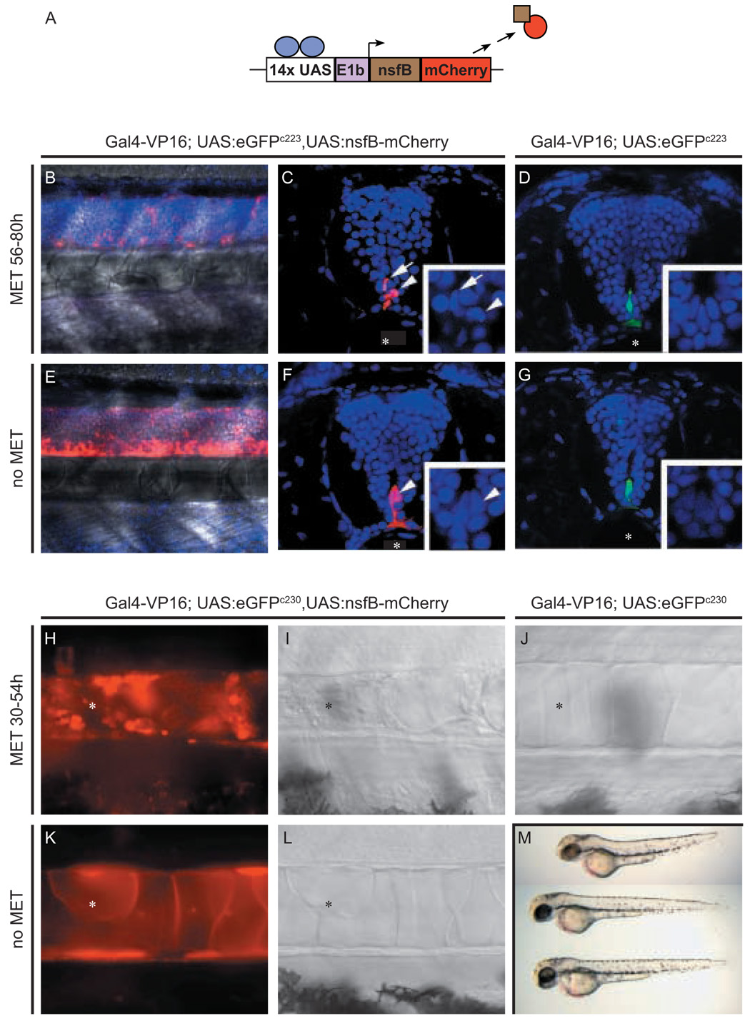
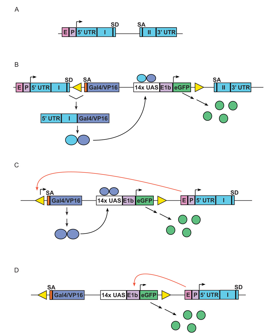
Prior studies with transgenic zebrafish confirmed the functionality of the transcription factor Gal4 to drive expression of other genes under the regulation of upstream activator sequences (UAS). However, widespread application of this powerful binary system has been limited, in part, by relatively inefficient techniques for establishing transgenic zebrafish and by the inadequacy of Gal4 to effect high levels of expression from UAS-regulated genes. We have used the Tol2 transposition system to distribute a self-reporting gene/enhancer trap vector efficiently throughout the zebrafish genome. The vector uses the potent, hybrid transcription factor Gal4-VP16 to activate expression from a UAS:eGFP reporter cassette. In a pilot screen, stable transgenic lines were established that express eGFP in reproducible patterns encompassing a wide variety of tissues, including the brain, spinal cord, retina, notochord, cranial skeleton and muscle, and can transactivate other UAS-regulated genes. We demonstrate the utility of this approach to track Gal4-VP16 expressing migratory cells in UAS:Kaede transgenic fish, and to induce tissue-specific cell death using a bacterial nitroreductase gene under UAS control. The Tol2-mediated gene/enhancer trapping system together with UAS transgenic lines provides valuable tools for regulated gene expression and for targeted labeling and ablation of specific cell types and tissues during early zebrafish development.


Figures

Fig. 1
Fig. 1. Tissue-specific eGFP expression in stable transgenic Gal4-VP16;UAS:eGFP lines
(A) Schematic depiction of SAGVG vector, containing sequence encoding Gal4-VP16 fusion protein, preceded by splice acceptor (orange), and followed by Gal4-VP16 responsive eGFP reporter cassette consisting of Gal4-responsive upstream activating sequence (UAS), adenoviral E1b minimal promoter element (E1b). The entire construct is flanked by right and left arms of Tol2 (yellow triangles). (B) Line c218 with variable expression in somitic muscle fibers at 3 dpf. (C) Line c220 with expression in forebrain and retina at 2 dpf. (D) Line c223 with expression in midbrain, hindbrain and ventral spinal cord at 5 dpf. (E) Line c229 with expression in the pineal (arrow) and early lens expression at 5 dpf. (F) Line c233 with expression in ventral cells dorsal to cardinal vein at 5 dpf. (G) Line c247 with expression in the caudal notochord at 1.5 dpf. (H) Line c236 with expression in subregions of the brain and in the heart at 6 dpf. (I) Line c237 with expression in putative sensory neurons anterior to the otocyst, subsets of spinal cord interneurons, and the ventral tail fin at 6 dpf. (J) Line c240 with expression throughout the skeletal and head musculature at 5 dpf. (K) Line c228 with expression in hindbrain rhombomeres 5 and 6 at 1.5 dpf. (L) Line c223 with expression in retina, dorsal midbrain and hindbrain floor plate at 1.5 dpf. (M) Higher magnification of line c229 demonstrating selective labeling of the pineal at 5 dpf (earlier expression, not shown). (N) Labeling of cranial musculature in c240 at 5 dpf. (O) Higher magnification image of c233 highlighting ventral cells shown in F. (P) c223 also exhibits labeling of the exocrine pancreas at 5 dpf (earlier expression, not shown). (Q) Higher magnification of the heart labeling in line c236 at 6 dpf. High levels of eGFP are observed in heart valves (arrow and refer to supplemental movie). (R) Higher magnification image of putative sensory neurons in line c237. (B–J) lateral views of whole larvae. (K, L, O, Q, and R) lateral views with anterior to the left, (P) is a lateral view with 1 anterior to the right. (M) dorsal and (N) ventral view. All larvae were imaged on a Leica MZFlII stereomicroscope.
Fig. 2
Fig. 2. Transient activation of UAS:mCherry in Gal4-VP16:UAS:eGFP transgenics
(A) Schematic of transient transactivation experiment. Tg(Gal4-VP16;UAS:eGFP) embryos were injected with non-linearised UAS:mCherry plasmid and then screened for transient expression of mCherry. Because of the mosaic nature of plasmid transmission, activation of UAS:mCherry is observed in a mosaic tissue-specific pattern, entirely restricted to the field of eGFP expression. (B) eGFP expression in skeletal muscle fibers of c218 embryo at 72 hpf. (C) Transient expression of mCherry is confined to eGFP-positive muscle fibers. (D) eGFP expression in retina, midbrain, hindbrain and floorplate of c223 embryo. (E) Transient expression of mCherry in eGFP-positive neuroepithilum.
Fig. 3
Fig. 3. Tissue-specific transactivation of UAS:Kaede transgene
(A) Schematic of UAS:Kaede transactivation experiment. Gal4-VP16 expression from a chromosomal vector insertion site results in vector derived eGFP expression as well as transactivation of Kaede expression. Kaede expression is detected by exposure of embryos to UV light and observing the emergence of red fluorescence in cells in which Gal4-VP16 is expressed. In these experiments Tg(Gal4-VP16;UAS:eGFP) F2 or F3 fish were mated with a stable UAS:Kaede transgenic line. Resultant larvae were imaged for green fluorescent cells potentially resulting from expression of both eGFP and Kaede prior to UV exposure (shown in images B–I) and following exposure to UV light (365 nm) for durations ranging from 30 to 60 sec (shown in images B’–I’). Shown are photoconverted muscle fibers in c218 (B,B’), neurons and floor plate cells in c223 (C,C’), jaw musculature in c240 (D,D’), interneurons in c237 (E,E’), notochord cells in c247 (F,F’), pinealocytes in c229 (G,G’), retinal cells in c220 (H,H’), and ventral migratory cells in c233 (I,I’). All are lateral views except D (ventral) and G (coronal). Images captured before and after UV exposure were digitally processed in an identical manner. Scale bars=50 µm.
Fig. 4
Fig. 4. Tracking of a migratory cell population by Kaede photoconversion
(A, A’) Images of a 6 dpf c233 larva immediately after Kaede photoconversion. Photoconverted red Kaede is restricted to the caudal tail. (B, B’) Same larva at 8 dpf showing the red cells have migrated rostrally (arrow). (C, C’, C”) Higher magnification of same larva as (A) with presumptive liver showing green, but not red cells. (D, D’, D”) At 8 dpf both green and red cells are observed (arrow). (E, E’, E”) Control transgenic larva that was not photoactivated shows only green, around the liver at 8 dpf. All images are lateral views, anterior to the left. Red fluorescent images were taken with the same exposure settings (40 seconds at low magnification, 12 seconds at higher magnification) and digitally processed in an identical manner. (C–E) Scale bar = 100µm. (F) Cryostat section through the viscera of a c233 heterozygous adult showing location of eGFP positive cells on the surface of the liver (arrow). Nuclei are blue due to Hoescht staining. (G) Haemotoxilin and eosin staining of adjacent section to show histology and location of eGFP positive cells. Intestinal smooth muscle (arrowhead) and the coelomic space between the intestines and liver are indicated (*)
Fig. 5
Fig. 5. Drug dependent cell ablation following transactivation of nitroreductase expressing transgene
(A) Schematic of NTR-mCherry fusion protein generated by Gal4-VP16 transactivation of UAS:nfsB-mCherry transgene. Transgenic c223 larvae at 80 hpf (B–G) and c230 larvae at 54 hpf (H-M) were treated with Met from 56 to 80 hpf (B, C, D) and 30 to 54 hpf (H, I, J), respectively. Negative, age-matched controls had either one (E, F, K, L) or no copies (D, G, J) of the UAS:nfsB-mCherry. Transactivation of the NTR-mCherry fusion protein in c223 in Z-stack projections of the trunk neural tube superimposed over bright field images (B and E) or in transverse vibratome sections of the neural tube (C and F), and in the c230 (H, K) notochord (asterisks). Larvae expressing NTR-mCherry show a prodrug dependent loss of fluorescent cells in the developing CNS (B, C) and notochord (H, I corresponding DIC image) when compared to untreated controls (E, F and K, L corresponding DIC image, respectively). In B to G, nuclei are counterstained with Hoechst dye. Inset panels show high magnification of the nuclei in cells of the ventral neural tube overlaying the notochord (asterisks) Arrows and arrowheads (C, D and F, G) mark individual fluorescent floor plate cells corresponding to identical cells in high magnification inserts. Following prodrug treatment, the few remaining NTR-mCherry expressing floor plate cells possess fragmented nuclei indicative of being apoptotic (inset in F). (M) Morphology of c230 larvae at 54 hpf either treated with Met (upper, as in H,I) or untreated (middle as in J, or lower as in K,L) demonstrating that ablating notochord cells leads to reduced body length.
Fig. 6
Fig. 6. Potential mechanisms of eGFP expression
(A) Schematic representation of a target endogenous gene, with two exons (Roman numerals); E, endogenous enhancer; P, endogenous promoter. (B) Insertion of SAGVG into the intron of gene, leading to activation of eGFP expression through traditional gene trap event; SA, splice acceptor; UAS, Gal4-responsive upstream activating sequence; E1b, adenoviral E1b minimal promoter element. After splicing of Gal4-VP16 to upstream endogenous coding sequence, the resulting fusion protein provides in cis activation of UAS:eGFP expression. (C and D) two methods by which enhancer trapping can lead to expression of eGFP expression (nb in reality both the orientation and distance from endogenous enhancer can be varied due to the nature of enhancer activity). (C) Gal4-VP16-dependent activation of eGFP expression by enhancer trapping. Endogenous enhancer “E” drives Gal4-VP16 expression through interaction with cryptic promoter elements in either Tol2 vector or in genomic DNA adjacent to insertion. (D) Gal4-VP16-independent activation of eGFP expression by enhancer trapping. Endogenous enhancer “E” drives eGFP expression directly through interaction with E1b minimal promoter in UAS:eGFP cassette.

References

    1. Ahmad K, Henikoff S. Modulation of a transcription factor counteracts heterochromatic gene silencing in Drosophila. Cell. 2001;104:839–847. - PubMed
    1. Amsterdam A, Nissen RM, Sun Z, Swindell EC, Farrington S, Hopkins N. Identification of 315 genes essential for early zebrafish development. Proc Natl Acad Sci U S A. 2004;101:12792–12797. - PMC - PubMed
    1. Ando R, Hama H, Yamamoto-Hino M, Mizuno H, Miyawaki A. An optical marker based on the UV-induced green-to-red photoconversion of a fluorescent protein. Proc Natl Acad Sci U S A. 2002;99:12651–12656. - PMC - PubMed
    1. Balciunas D, Davidson AE, Sivasubbu S, Hermanson SB, Welle Z, Ekker SC. Enhancer trapping in zebrafish using the Sleeping Beauty transposon. BMC Genomics. 2004;5:62. - PMC - PubMed
    1. Brand AH, Dormand EL. The GAL4 system as a tool for unravelling the mysteries of the Drosophila nervous system. Curr Opin Neurobiol. 1995;5:572–578. - PubMed

Publication types

MeSH terms

LinkOut - more resources