. 2007 Nov;132(1-2):53-66.
doi: 10.1016/j.pain.2007.01.024. Epub 2007 Mar 1.

Spinal CGRP1 receptors contribute to supraspinally organized pain behavior and pain-related sensitization of amygdala neurons

Affiliations

Spinal CGRP1 receptors contribute to supraspinally organized pain behavior and pain-related sensitization of amygdala neurons

Hita Adwanikar et al. Pain. 2007 Nov.

Abstract

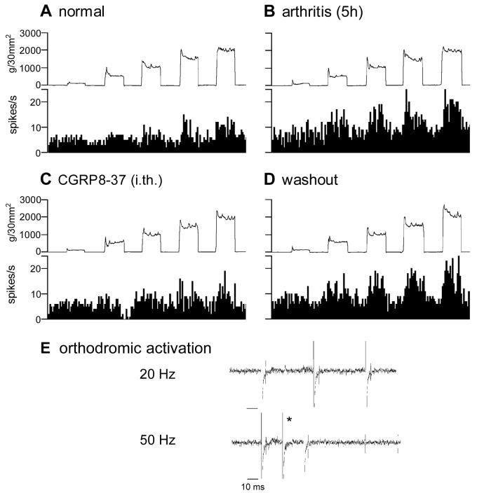
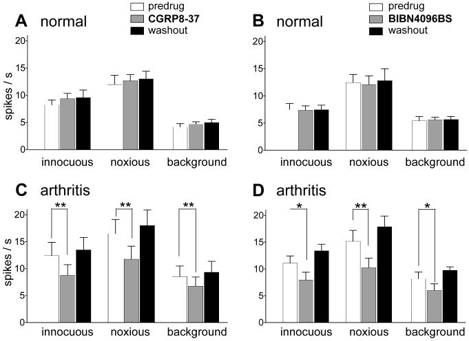
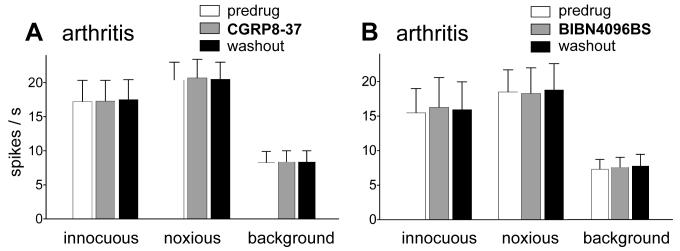
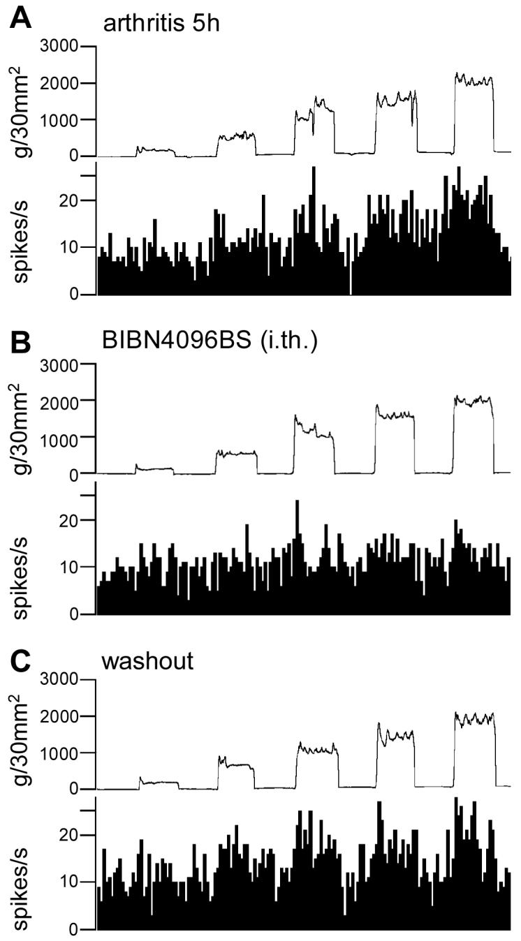
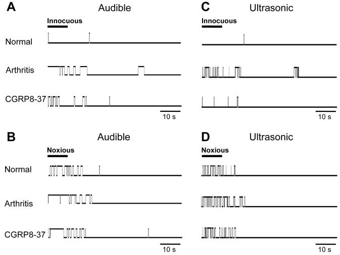
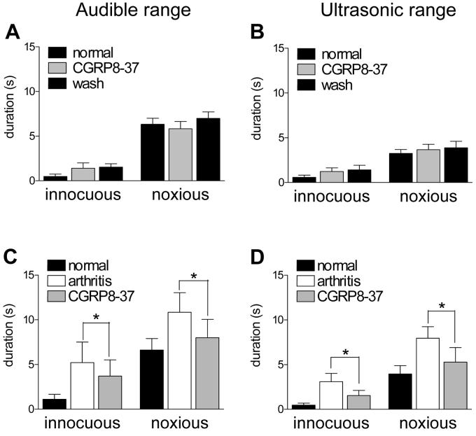
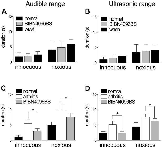
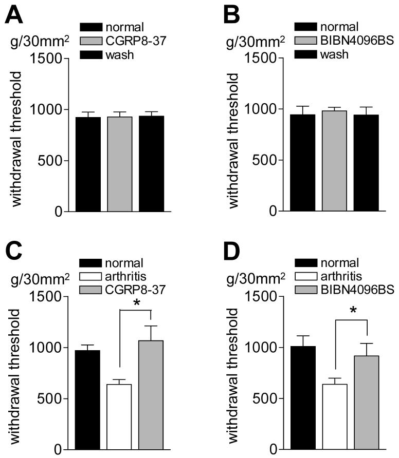
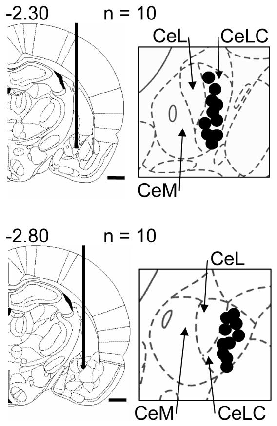
CGRP receptor activation has been implicated in peripheral and central sensitization. The role of spinal CGRP receptors in supraspinal pain processing and higher integrated pain behavior is not known. Here we studied the effect of spinal inhibition of CGRP1 receptors on supraspinally organized vocalizations and activity of amygdala neurons. Our previous studies showed that pain-related audible and ultrasonic vocalizations are modulated by the central nucleus of the amygdala (CeA). Vocalizations in the audible and ultrasonic range and hindlimb withdrawal thresholds were measured in awake adult rats before and 5-6h after induction of arthritis by intra-articular injections of kaolin and carrageenan into one knee. Extracellular single-unit recordings were made from neurons in the latero-capsular division of the CeA (CeLC) in anesthetized rats before and after arthritis induction. CGRP1 receptor antagonists were applied to the lumbar spinal cord intrathecally (5 microl/min) 6h postinduction of arthritis. Spinal administration of peptide (CGRP8-37, 1 microM) and non-peptide (BIBN4096BS, 1 microM) CGRP1 receptor antagonists significantly inhibited the increased responses of CeLC neurons to mechanical stimulation of the arthritic knee but had no effect under normal conditions. In arthritic rats, the antagonists also inhibited the audible and ultrasonic components of vocalizations evoked by noxious stimuli and increased the threshold of hindlimb withdrawal reflexes. The antagonists had no effect on vocalizations and spinal reflexes in normal rats. These data suggest that spinal CGRP1 receptors are not only important for spinal pain mechanisms but also contribute significantly to the transmission of nociceptive information to the amygdala and to higher integrated behavior.


Figures

Figure 1
Figure 1. Spinal administration of CGRP8-37 inhibits the enhanced responses of an amygdala neuron in the arthritis pain state
Extracellular single-unit recordings of the responses (spikes/s) of one neuron in the latero-capsular part of the central nucleus of the amygdala (CeLC) to brief (15 s) graded mechanical stimulation of the knee joint before (A) and 5 h after induction of the knee joint arthritis (B). Intrathecal application of the CGRP1 receptor antagonist CGRP8-37 (1 μM, 15 min; dissolved in ACSF) inhibited the evoked responses and background activity of the neuron (C). This effect was reversed during washout with ACSF (D). Bin width of peristimulus time histograms is 1 s. Top traces in A-D show the actual stimulus intensities (force); they are online recordings of the calibrated output of the force transducer on the forceps used to compress the knee with graded intensities (see 2.2.3). (E) Electrical stimulation in the pontine parabrachial nucleus activated the neuron orthodromically, but not antidromically (see 2.2.2 and Neugebauer and Li 2002). The asterisk indicates where an evoked action potential would be expected if the spike followed high-frequency stimulation.
Figure 2
Figure 2. Inhibitory effects of spinally administered CGRP antagonists on the responses of amygdala neurons in the arthritis pain state but not under normal conditions
(A) and (B) Intrathecally (i.th.) administered CGRP8-37 (1 μM, 15 min, n = 5 neurons; A) and BIBN4096BS (1 μM, 15 min, n = 5 neurons; B) had not effect on the evoked responses and background activity of CeA neurons under normal conditions (no arthritis). (C) and (D) Intrathecal (i.th.) administration of CGRP8-37 (1 μM, 15 min, n = 5 neurons; C) and BIBN4096BS (1 μM, 15 min, n = 5 neurons; D) inhibited the activity of CeLC neurons 5-6 h postinduction of arthritis in the knee. Background activity and responses to normally innocuous (500 g/30 mm2) and noxious (2000 g/30 mm2) stimulation of the arthritic knee were recorded before and during (15 min) drug administration and 30 min after washout in ACSF (see examples in Fig. 1 and 3). (A-D) Original data (spikes/s) were averaged across the sample of neurons. * P < 0.05, ** P < 0.01, paired t-test.
Figure 3
Figure 3. Placement control. Drug administration onto the cervical enlargement had no effect on the responses of amygdala neurons in arthritis
As a control for the potential spread of drugs to rostral sites, CGRP8-37 (A) and BIBN4096BS (B) were administered intrathecally onto the cervical enlargement 5-6 h postinduction of arthritis. Cervical application of CGRP8-37 (1 μM, 15 min, n = 4) or BIBN4096BS (1 μM, 15 min, n = 4) had no significant effect on the evoked and background activity of amygdala (CeLC) neurons. Background activity and responses to normally innocuous (500 g/30 mm2) and noxious (2000 g/30 mm2) stimulation of the arthritic knee were recorded before and during (15 min) drug administration and 30 min after washout in ACSF.
Figure 4
Figure 4. Spinal administration of BIBN4096BS inhibits the enhanced responses of an amygdala neuron in the arthritis pain state
Extracellular single-unit recordings of the responses (spikes/s) of one CeLC neuron to brief (15 s) graded mechanical stimulation of the knee joint 5 h postinduction of arthritis in the knee (A). Intrathecal application of the CGRP1 receptor antagonist BIBN4096BS (1 μM, 15 min; dissolved in ACSF) inhibited the evoked responses of the neuron (B). This effect was reversed during 30 min washout with ACSF (C). Bin width of peristimulus time histograms is 1 s. Top traces in A-C show the actual stimulus intensities (force) (see Fig. 1 and 2.2.3).
Figure 5
Figure 5. Increased audible and ultrasonic components of vocalizations of an arthritic animal are inhibited by spinal administration of CGRP8-37
Original recording records of vocalization in the audible (< 16 kHz; A, B) and ultrasonic (25 ± 4 kHz; C, D) ranges in one animal using an adjustable recording chamber and computerized analysis system as described before (Han et al., 2005a). Vocalizations were measured in response to innocuous (500 g/30 mm2; A, C) and noxious (2000 g/30 mm2; B, D) stimulation of the knee (10 s) for a period of 1 min starting with the onset of the stimulus. Stimulus duration is indicated by the horizontal bar in A-D. Each upward deflection from baseline indicates a vocalization response (“event”) and its duration. Under normal conditions, noxious, but not innocuous, stimulation evoked a vocalization response. 5-6 h postinduction of arthritis, a normally innocuous stimulus now evoked vocalizations and vocalizations to the noxious stimulus increased. Intrathecal application of CGRP8-37 (1 μM; 15 min) inhibited the vocalizations of the arthritic animal. Arthritis and drug application affected both vocalizations during stimulation and vocalizations that continued after stimulation (“vocalization afterdischarges”). No vocalizations were detected in a control period of 5-10 min before stimulation.
Figure 6
Figure 6. Spinal administration of CGRP8-37 inhibits increased vocalizations of arthritic animals
Vocalizations were recorded in awake animals as described before (Han et al., 2005a). The duration of vocalizations in the audible (A, C) and ultrasonic (B, D) ranges evoked by innocuous (500 g/30 mm2) and noxious (2000 g/30 mm2) stimulation of the knee was measured for a period of 1 min starting with the onset of the stimulus. Total duration (A, B) represents the arithmetic sum of individual vocalization events. (A) and (B) In normal animals intrathecal application of CGRP8-37 (1 μM; 15 min) had no effect on vocalizations evoked by innocuous or noxious stimuli. (C) and (D) To determine drug effects in the arthritis pain model, vocalizations were measured in another group of animals before (normal) and 5-6 h after arthritis induction (arthritis) and during intrathecal application of CGRP8-37 (1 μM; 15 min). Increased audible and ultrasonic vocalizations of arthritic animals were significantly inhibited by CGRP8-37 (n = 5; paired t-test). * P < 0.05 (paired t-test).
Figure 7
Figure 7. Spinal administration of BIBN4096BS inhibits increased vocalizations of arthritic animals
(A) and (B) In normal animals, BIBN4096BS (1 μM; 15 min) had no effect on the vocalizations in the audible (A, C) and ultrasonic (B, D) ranges evoked by innocuous (500 g/30 mm2) and noxious (2000 g/30 mm2) stimulation of the knee (n = 3). (C) and (D) BIBN4096BS (1 μM; 15 min) significantly (P < 0.05) decreased the vocalizations of arthritic animals (n = 8). In this group of animals, vocalizations were recorded before (normal) and 5-6 h after arthritis induction (arthritis) and during intrathecal application of BIBN4096BS (1 μM; 15 min). * P < 0.05 (paired t-test).
Figure 8
Figure 8. Spinal administration of CGRP1 antagonists reverses the decreased hindlimb withdrawal thresholds in arthritic animals
(A) and (B) In normal animals, intrathecal application of CGRP8-37 (1 μM, 15 min; n = 4) or BIBN4096BS (1 mM, 15 min; n = 3) had no effect on the mechanical thresholds (g/30 mm2) for hindlimb withdrawal reflexes in response to stimulation of the knee. To determine the effects of CGRP1 receptor antagonists in the arthritis pain model, mechanical reflex thresholds were measured in another group of rats before (normal) and 5-6 h after arthritis induction (arthritis) and during intrathecal application of CGRP8-37 (C) or BIBN4096BS (D). Intrathecal application of CGRP8-37 (1 μM, 15 min; n = 4) or BIBN4096BS (1 mM, 15 min; n = 8) increased (reversed) the withdrawal thresholds in arthritic rats significantly (P < 0.05, paired t-test). Mechanical stimuli of increasing intensity were applied to the knee joint by means of a forceps pressure transducer (see Methods). * P < 0.05 (paired t-test).
Figure 9
Figure 9
Histologically verified sites (black dots) of the recording electrodes in the CeLC (see 2.5) in the electrophysiological experiments. The boundaries of the different amygdala nuclei are easily identified under the microscope. Diagrams adapted from (Paxinos and Watson, 1998) show coronal sections through the right hemisphere at different levels posterior to bregma (−2.30 and −2.80). Next to each diagram is shown in detail the CeA and its subdivisions, the medial (CeM), lateral (CeL) and latero-capsular (CeLC) part. Calibration bars for diagrams are 1 mm.

Similar articles

Cited by

References

    1. Bernard J-F, Bester H, Besson JM. Involvement of the spino-parabrachio -amygdaloid and -hypothalamic pathways in the autonomic and affective emotional aspects of pain. Prog Brain Res. 1996;107:243–255. - PubMed
    1. Borszcz GS. Increases in vocalization and motor reflex thresholds are influenced by the site of morphine microinjection: comparisons following administration into the periaqueductal gray, ventral medulla, and spinal subarachnoid space. Behav Neurosci. 1995;109:502–522. - PubMed
    1. Borszcz GS, Johnson CP, Williams DH. Increases in vocalization and motor reflex thresholds generated by the intrathecal administration of serotonin or norepinephrine. Behav Neurosci. 1996;110:809–822. - PubMed
    1. Borszcz GS, Leaton RN. The effect of amygdala lesions on conditional and unconditional vocalizations in rats. Neurobiol Learn and Mem. 2003;79:212–225. - PubMed
    1. Borta A, Wohr M, Schwarting RKW. Rat ultrasonic vocalization in aversively motivated situations and the role of individual differences in anxiety-related behavior. Behavioural Brain Research. 2006;166:271–280. - PubMed

Publication types

MeSH terms

Substances