

Increased hemodynamic response in the hippocampus, thalamus and prefrontal cortex during abnormal sensory gating in schizophrenia
- PMID: 17336502
- PMCID: PMC2726714
- DOI: 10.1016/j.schres.2006.12.033
Increased hemodynamic response in the hippocampus, thalamus and prefrontal cortex during abnormal sensory gating in schizophrenia
Abstract
Objective: Deficits in sensory gating are a common feature of schizophrenia. Failure of inhibitory gating mechanisms, shown by poor suppression of evoked responses to repeated auditory stimuli, has been previously studied using EEG methods. These methods yield information about the temporal characteristics of sensory gating deficits, but do not identify brain regions involved in the process. Hence, the neuroanatomical substrates of poor sensory gating in schizophrenia remain largely unknown. This study used functional magnetic resonance imaging (fMRI) to investigate the functional neuroanatomy of sensory gating deficits in schizophrenia.
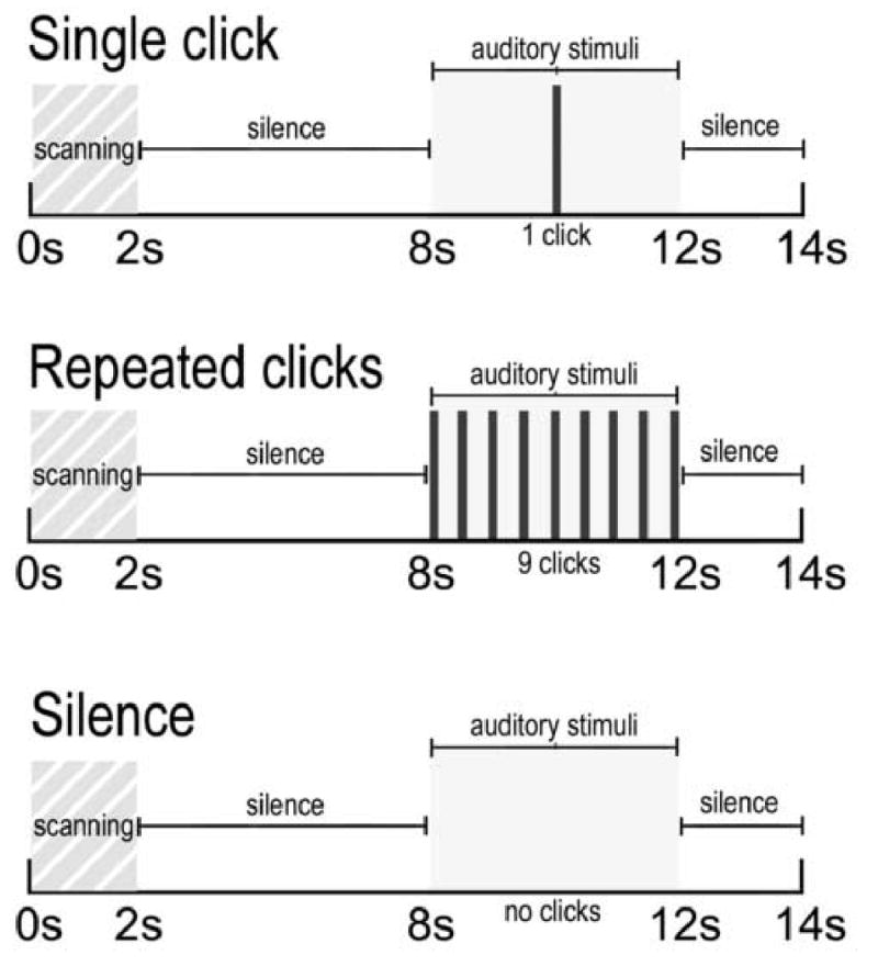
Methods: Twelve patients with schizophrenia and 12 healthy comparison subjects were scanned at 3 Tesla while performing a sensory gating task developed for fMRI. P50 EEG evoked potential recordings from a paired-stimulus conditioning-test paradigm were obtained from the same subjects.
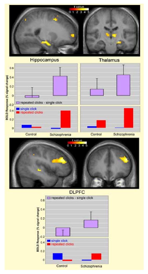
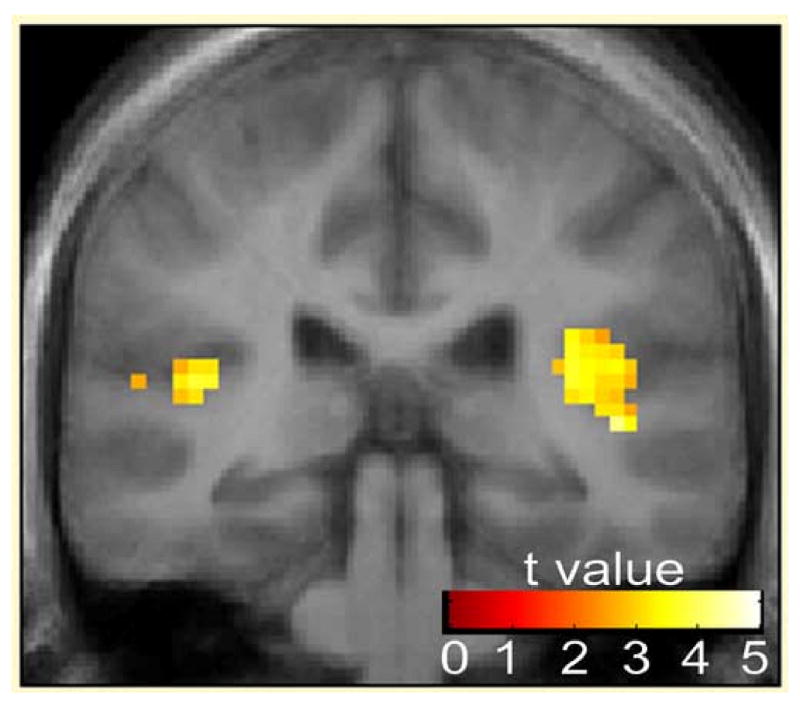
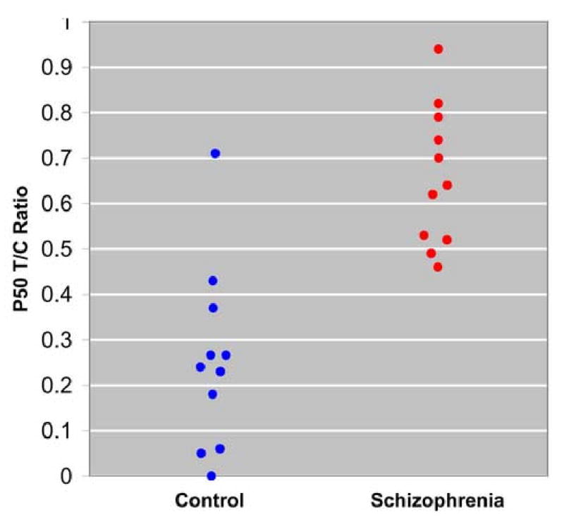
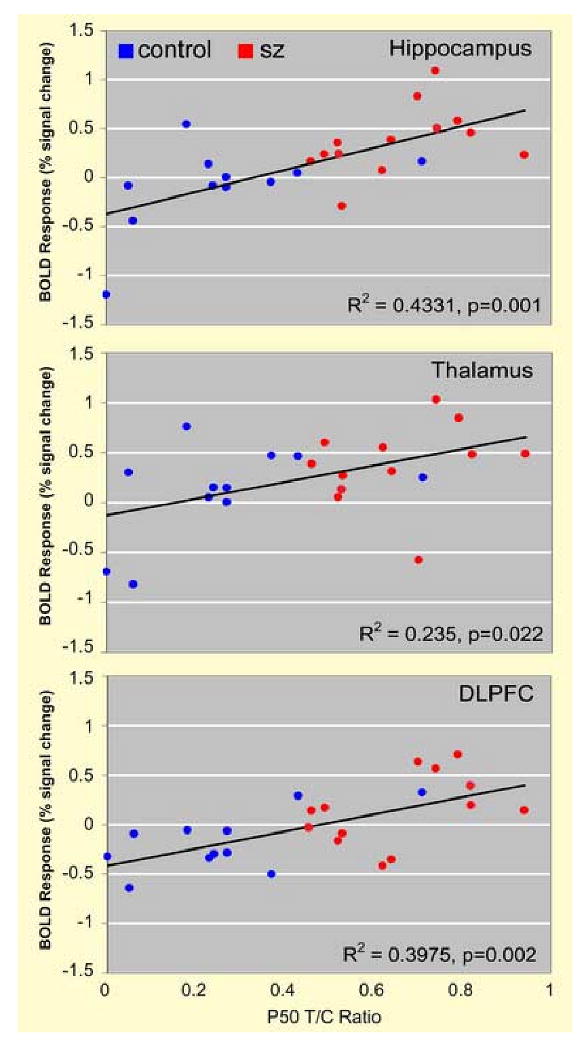
Results: Compared to healthy comparison subjects, patients with schizophrenia exhibited greater activation in the hippocampus, thalamus, and dorsolateral prefrontal cortex (DLPFC) during the fMRI sensory gating task. No group difference was observed in the superior temporal gyrus. Schizophrenia subjects also showed decreased P50 suppression as measured with EEG. Hemodynamic response in the fMRI measure was positively correlated with test/conditioning ratios from the EEG sensory gating measure.
Conclusions: Poor sensory gating in schizophrenia is associated with dysfunction of an apparent network of brain regions, including the hippocampus, thalamus and DLPFC. Greater activation of these regions is consistent with evidence for diminished inhibitory function in schizophrenia.
Figures
References
-
- Adler LE, Olincy A, Cawthra EM, McRae KA, Harris JG, Nagamoto HT, Waldo MC, Hall MH, Bowles A, Woodward L, Ross RG, Freedman R. Varied effects of atypical neuroleptics on P50 auditory gating in schizophrenia patients. Am J Psychiatry. 2004;161:1822–1828. - PubMed
-
- Adler LE, Pachtman E, Franks RD, Pecevich M, Waldo MC, Freedman R. Neurophysiological evidence for a defect in neuronal mechanisms involved in sensory gating in schizophrenia. Biol Psychiatry. 1982;17:639–654. - PubMed
-
- Adler LE, Waldo MC, Freedman R. Neurophysiologic studies of sensory gating in schizophrenia: comparison of auditory and visual responses. Biol Psychiatry. 1985;20:1284–1296. - PubMed
-
- Andrews J, Wang L, Csernansky JG, Gado MH, Barch DM. Abnormalities of thalamic activation and cognition in schizophrenia. Am J Psychiatry. 2006;163:463–469. - PubMed
-
- Benes FM, McSparren J, Bird ED, SanGiovanni JP, Vincent SL. Deficits in small interneurons in prefrontal and cingulate cortices of schizophrenic and schizoaffective patients. Arch Gen Psychiatry. 1991;48:996–1001. - PubMed
Publication types
MeSH terms
Grants and funding
LinkOut - more resources
Full Text Sources
Medical
Research Materials
