. 2007 Apr 15;35(3):1201-10.
doi: 10.1016/j.neuroimage.2007.01.024. Epub 2007 Feb 4.

Mapping the spinal and supraspinal pathways of dynamic mechanical allodynia in the human trigeminal system using cardiac-gated fMRI

Affiliations

Mapping the spinal and supraspinal pathways of dynamic mechanical allodynia in the human trigeminal system using cardiac-gated fMRI

Caterina Mainero et al. Neuroimage. .

Abstract

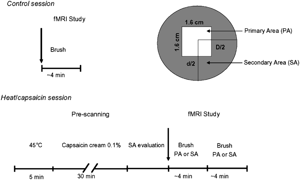
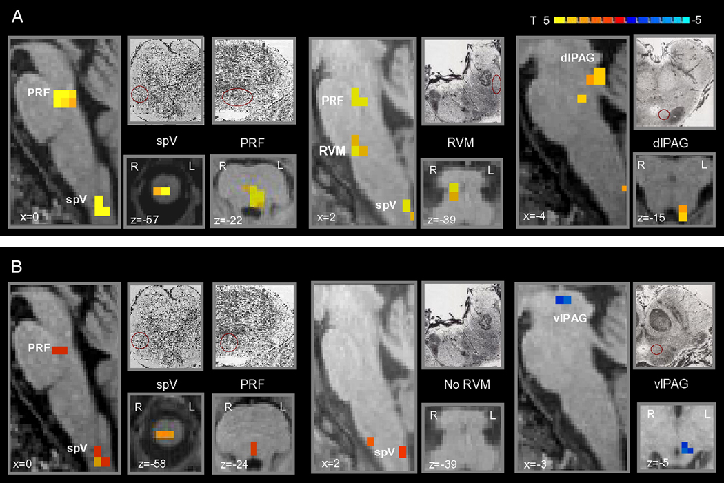
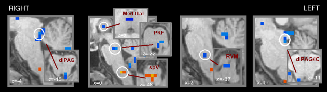
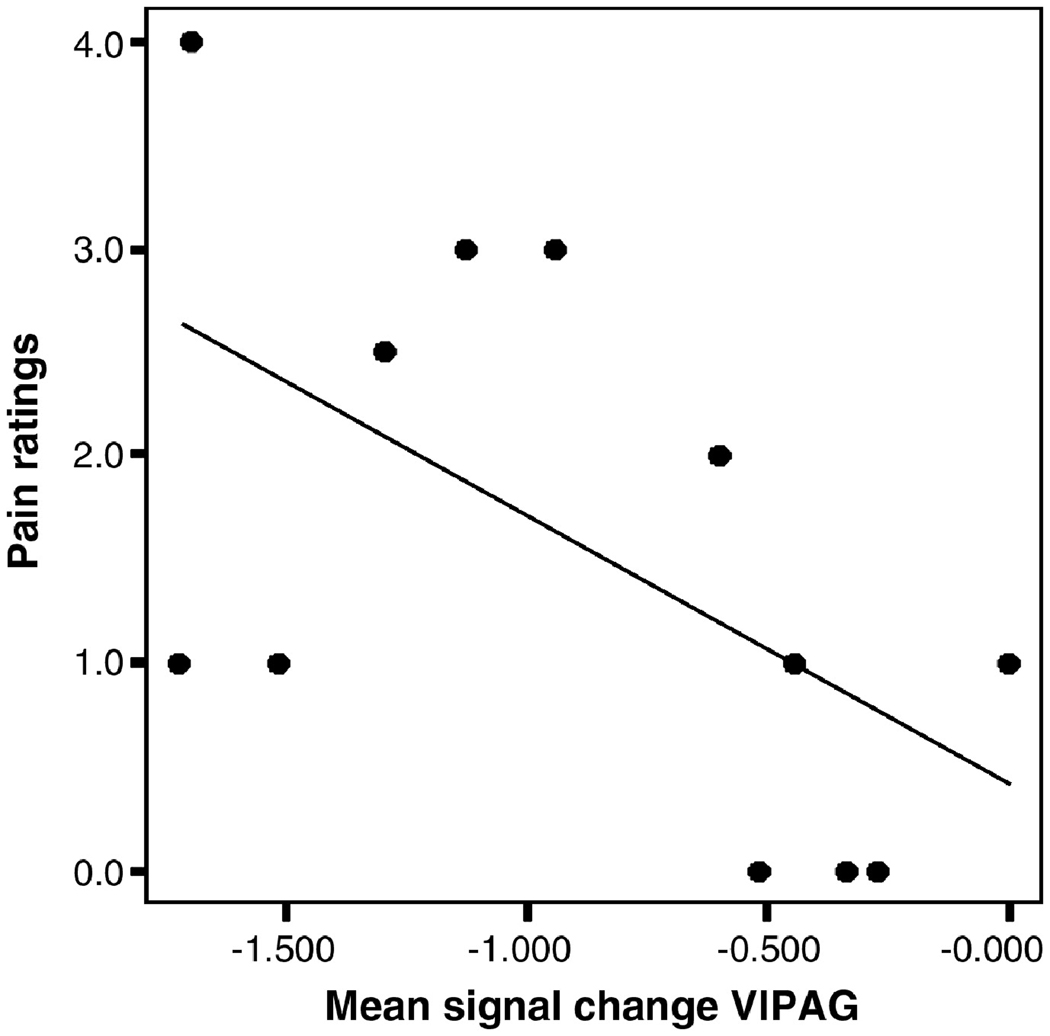
Following injury and inflammation, pain to light stroking (dynamic mechanical allodynia) might develop at the damaged site (primary area) or in adjacent normal tissue (secondary area). Using fMRI we mapped changes in the spinal trigeminal nucleus (spV), and supraspinal brainstem nuclei following heat/capsaicin-induced primary and secondary dynamic mechanical allodynia in the human trigeminal system. The role of these structures in dynamic mechanical allodynia has not been clarified yet in humans. During the control session we applied the same mechanical stimuli to the same untreated trigeminal area. Primary and secondary mechanical allodynia showed equal levels of perceived pain intensity, and compared to control mechanical stimulation exhibited similar responses in the ipsilateral spV and contralateral ventrolateral periaqueductal gray (vlPAG). Activity in the spV was significantly higher during both conditions versus the control mechanical stimulation, indicating that central sensitization of second-order neurons is similar for primary and secondary mechanical allodynia. The vlPAG showed decreased activity that inversely correlated with pain ratings during primary allodynia, i.e. the more deactivated the vlPAG the higher the pain intensity (p<0.05, Pearson's correlation). Primary and secondary dynamic mechanical allodynia were also characterized by significant differences involving distinct supraspinal structures mainly involved in pain modulation and including the rostroventromedial medulla, pons reticular formation, dorsolateral PAG, all more active during primary versus secondary allodynia, and the medial reticular formation of the caudal medulla that was more active during secondary versus primary allodynia. These results indicate that the pain modulatory system is involved to a different extent during primary versus secondary mechanical allodynia.


Figures

Figure 1
Figure 1. Experimental design
Twelve healthy subjects were randomly assigned to one of two counterbalanced groups to undergo two fMRI sessions, separated by an interval of at least a week, one during innocuous brush to the right ophthalmic division (V1) of the trigeminal nerve immediately after heat/capsaicin-induced allodynia (heat/capsaicin session), and one during the same stimuli to the untreated right V1 (control session). The order of the two sessions was randomized. During the heat/capsaicin session, we first marked in each subject the treatment site on the right V1 (1.6 cm2). The premarked skin was then heated for 5 minutes at 45°C using a 1.6 cm2 Peltier thermode (Medoc, Haifa Israel) after which 0.1% of capsaicin cream (Capzasin-HP, Chattem, Inc, USA) was applied over the same area for other 30 minutes. After capsaicin removal, the borders of the area of secondary allodynia were delineated with a brush. Then, during fMRI innocuous mechanical stimuli were administered to the premarked sites of interest (primary area, PA; secondary area, SA) on the right V1. In the control session, the same stimuli were administered to the normal skin of the same cutaneous area.
Figure 2
Figure 2. Brainstem anatomical outlines o f study subjects
Sagittal view of brainstem anatomical outlines of all study subjects. Individual outlines are displayed in different colors. To quantify the accuracy of brainstem co-registration, we defined in the mid-sagittal plane the angle between the line connecting the anterior and posterior commissure and the line connecting the posterior commissure and the obex in the medulla. The angles of all subjects ranged from 112.17 to 115.68 degrees (114.02±1.19, mean±SD), which corresponds to the SD of 1–1.5 mm at the obex.
Figure 3
Figure 3. Brainstem differences of brush to either the primary or secondary area versus the normal skin
Group statistical comparison t maps (radiological convention; p<0.05, corrected), overlaid on high resolution anatomical images in Talairach space, showing in 11 healthy subjects significant differences in the brainstem during brush to either (A) the primary or (B) secondary area following heat/capsaicin application to the right ophthalmic division of the trigeminal nerve versus brush to the untreated skin of the same cutaneous territory. Sketches and micrographs adapted from the atlas of Duvernoy (1995) were used to help identify the location of activation clusters. (spV = spinal trigeminal nucleus; PRF = pons reticular formation; RVM = rostroventromedial medulla; dlPAG = dorsolateral PAG; vlPAG = ventrolateral PAG).
Figure 4
Figure 4. Brainstem differences of brush to the secondary area versus brush to the primary area
Group statistical comparison t maps (radiological convention; p<0.05, corrected), overlaid on high resolution anatomical images in Talairach space, showing in 11 healthy subjects significant differences in the brainstem during brush to either secondary area versus brush to the primary following heat/capsaicin application to the right ophthalmic division of the trigeminal nerve versus brush to the untreated skin of the same cutaneous territory. In blue are represented the clusters more activated during primary versus secondary dynamic mechanical allodynia, the opposite is true for the activation in the caudal medulla shown in yellow/red. (MRF = medulla reticular formation; PRF = pons reticular formation; dlPAG = dorsolateral PAG; Med Thal = medial thalamus; IC = inferior colliculus).
Figure 5
Figure 5. Correlation between the mean signal change in the contralateral ventrolateral periaqueductal gray and pain intensity ratings during primary mechanical allodynia
Correlation analysis showing a significant inverse correlation (p<0.05, Pearson’s correlation coefficient = −0.6) between the mean signal change in the contralateral ventrolateral periaqueductal gray and pain intensity ratings during primary dynamic mechanical allodynia, i.e. the more deactivated the vlPAG the higher the pain intensity.

Comment in

References

    1. Ali Z, Meyer RA, Campbell JN. Secondary hyperalgesia to mechanical but not heat stimuli following a capsaicin injection in hairy skin. Pain. 1996;68:401–411. - PubMed
    1. Arthurs OJ, Boniface S. How well do we understand the neural origins of the fMRI BOLD signal? Trends Neurosci. 2002;25:27–31. - PubMed
    1. Bago M, Dean C. Sympathoinhibition from ventrolateral periaqueductal gray mediated by 5-HT(1A) receptors in the RVLM. Am J Physiol Regul Integr Comp Physiol. 2001;280:R976–R984. - PubMed
    1. Beckett S, Marsden CA. The effect of central and systemic injection of the 5-HT1A receptor agonist 8-OHDPAT and the 5-HT1A receptor antagonist WAY100635 on periaqueductal grey-induced defence behaviour. J Psychopharmacol. 1997;11:35–40. - PubMed
    1. Beckett SR, Lawrence AJ, Marsden CA, Marshall PW. Attenuation of chemically induced defence response by 5-HT1 receptor agonists administered into the periaqueductal gray. Psychopharmacology (Berl) 1992;108:110–114. - PubMed

Publication types