. 2007 Apr 15;304(2):771-85.
doi: 10.1016/j.ydbio.2007.01.029. Epub 2007 Jan 25.

The axon guidance gene lola is required for programmed cell death in the Drosophila ovary

Affiliations

The axon guidance gene lola is required for programmed cell death in the Drosophila ovary

B Paige Bass et al. Dev Biol. .

Abstract

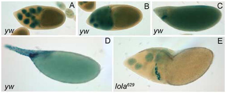
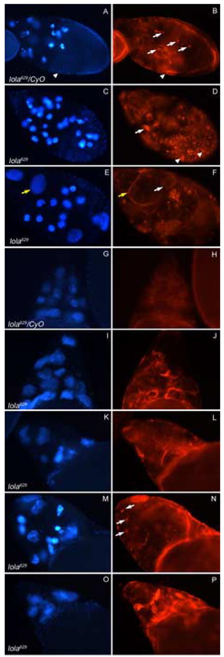
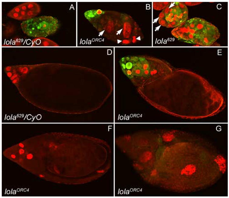
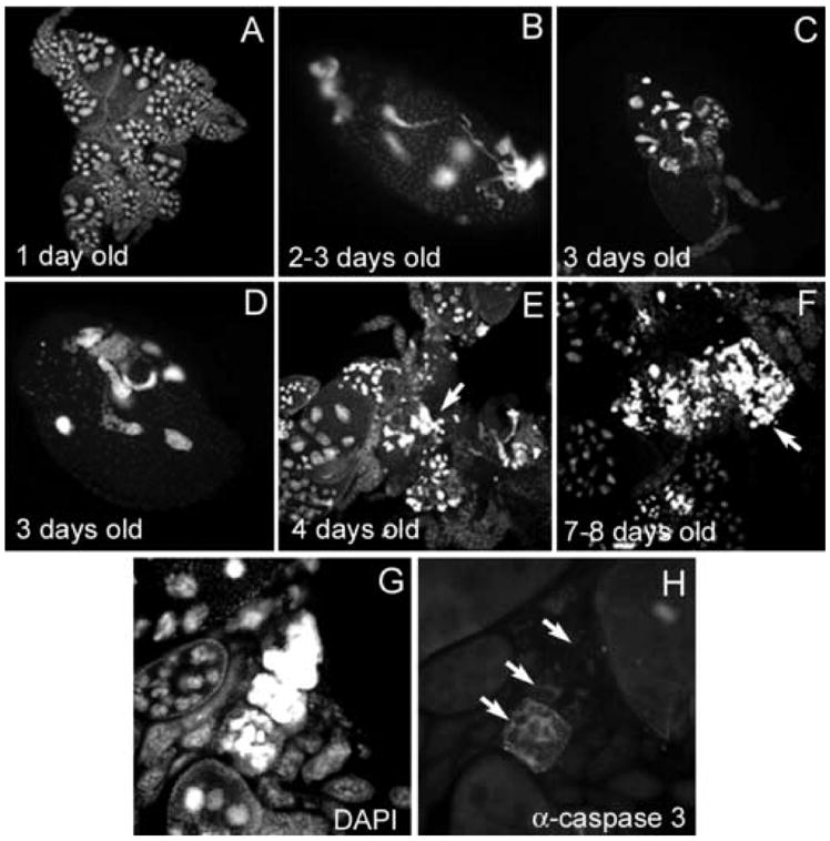
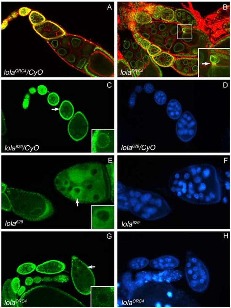
longitudinals-lacking (lola) was identified in Drosophila as a gene encoding several alternatively spliced transcription factors involved in axon guidance. Here we report that lola also plays a critical role in programmed cell death in the ovary. lola mutant germline clones show a high percentage of egg chambers with nurse cell nuclei persisting past stage 13, indicating a block in developmental nurse cell death. Mutants also show a disruption in the induced programmed cell death that occurs during mid-oogenesis in response to starvation. Further characterization revealed that lola germline clones exhibit abnormal nuclear organization which becomes increasingly severe with age. Chromatin appears diffuse and fails to condense properly or undergo DNA fragmentation in dying nurse cells. Masses of nuclear material accumulate in the ovaries of older flies containing lola germline clones. We propose that lola is necessary for complete chromatin condensation which occurs during programmed cell death in the ovary. Alleles differed in the strength of their phenotypes but interestingly, the severity of their ovarian phenotypes was independent of the strength of their neuronal phenotypes, suggesting a differential requirement for individual lola isoforms in the ovary and nervous system.


Figures

Fig. 1
Fig. 1
lola alleles vary in the severity of their GLC phenotypes. All egg chambers have been DAPI-stained. (A) Control, yw stage 14 egg chambers do not contain any remaining nurse cell nuclei. (B–H) Representative stage 14 egg chambers containing GLCs of the indicated lola alleles show that most cause moderate to severe defects during oogenesis including dumpless egg chambers. However, lola4E4 (C) looks wild-type.
Fig. 2
Fig. 2
Intragenic complementation analysis. (A) Complementation results with different lethal alleles of lola helped narrow down the location of the lethal mutation in lola629. Offspring from the F1 generation of the indicated cross were counted. Percent viability is calculated as 100 x (observed number of straight winged flies/ expected number of straight winged flies). Expected number of straight winged flies = 1/2(observed number of curly winged flies) (Horiuchi et al., 2003). (B) Locations of P-element insertions in lola4E4, lola00642, and lola5D2, and EMS-induced mutations in lolaORE120, lolaORC4, and lolaORE50. Only relevant exons are depicted and drawing is not to scale (Horiuchi et al., 2003).
Fig. 3
Fig. 3
Quantification of ovarian phenotypes of different lola alleles in order of increasing severity. Stage 14 egg chambers were classified as one of four indicated categories. Those classified as other had dorsal appendage defects or were cup-shaped but did not contain persisting nurse cell nuclei and were not dumpless. FRTG13 germline clones were analyzed as a control. n= the total number of stage 14 germline clone egg chambers classified.
Fig. 4
Fig. 4
Lola isoform K expression is necessary for normal oogenesis. (A) Extracts from embryos of the genotypes indicated above were separated by SDS-PAGE and subjected to western analysis using an antibody that recognizes the constant region of Lola. The positions of standard protein molecular weight markers are indicated to the right. Compared with yw control embryos, those heterozygous and homozygous for lola629, as well as lolaORC4, and lolaORE50 embryos express truncated versions of isoform K (arrows). lola629, lolaORC4, lolaORE50, and lola5D2 homozygotes lack expression of the normal sized isoform K while lola4E4 retains expression (arrowhead). (B) The same membrane used for (A) was probed with an antibody for Lamin Dm0 to verify that comparable amounts of protein were added to each lane. (C) Protein extracts from ovaries of the genotypes indicated above were subjected to western analysis as in (A). Ovaries from lola629 heterozygotes and those containing GLCs of lola629 express a truncated version of isoform K (arrow), while only the GLCs lack expression of the normal sized isoform (arrowhead).
Fig. 5
Fig. 5
lola GLCs do not undergo DNA fragmentation during checkpoint or developmental PCD. TUNEL analysis (dark purple) and DAPI staining (blue) was performed on all egg chambers. (A) Heterozygous mid-stage egg chambers from nutrient deprived females undergo checkpoint PCD and show TUNEL positive nurse cell nuclei. (B) As an internal control, ovoD tissue, surrounding lolaORC4 GLCs, did contain TUNEL positive nuclei (arrows). (C) Mid-stage degenerating egg chambers containing lolaORC4 GLCs are not TUNEL positive. Arrows indicate three separate egg chambers. (D) Late stage 12 and (E) early stage 13 heterozygous control egg chambers display TUNEL positive nurse cells during developmental PCD. (F) lolaORC4 GLCs in a dumpless stage 14 egg chamber are not TUNEL positive.
Fig. 6
Fig. 6
Nurse cell nuclear membranes do not break down in lola629 GLCs. (A–D) y w hsflp control and (E) lola629 GLC egg chambers contain the BB127 enhancer trap encoding nuclear β-galactosidase and are X-gal stained (blue). (A) A stage 10 yw egg chamber has intact nurse cell nuclear membranes. (B) By stage 10B, the nurse cell nuclear membranes break down and X-gal staining leaks into the cytoplasm. (C) A stage 12 yw egg chamber has undergone cytoplasmic transfer and X-gal staining is seen in the oocyte. (D) By stage 14, no nurse cells remain and all the X-gal staining is seen in the oocyte. (E) A dumpless egg chamber containing lola629 GLCs retains X-gal staining in the nurse cell nuclei.
Fig. 7
Fig. 7
Abnormal actin structure is seen in degenerating lola629 GLC egg chambers. Panels depict DAPI staining (blue) on the left and rhodamine-phalloidin staining (red) on the right of the same egg chambers. All images were taken at the same magnification and rhodamine-phalloidin pictures were taken at the same exposure. (A–B) A heterozygous mid-stage egg chamber undergoing checkpoint PCD, induced through nutrient deprivation, contains condensed nurse cell nuclei, small actin clumps (B, arrows) and follicle cells with cellular membranes still intact (arrowheads). (C–F) lola629 GLC egg chambers undergoing checkpoint PCD, without starvation, contain abnormal actin structures (D and F, white arrows), degenerating follicle cells (D, arrowheads), and uncondensed nurse cell nuclei with intact cellular membranes (E and F, yellow arrows). (G–H) A stage 10B heterozygous egg chamber shows formation of organized actin bundles in all nurse cells. (I–J) A stage 10B egg chamber containing lola629 GLCs exhibits actin bundle formation that is disorganized and not uniform throughout the nurse cells. (K–P) Dumpless egg chambers containing lola629 GLCs continue to show disorganized actin structure. Some contain small actin clumps like those seen during mid-stage PCD (N, arrows).
Fig. 8
Fig. 8
Variable levels of caspase activity are seen during checkpoint and developmental PCD in lola629 and lolaORC4 GLCs. Cleaved Caspase-3 antibody staining (green) depicts caspase activity and propidium iodide staining (red) shows nuclei. All images were taken at the same exposure and magnification. (A) A heterozygous mid-stage egg chamber undergoing checkpoint PCD, induced by nutrient deprivation, shows condensed nurse cell nuclei and high levels of caspase activity throughout the egg chamber. (B) Mid-stage egg chambers containing lolaORC4 or (C) lola629 GLCs, undergoing checkpoint PCD, also show high levels of caspase activity, but it is not uniformly found throughout the entire egg chamber. Nurse cells are not always condensed (B and C, arrows) and a lack of caspase activity is seen both with (B, arrowheads) and without (B and C, arrows) nuclear condensation. (D) A heterozygous stage 12 egg chamber undergoing developmental PCD does not show significant levels of caspase activity with this antibody. (E–G) Dumpless egg chambers containing lolaORC4 GLCs show either high levels of caspase activity (E), none (F), or very low levels (G). Lack of proper nuclear condensation can also be seen in some of these dumpless egg chambers (G).
Fig. 9
Fig. 9
lola629 GLCs exhibit chromatin defects that increase with age of the female. Egg chambers in (A–G) were stained with DAPI and (H) was stained with Cleaved Caspase-3 antibody. (A) lola629 GLC tissue from a one day old female appears normal. (B–D) By two-three days, abnormally-shaped nuclei, along with nuclear condensation defects can be seen. (E) At four days, masses of nuclear material accumulate between egg chambers (arrow) and (F) by seven-eight days, these masses become larger (arrow). (G–H) A group of egg chambers stained with DAPI (G) and Cleaved Caspase-3 antibody (H) show loss of caspase activity after the follicle cells die and the nurse cell cytoplasm disperses (arrows). Remaining nuclear material in (G) may contribute to the masses in (E–F). (A), (C), and (E–F) were taken at the same magnification. (B), (D), and (G–H) were taken at higher magnification.
Fig. 10
Fig. 10
lola629 and lolaORC4 GLCs exhibit abnormal nuclear lamina morphology. Egg chambers in (A–B) were double-labeled with lamin Dm0 ADL84.12 antibody (green) and rhodamine-phalloidin (red). (A) Heterozygous control egg chambers exhibit normal nuclear lamina morphology. (B) A lolaORC4 GLC egg chamber exhibits a protrusion of the nurse cell nuclear lamina through the ring canal (arrow, higher magnification inset). Egg chambers in (C–H) were stained with lamin Dm0 ADL101 antibody (green, C, E, and G) and DAPI to visualize DNA (blue, D, F, and H). (C) Nuclear lamin Dm0 surrounds the nuclei in heterozygous control egg chambers (higher magnification inset of nucleus indicated by arrow). (E and G) lola629 and lolaORC4 GLCs exhibit mislocalization of lamin Dm0 to the cytoplasm ((higher magnification insets of nuclei indicated by arrows). (F and H) No abnormal chromatin morphology is seen in the egg chambers with mislocalized lamin Dm0 (compare with neighboring egg chambers and heterozygous controls in D).

References

    1. Bao X, Zhang W, Krencik R, Deng H, Wang Y, Girton J, Johansen J, Johansen KM. The JIL-1 kinase interacts with lamin Dm0 and regulates nuclear lamina morphology of Drosophila nurse cells. J Cell Sci. 2005;118:5079–87. - PubMed
    1. Baum JS, St George JP, McCall K. Programmed cell death in the germline. Semin Cell Dev Biol. 2005;16:245–59. - PubMed
    1. Cavaliere V, Taddei C, Gargiulo G. Apoptosis of nurse cells at the late stages of oogenesis. Dev Genes Evol. 1998;208:106–112. - PubMed
    1. Cooley L, Verheyen E, Ayers K. chickadee encodes a profilin required for intercellular cytoplasm transport during Drosophila oogenesis. Cell. 1992;69:173–184. - PubMed
    1. Crowner D, Madden K, Goeke S, Giniger E. Lola regulates midline crossing of CNS axons in Drosophila. Development. 2002;129:1317–25. - PubMed

Publication types

LinkOut - more resources