

First evidence of division and accumulation of viable but nonculturable Pseudomonas fluorescens cells on surfaces subjected to conditions encountered at meat processing premises
- PMID: 17337551
- PMCID: PMC1892859
- DOI: 10.1128/AEM.02267-06
First evidence of division and accumulation of viable but nonculturable Pseudomonas fluorescens cells on surfaces subjected to conditions encountered at meat processing premises
Abstract
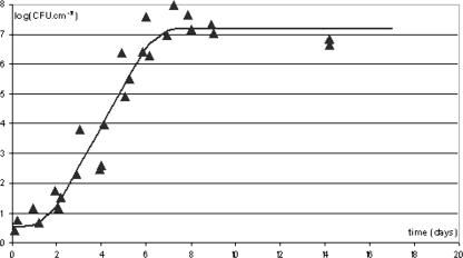
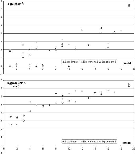
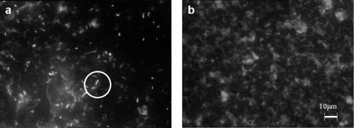
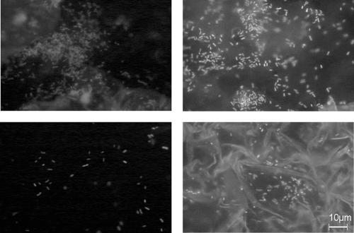
Cleaning and disinfection of open surfaces in food industry premises leave some microorganisms behind; these microorganisms build up a resident flora on the surfaces. Our goal was to explore the phenomena involved in the establishment of this biofilm. Ceramic coupons were contaminated, once only, with Pseudomonas fluorescens suspended in meat exudate incubated at 10 degrees C. The mean adhering population after 1 day was 10(2) CFU x cm(-2) and 10(3) total cells x cm(-2), i.e., the total number of cells stained by DAPI (4',6'-diamidino-2-phenylindole). The coupons were subjected daily to a cleaning product, a disinfectant, and a further soiling with exudate. The result was a striking difference between the numbers of CFU, which reached 10(4) CFU x cm(-2), and the numbers of total cells, which reached 2 x 10(6) cells x cm(-2) in 10 days. By using hypotheses all leading to an overestimation of the number of dead cells, we showed that the quantity of nonculturable cells (DAPI-positive cells minus CFU) observed cannot be accounted for as an accumulation of dead cells. Some nonculturable cells are therefore dividing on the surface, although cell division is unable to continue to the stage of macrocolony formation on agar. The same phenomenon was observed when only a chlorinated alkaline product was used and the number of cells capable of reducing 5-cyano-2,3-ditolyl tetrazolium chloride was close to the number of total cells, confirming that most nonculturable cells are viable but nonculturable. Furthermore, the daily shock applied to the cells does not prompt them to enter a new lag phase. Since a single application of microorganisms is sufficient to produce this accumulation of cells, it appears that the phenomenon is inevitable on open surfaces in food industry premises.
Figures
Similar articles
-
Potential of Escherichia coli O157:H7 to persist and form viable but non-culturable cells on a food-contact surface subjected to cycles of soiling and chemical treatment.Int J Food Microbiol. 2010 Nov 15;144(1):96-103. doi: 10.1016/j.ijfoodmicro.2010.09.002. Epub 2010 Sep 7. Int J Food Microbiol. 2010. PMID: 20888655
-
Biofilm formation and desiccation survival of Listeria monocytogenes with microbiota on mushroom processing surfaces and the effect of cleaning and disinfection.Int J Food Microbiol. 2024 Feb 2;411:110509. doi: 10.1016/j.ijfoodmicro.2023.110509. Epub 2023 Nov 30. Int J Food Microbiol. 2024. PMID: 38101188
-
Impact of cleaning and disinfection agents on biofilm structure and on microbial transfer to a solid model food.J Appl Microbiol. 2004;97(2):262-70. doi: 10.1111/j.1365-2672.2004.02296.x. J Appl Microbiol. 2004. PMID: 15239692
-
Dynamics of biofilm formation by Listeria monocytogenes on stainless steel under mono-species and mixed-culture simulated fish processing conditions and chemical disinfection challenges.Int J Food Microbiol. 2018 Feb 21;267:9-19. doi: 10.1016/j.ijfoodmicro.2017.12.020. Epub 2017 Dec 19. Int J Food Microbiol. 2018. PMID: 29275280
-
Direct estimate of active bacteria: CTC use and limitations.J Microbiol Methods. 2003 Jan;52(1):19-28. doi: 10.1016/s0167-7012(02)00128-8. J Microbiol Methods. 2003. PMID: 12401223 Review.
Cited by
-
Current Perspectives on Viable but Non-culturable State in Foodborne Pathogens.Front Microbiol. 2017 Apr 4;8:580. doi: 10.3389/fmicb.2017.00580. eCollection 2017. Front Microbiol. 2017. PMID: 28421064 Free PMC article. Review.
-
Quantitative approach to determining the contribution of viable-but-nonculturable subpopulations to malolactic fermentation processes.Appl Environ Microbiol. 2009 May;75(9):2977-81. doi: 10.1128/AEM.01707-08. Epub 2009 Mar 6. Appl Environ Microbiol. 2009. PMID: 19270138 Free PMC article.
-
Isolation and characterization of a T7-like lytic phage for Pseudomonas fluorescens.BMC Biotechnol. 2008 Oct 27;8:80. doi: 10.1186/1472-6750-8-80. BMC Biotechnol. 2008. PMID: 18954452 Free PMC article.
-
Methods for recovering microorganisms from solid surfaces used in the food industry: a review of the literature.Int J Environ Res Public Health. 2013 Nov 14;10(11):6169-83. doi: 10.3390/ijerph10116169. Int J Environ Res Public Health. 2013. PMID: 24240728 Free PMC article. Review.
-
Abundance and Distribution of Enteric Bacteria and Viruses in Coastal and Estuarine Sediments-a Review.Front Microbiol. 2016 Nov 1;7:1692. doi: 10.3389/fmicb.2016.01692. eCollection 2016. Front Microbiol. 2016. PMID: 27847499 Free PMC article. Review.
References
-
- Alliot, M. 1999. Ecologie microbienne des sites industriels fromagers: identification, caractérisation des flores isolées et étude des facteurs régissant leur implantation. Ph.D. thesis. Institut National Agronomique de Paris-Grignon, Paris, France.
-
- Anonymous. 1997. Geometrical product specification—surface texture: profile method—terms, definitions and surface texture parameters. ISO 4287. International Organization for Standardization, Geneva, Switzerland.
-
- Asakura, H., S. Makino, T. Takagi, A. Kuri, T. Kurazono, M. Watarai, and T. Shirahata. 2002. Passage in mice causes a change in the ability of Salmonella enterica serovar Oranienburg to survive NaCl osmotic stress: resuscitation from the viable but non-culturable state. FEMS Microbiol. Lett. 212:87-93. - PubMed
-
- Asséré, A., N. Oulahal-Lagsir, and B. Carpentier. Comparative evaluation of methods for counting surviving biofilm cells adhering to a polyvinyl chloride surface exposed to chlorine or dehydration. J. Appl. Microbiol., in press. - PubMed
Publication types
MeSH terms
Substances
LinkOut - more resources
Full Text Sources
