

doi: 10.1128/JB.01662-06.
Epub 2007 Mar 2.
HpdR is a transcriptional activator of Sinorhizobium meliloti hpdA, which encodes a herbicide-targeted 4-hydroxyphenylpyruvate dioxygenase
Affiliations
- PMID: 17337579
- PMCID: PMC1855912
- DOI: 10.1128/JB.01662-06
Item in Clipboard
HpdR is a transcriptional activator of Sinorhizobium meliloti hpdA, which encodes a herbicide-targeted 4-hydroxyphenylpyruvate dioxygenase
J Bacteriol.
2007 May.
Abstract
Sinorhizobium meliloti hpdA, which encodes the herbicide target 4-hydroxyphenylpyruvate dioxygenase, is positively regulated by HpdR. Gel mobility shift and DNase I footprinting analyses revealed that HpdR binds to a region that spans two conserved direct-repeat sequences within the hpdR-hpdA intergenic space. HpdR-dependent hpdA transcription occurs in the presence of 4-hydroxyphenylpyruvate, tyrosine, and phenylalanine, as well as during starvation.
Figures
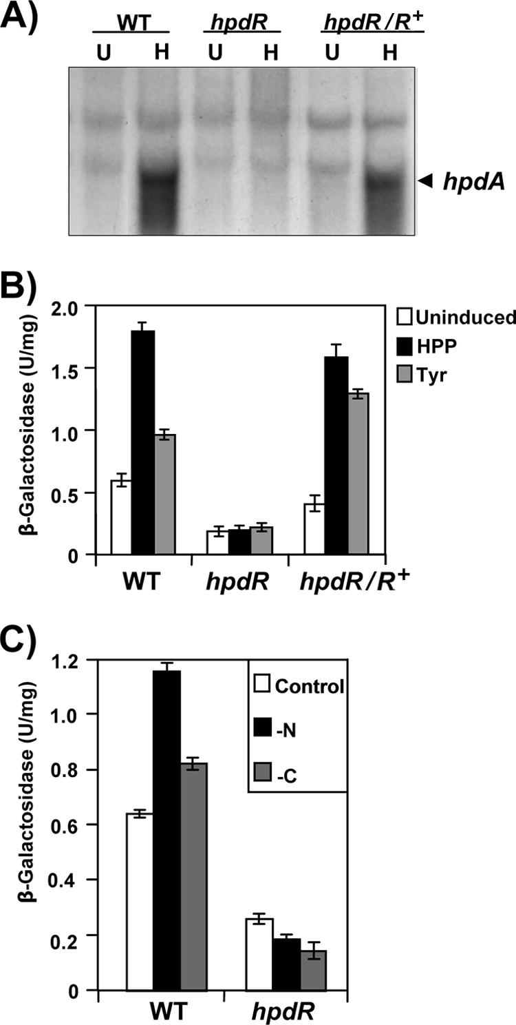
HpdR-dependent regulation of S. meliloti hpdA expression in response to tyrosine and HPP. (A) An autoradiogram of a Northern blot of total RNA extracted from wild-type S. meliloti, the S. meliloti hpdR insertion mutant (hpdR), and the S. meliloti hpdR insertion mutant strain complemented with a functional hpdR (hpdR/hpdR+) and probed with a 32P-labeled internal fragment of S. meliloti hpdA. Cultures were either induced for 30 min with 5 mM HPP (H) or uninduced (U) prior to RNA extraction. (B) Results of β-galactosidase assays of wild-type S. meliloti and hpdR and hpdR/hpdR+ strains carrying the hpdA promoter-lacZ fusion plasmid pMPA1 are shown. Cultures were either uninduced or induced for 30 min with 5 mM HPP or tyrosine prior to the assay. (C) HpdR-dependent regulation of hpdA expression in response to nutrient starvation. The results of β-galactosidase assays of wild-type S. meliloti and the hpdR strain carrying the hpdA promoter-lacZ fusion plasmid pMPA1 are shown. Cultures were either uninduced or incubated in M-9 minimal medium lacking either a carbon (sucrose) or a nitrogen (NH4Cl) source for 30 min prior to the assay. Error bars show standard deviations. WT, wild type.
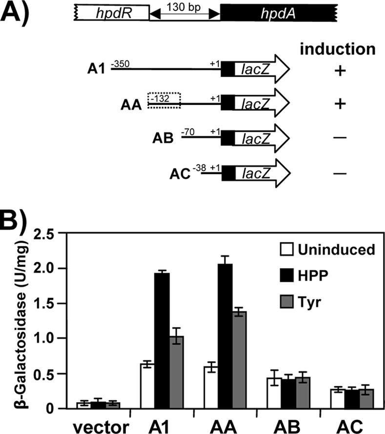
Deletion mapping of the S. meliloti hpdA promoter. (A) Diagram of the various hpdA promoter-lacZ transcriptional fusion constructs used. The numbers represent the location of the hpdA translation start (+1) and the amount of upstream sequence contained in each fusion plasmid. The ability of each fusion to be induced in the presence of HPP or tyrosine is indicated. The dotted box delimits a region important for HpdA expression. (B) Results of β-galactosidase assays of extracts of wild-type S. meliloti strains carrying the various hpdA promoter-lacZ fusion plasmids shown in Fig. 3A. Cultures were either uninduced or induced for 30 min with 5 mM HPP or tyrosine prior to the assay. Error bars show standard deviations.
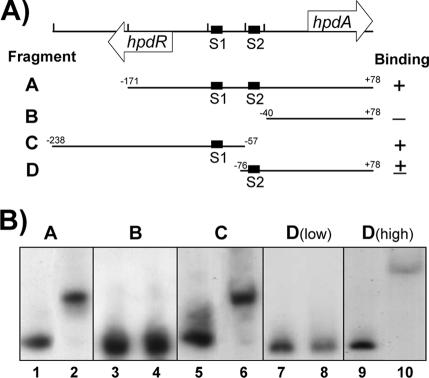
Binding of HpdR to the S. meliloti hpdA promoter region. (A) A map of the hpdA intergenic region is shown along with the PCR fragments, A through D, used as probes in the gel mobility shift assays. The numbers indicate the positions of the ends of each probe relative to the hpdA translation start. The dark boxes indicate the positions of the S1 and S2 direct repeat sequences. The ability of HpdR to bind to each probe is also indicated (+, −, or ±). (B) A compilation of autoradiograms of gel mobility shift assays performed using recombinant S. meliloti HpdR-His6 and the 32P-labeled PCR fragments shown in Fig. 3A. The probe used is shown at the top of each panel. The odd lanes contain gel shift reaction mixtures with no protein added. Lanes 2, 6, and 8 contain reaction mixtures to which HpdR-His6 was added to a final concentration of 0.16 mg/ml. The reaction mixtures whose results are shown in lanes 4 and 10 contained 0.48 mg/ml HpdR-His6. D(low), low concentration of HpdR-His6 (0.16 mg/ml); D(high), high concentration of HpdR-His6 (0.48 mg/ml).
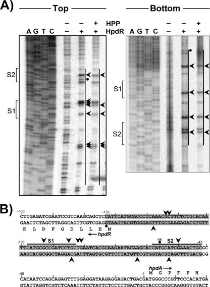
DNase I footprint of HpdR bound to the HpdA promoter. (A) Autoradiograms of DNase I protection assays of HpdR using an hpdR-hpdA intergenic probe, labeled on either the top or bottom strand, in the presence or absence of the inducer, HPP. Single end-labeled probes were generated by PCR amplification of the S1- and S2-spanning region in pICHA1, in which one of the primers had been end labeled using T4 polynucleotide kinase and [γ-32P]ATP. 32P-dideoxy sequencing ladders, generated using the same oligonucleotides as those used to amplify the probe, are included as sizing standards. The brackets indicate the positions of the conserved S1 and S2 repeats. The vertical lines delimit the extents of the protected regions. The arrowheads denote DNase I-hypersensitive sites, while the dots indicate bands that display increased protection in the presence of HPP. (B) Sequence of the hpdR-hpdA intergenic region summarizing the DNase I footprint data. The DNase I-protected region is shaded, and the horizontal lines indicate the S1 and S2 repeats. The arrowheads identify DNase I-hypersensitive sites, while the dots indicate bands that show increased protection in the presence of HPP.
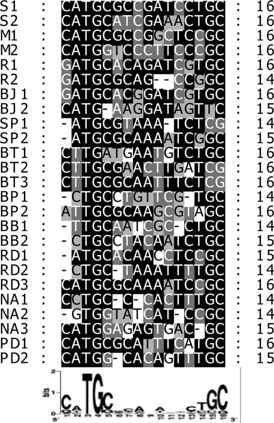
Comparison of HpdR binding site repeat sequences. A comparison of the S. meliloti hpdA promoter S1 and S2 repeat sequences with repeats present in the hpdA promoters of; Mesorhizobium loti MAFF303099 (M1 and M2), Bradyrhizobium japonicum USDA110 (BJ1 and BJ2), Rhodopseudomonas palustris CGA009 (R1 and R2), Silicibacter pomeroyi DSS-3 (SP1 and SP2), Burkholderia pseudomallei 1710b (BP1 and BP2), Bordetella bronchiseptica RB50 (BB1 and BB2), Burkholderia thailandensis E264 (BT1, BT2, and BT3), Roseobacter denitrificans OCH114 (RD1, RD2, and RD3), Novosphingobium aromaticivorans DSM12444 (NA1, NA2, and NA3), and Paracoccus denitrificans PD1222 (PD1 and PD2). A consensus sequence in which the height of each letter is proportional to the frequency of the residue at that position is shown at the bottom (4). Numbers on the right are sequence lengths, in base pairs.
Similar articles
-
The tyrosine degradation gene hppD is transcriptionally activated by HpdA and repressed by HpdR in Streptomyces coelicolor, while hpdA is negatively autoregulated and repressed by HpdR.Mol Microbiol. 2007 Aug;65(4):1064-77. doi: 10.1111/j.1365-2958.2007.05848.x. Epub 2007 Jul 19. Mol Microbiol. 2007. PMID: 17640269
-
Rem, a new transcriptional activator of motility and chemotaxis in Sinorhizobium meliloti.J Bacteriol. 2006 Oct;188(19):6932-42. doi: 10.1128/JB.01902-05. J Bacteriol. 2006. PMID: 16980496 Free PMC article.
-
The upstream region of the nodD3 gene of Sinorhizobium meliloti carries enhancer sequences for the transcriptional activator NtrC.FEMS Microbiol Lett. 1999 Oct 15;179(2):491-9. doi: 10.1111/j.1574-6968.1999.tb08768.x. FEMS Microbiol Lett. 1999. PMID: 10518756
-
Negative Regulation of Ectoine Uptake and Catabolism in Sinorhizobium meliloti: Characterization of the EhuR Gene.J Bacteriol. 2016 Dec 13;199(1):e00119-16. doi: 10.1128/JB.00119-16. Print 2017 Jan 1. J Bacteriol. 2016. PMID: 27795315 Free PMC article.
-
Identification of direct transcriptional target genes of ExoS/ChvI two-component signaling in Sinorhizobium meliloti.J Bacteriol. 2009 Nov;191(22):6833-42. doi: 10.1128/JB.00734-09. Epub 2009 Sep 11. J Bacteriol. 2009. PMID: 19749054 Free PMC article.
Cited by
-
The hdhA gene encodes a haloacid dehalogenase that is regulated by the LysR-type regulator, HdhR, in Sinorhizobium meliloti.Mol Biotechnol. 2013 Jun;54(2):148-57. doi: 10.1007/s12033-012-9556-1. Mol Biotechnol. 2013. PMID: 22638965
-
Comparative transcriptomic analysis reveals the significant pleiotropic regulatory effects of LmbU on lincomycin biosynthesis.Microb Cell Fact. 2020 Feb 12;19(1):30. doi: 10.1186/s12934-020-01298-0. Microb Cell Fact. 2020. PMID: 32050973 Free PMC article.
References
-
- Alexeyev, M. F. 1999. The pKNOCK series of broad-host-range mobilizable suicide vectors for gene knockout and targeted DNA insertion into the chromosome of gram-negative bacteria. BioTechniques. 26:824-828. - PubMed
-
- Barran, L. R., E. S. Bromfield, and D. C. Brown. 2002. Identification and cloning of the bacterial nodulation specificity gene in the Sinorhizobium meliloti-Medicago laciniata symbiosis. Can. J. Microbiol. 48:765-771. - PubMed
-
- Brinkman, A. B., T. J. Ettema, W. M. de Vos, and J. van der Oost. 2003. The Lrp family of transcriptional regulators. Mol. Microbiol. 48:287-294. - PubMed
Publication types
MeSH terms
Substances
LinkOut - more resources
Full Text Sources
Other Literature Sources
Molecular Biology Databases
