. 2007 May;27(9):3441-55.
doi: 10.1128/MCB.01643-06. Epub 2007 Mar 5.

mRNAs encoding polarity and exocytosis factors are cotransported with the cortical endoplasmic reticulum to the incipient bud in Saccharomyces cerevisiae

Affiliations

mRNAs encoding polarity and exocytosis factors are cotransported with the cortical endoplasmic reticulum to the incipient bud in Saccharomyces cerevisiae

Stella Aronov et al. Mol Cell Biol. 2007 May.

Abstract

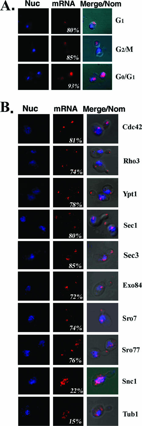
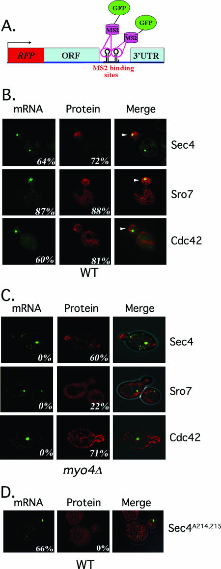
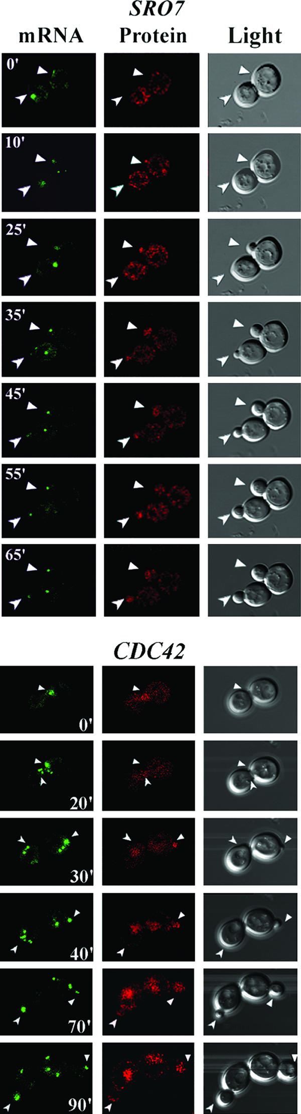
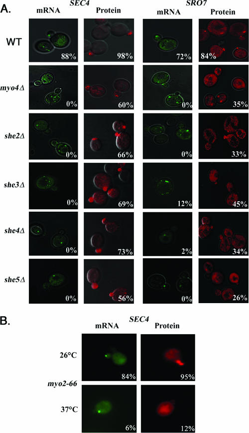
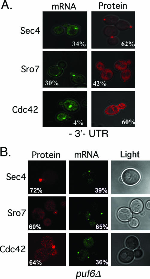
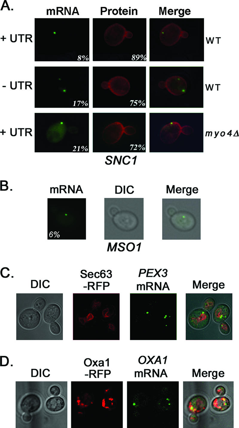
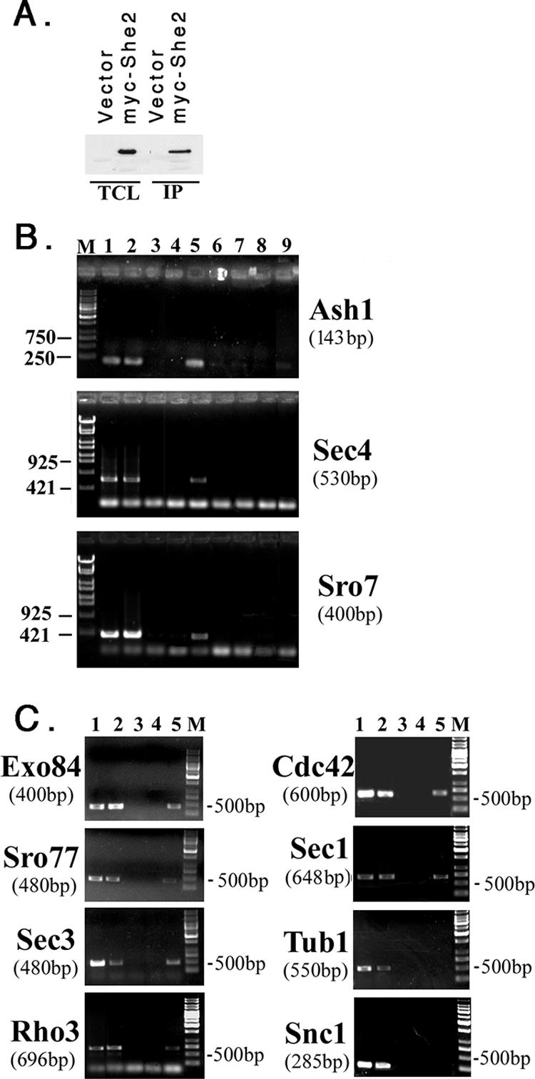
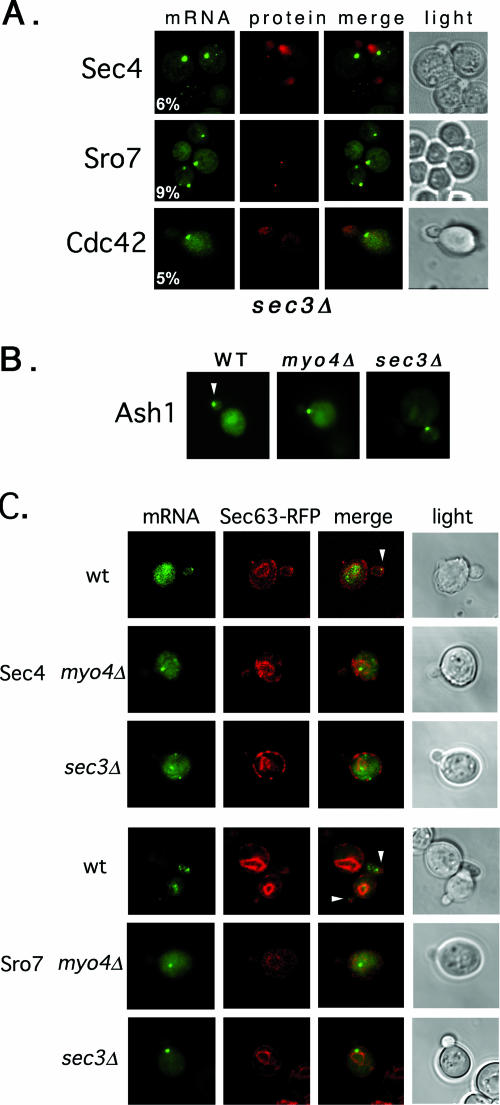
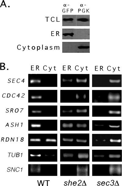
Polarized growth in the budding yeast Saccharomyces cerevisiae depends upon the asymmetric localization and enrichment of polarity and secretion factors at the membrane prior to budding. We examined how these factors (i.e., Cdc42, Sec4, and Sro7) reach the bud site and found that their respective mRNAs localize to the tip of the incipient bud prior to nuclear division. Asymmetric mRNA localization depends upon factors that facilitate ASH1 mRNA localization (e.g., the 3' untranslated region, She proteins 1 to 5, Puf6, actin cytoskeleton, and a physical association with She2). mRNA placement precedes protein enrichment and subsequent bud emergence, implying that mRNA localization contributes to polarization. Correspondingly, mRNAs encoding proteins which are not asymmetrically distributed (i.e., Snc1, Mso1, Tub1, Pex3, and Oxa1) are not polarized. Finally, mutations which affect cortical endoplasmic reticulum (ER) entry and anchoring in the bud (myo4Delta, sec3Delta, and srp101) also affect asymmetric mRNA localization. Bud-localized mRNAs, including ASH1, were found to cofractionate with ER microsomes in a She2- and Sec3-dependent manner; thus, asymmetric mRNA transport and cortical ER inheritance are connected processes in yeast.


Figures

FIG. 1.
FIG. 1.
In situ localization of endogenous POL mRNAs. (A) Endogenous SEC4 mRNA localizes to the bud tip prior to nuclear division and segregation. WT yeast cells were grown on rich medium at 26°C prior to fixation and in situ hybridization with a specific digoxigenin-labeled RNA antisense probe for SEC4. The panels show nuclear staining performed with propidium iodide (Nuc), mRNA visualized using Cy5-conjugated antidigoxigenin antibodies (mRNA), and merging of the Nuc/mRNA windows with Nomarski-visualized cells (Merge/Nom). Numbers represent the percentage of small-budded cells having mRNA present in the bud prior to nuclear division. The phase of the cell cycle is listed, based upon the observed cellular morphology. (B) Endogenous mRNAs encoding bud-localized proteins also localize to the bud tip prior to nuclear division. WT yeast cells were treated as above and hybridized in situ with specific digoxigenin-labeled RNA antisense probes for different POL genes (as labeled). Representative small-budded (early G2/M) cells are shown.
FIG. 2.
FIG. 2.
POL mRNA and protein localize to the bud tip. (A) A schematic of the expression construct used for dual mRNA and protein detection. This construct expresses monomeric RFP fused to the amino terminus of a given POL gene (ORF, representing CDC42, SEC4, or SRO7). A hemagglutinin epitope tag is present upstream of RFP (not shown). Downstream to the termination codon and upstream of the 3′ UTR are 12 MS2 coat protein binding sites. Full-length 3′ UTRs (CDC42, +435 bp; SEC4, +358 bp; SRO7, +500 bp) were placed downstream of the MS2 binding sites. Gene expression is under control of the ADH1 promoter. (B) POL mRNAs localize to thebud tip. WT yeast cells expressing MS2-GFP and the above RFP-CDC42, RFP-SEC4, or RFP-SRO7 expression constructs were examined by confocal fluorescence microscopy. GFP fluorescence (mRNA) and RFP-POL protein fluorescence (protein) are shown, and images are merged in the last row. Numbers indicate the percentage of cells bearing mRNA or protein at the bud tip. (C) POL mRNAs mislocalize to the mother in myo4Δ cells. myo4Δ yeast cells expressing MS2-GFP and the above CDC42, SEC4, or SRO7 expression constructs were examined by confocal microscopy. Numbers indicate the percentage of cells bearing mRNA or protein at the bud tip. (D) Sec4 lacking its geranylgeranylation site mislocalizes to the cytoplasm. WT yeast cells expressing MS2-GFP and RFP-SEC4A214,215, which lacks the C-terminal residues necessary for anchor attachment, were examined by confocal microscopy. Numbers indicate the percentage of cells bearing mRNA or protein at the bud tip.
FIG. 3.
FIG. 3.
POL mRNA localization precedes POL protein enrichment and bud emergence. WT yeast cells expressing MS2-GFP and either the SRO7 or CDC42 mRNA and protein detection constructs were examined by time-lapse confocal microscopy. GFP fluorescence (mRNA), RFP-POL protein fluorescence (protein), and differential interference contrast microscopy (light) are shown. Numbers indicate time in minutes. Arrows in the respective windows indicate the localization of mRNA, protein, or the selected bud site(s). Full-length movies of the time-lapse experiments for SRO7 (Movie S2) and CDC42 (Movie S3) are available in the supplemental material.
FIG. 4.
FIG. 4.
POL mRNAs are mislocalized in sheΔ mutants and in temperature-shifted myo2-66 cells. (A) POL mRNAs are mislocalized in sheΔ cells. To localize mRNA, sheΔ yeast cells were transformed with plasmids expressing MS2-GFP and either an SEC4 or SRO7 single-detection construct bearing MS2 binding sites upstream of the 3′ UTR (+136 bp and +500 bp, respectively). To localize protein, sheΔ yeast cells were transformed in parallel with plasmids expressing RFP fusions with either SEC4 (with the SEC4 3′ UTR +136 bp) or SRO7 (with the SRO7 3′ UTR +500 bp). Cells were grown at 26°C prior to scoring for mRNA localization to the bud tip (mRNA) or RFP protein enrichment (protein) in the bud (see also Tables 3 and 4). (B) SEC4 mRNA is mislocalized in temperature-shifted myo2-66 cells. To localize mRNA, myo2-66 yeast cells were transformed with plasmids expressing MS2-GFP and a SEC4 single detection construct bearing MS2 binding sites upstream of the 3′ UTR (+136 bp). To localize protein, myo2-66 yeast cells were transformed in parallel with plasmids expressing an RFP fusion with SEC4 (3′ UTR +136 bp). Cultures were grown and maintained at 26°C or shifted to 37°C for 2.5 h prior to scoring as above.
FIG. 5.
FIG. 5.
POL mRNAs partly mislocalize in the absence of their 3′ UTRs and in puf6Δ cells. (A) POL mRNAs mislocalize in the absence of their 3′ UTRs. WT yeast cells expressing MS2-GFP and either the RFP-SEC4, RFP-CDC42, or RFP-SRO7 mRNA and protein detection constructs lacking their 3′ UTRs were examined by confocal microscopy. GFP fluorescence (mRNA) and RFP-POL fluorescence (protein) are shown. Numbers indicate the percentages of cells showing mRNA or protein localization at the bud tip. (B) POL mRNAs are mislocalized in puf6Δ cells. WT and puf6Δ yeast cells expressing MS2-GFP and the RFP-SEC4, RFP-CDC42, or RFP-SRO7 dual-detection constructs were examined by confocal microscopy. GFP fluorescence (mRNA) and RFP-POL protein fluorescence (protein) are shown. Numbers indicate the percentages of cells showing mRNA or protein localization at the bud tip.
FIG. 6.
FIG. 6.
SNC1, MSO1, PEX3, and OXA1 mRNAs are not polarized in yeast. (A) SNC1 mRNA localizes to mother cells and not to the bud tip. WT and myo4Δ yeast cells expressing MS2-GFP and RFP-SNC1 bearing MS2 sites (dual-detection constructs) were examined by fluorescence microscopy. RFP-SNC1 bearing its 3′ UTR (+UTR) or lacking its 3′ UTR (−UTR) was examined in WT cells. In myo4Δ yeast, RFP-SNC1 bearing the 3′ UTR was examined. MS2-GFP fluorescence (mRNA) and RFP-Snc1 fluorescence (protein) are shown. Numbers indicate the percentages of cells showing either mRNA localization to the bud tip or protein localization to the plasma membrane of both mother and bud. The numbers of cells counted for mRNA and protein were as follows, respectively: 129 and 166 for WT cells with the UTR; 105 and 97 WT cells lacking the UTR; and 121 and 117 for myo4Δ cells with the UTR. (B) MSO1 mRNA localizes to mother cells. WT cells expressing MSO1 bearing MS2 binding sites and its native 3′ UTR and MS2-GFP were examined by fluorescence microscopy. Numbers indicate the percentages of cells showing mRNA localization to the bud tip. (C) PEX3 is not polarized and colocalizes with Sec63-RFP-labeled membranes. WT cells expressing PEX3 bearing MS2 binding sites upstream of its native 3′ UTR, SEC63-RFP, and MS2-GFP were examined by confocal microscopy. (D) OXA1 mRNA is not polarized and colocalizes with Oxa1-RFP. WT cells expressing OXA1-RFP bearing MS2 binding sites upstream of its native 3′ UTR and MS2-GFP were examined by confocal microscopy.
FIG. 7.
FIG. 7.
She2 binds to ASH1 and POL mRNAs. (A) IP of She2. WT yeast cells expressing myc-tagged She2 (myc-She2) or a control vector (vector) were lysed, subjected to IP with anti-myc antibodies, and detected by immunoblotting with anti-myc antibodies (dilution of 1:1,000). TCL, total cell lysate (50 μg of protein). (B) ASH1, SEC4, and SRO7 mRNAs coimmunoprecipitate with myc-She2. WT yeast cells expressing myc-She2 or a control vector were lysed and subjected to IP, RNA extraction, and RT-PCR with specific oligonucleotide pairs to ASH1, SEC4, or SRO7. Samples were electrophoresed on 1% agarose gels. Lane M, molecular mass markers (bp); lane 1, RNA derived from control lysate with RT-PCR; lane 2, RNA derived from myc-She2 lysate with RT-PCR; lane 3, No template with RT-PCR; lane 4, RNA derived from control IP with RT-PCR; lane 5, RNA derived from myc-She2 IP with RT-PCR; lane 6, RNA derived from control lysate without RT-PCR; lane 7, RNA derived from myc-She2 lysate without RT-PCR; lane 8, RNA derived from control IP with RT-PCR; and lane 9, RNA derived from myc-She2 IP with RT-PCR. (C) Other mRNAs coimmunoprecipitate with myc-She2, except SNC1 and TUB1. The experiment is the same as described in panel B, except that specific oligonucleotide pairs were used in the PCR to detect other POL mRNAs (i.e., EXO84, SRO77, SEC3, and SNC1) as well as TUB1. The same pairs were used for generating templates for FISH probes (see Materials and Methods), except for SNC1. Lane M, mass markers (bp); lane 1, RNA derived from control lysate with RT-PCR; lane 2, RNA derived from myc-She2 lysate with RT-PCR; lane 3, no template with RT-PCR; lane 4, RNA derived from control IP with RT-PCR; and lane 5, RNA derived from myc-She2 IP with RT-PCR.
FIG. 8.
FIG. 8.
SEC3 is required for ER inheritance and mRNA localization. (A) POL mRNA localization is Sec3 dependent. sec3Δ yeast cells expressing RFP-SEC4, RFP-CDC42, or RFP-SRO7 gene fusions bearing MS2 binding sites in their 3′ UTRs (dual-detection constructs) and MS2-GFP were examined by confocal microscopy. (B) ASH1 mRNA localization is Sec3 dependent. WT, myo4Δ, and sec3Δ yeast cells expressing an ASH1 gene fragment bearing MS2 binding sites prior to the ASH1 3′ UTR and MS2-GFP were examined by confocal microscopy. Only MS2-GFP fluorescence is shown. Arrowheads indicate localization of the mRNA granule to the bud tip. (C) POL mRNA localization correlates with ER localization. WT, sec3Δ, and myo4Δ yeast cells expressing either SEC4 or SRO7 bearing MS2 binding sites upstream of their 3′ UTRs (single-detection constructs), as well as MS2-GFP and RFP-SEC63 from plasmids, were examined by confocal microscopy. Arrowheads indicate localization of the mRNA granule to the bud tip.
FIG. 9.
FIG. 9.
POL mRNAs are enriched in the ER fraction of WT cells but not in she2Δ and sec3Δ cells. (A) Fractionation of lysates into ER microsome and cytosolic fractions. WT, she2Δ, and sec3Δ yeast cells expressing Sec63-GFP were lysed and subjected to sucrose density gradient centrifugation (see Materials and Methods). Aliquots from the total crude lysate (TCL) and different fractions obtained through centrifugation were subjected to SDS-polyacrylamide gel electrophoresis and detected in blots with antibodies against an ER marker, Sec63 (anti [α]-GFP), or a cytosolic marker, phosphoglycerate kinase (anti-PGK). Data from WT cells are shown; no differences were observed with the mutants. (B) mRNAs encoding ASH1 and POLs are enriched in the ER microsome fraction in WT but not she2Δ or sec3Δ cells. Total RNA isolated from the fractions obtained using density gradient centrifugation was subjected to DNase I treatment and RT-PCR with oligonucleotide pairs specific to ASH1, SEC4, CDC42, SRO7, RDN18, TUB1, and SNC1. Samples were electrophoresed on agarose gels and documented by ethidium bromide labeling.

References

    1. Aronov, S., and J. E. Gerst. 2004. Involvement of the late secretory pathway in actin regulation and mRNA transport in yeast. J. Biol. Chem. 279:36962-36971. - PubMed
    1. Barral, Y., V. Mermall, M. S. Mooseker, and M. Snyder. 2000. Compartmentalization of the cell cortex by septins is required for maintenance of cell polarity in yeast. Mol. Cell 5:841-851. - PubMed
    1. Baum, B. 2004. Animal development: crowd control. Curr. Biol. 14:R716—R718. - PubMed
    1. Beach, D. L., E. D. Salmon, and K. Bloom. 1999. Localization and anchoring of mRNA in budding yeast. Curr. Biol. 9:569-578. - PubMed
    1. Bertrand, E., P. Chartrand, M. Schaefer, S. M. Shenoy, R. H. Singer, and R. M. Long. 1998. Localization of ASH1 mRNA particles in living yeast. Mol. Cell 2:437-445. - PubMed

Publication types

MeSH terms

Substances

LinkOut - more resources