. 2007 May;27(9):3367-77.
doi: 10.1128/MCB.02238-06. Epub 2007 Mar 5.

Function of a conserved checkpoint recruitment domain in ATRIP proteins

Affiliations

Function of a conserved checkpoint recruitment domain in ATRIP proteins

Heather L Ball et al. Mol Cell Biol. 2007 May.

Abstract

The ATR (ATM and Rad3-related) kinase is essential to maintain genomic integrity. ATR is recruited to DNA lesions in part through its association with ATR-interacting protein (ATRIP), which in turn interacts with the single-stranded DNA binding protein RPA (replication protein A). In this study, a conserved checkpoint protein recruitment domain (CRD) in ATRIP orthologs was identified by biochemical mapping of the RPA binding site in combination with nuclear magnetic resonance, mutagenesis, and computational modeling. Mutations in the CRD of the Saccharomyces cerevisiae ATRIP ortholog Ddc2 disrupt the Ddc2-RPA interaction, prevent proper localization of Ddc2 to DNA breaks, sensitize yeast to DNA-damaging agents, and partially compromise checkpoint signaling. These data demonstrate that the CRD is critical for localization and optimal DNA damage responses. However, the stimulation of ATR kinase activity by binding of topoisomerase binding protein 1 (TopBP1) to ATRIP-ATR can occur independently of the interaction of ATRIP with RPA. Our results support the idea of a multistep model for ATR activation that requires separable localization and activation functions of ATRIP.


Figures
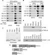
FIG. 1.
FIG. 1.
ATRIP N terminus binds RPA70N in vitro. HA-tagged, full-length ATRIP or ATRIP fragments generated using a coupled transcription/translation system were incubated with single-stranded DNA bound to Sepharose beads in the presence (+) or absence (−) of purified RPA (A) or His-RPA bound to nickel beads (B). After washing, the bound proteins were eluted, separated by SDS-PAGE, blotted, and probed with HA antibody. Input (In) (10%) data are included for comparison. (C and D) Purified, recombinant His-tagged RPA domains were added to in vitro translation reaction mixtures containing HA-ATRIP, HA-ATRIPΔN (C), or HA-ATRIP1-107 (D). Protein complexes were isolated using nickel beads, separated by SDS-PAGE, blotted, and probed using an HA antibody. (E) Diagram of RPA heterotrimer. Black bars above protein segments indicate protein interaction domains.
FIG. 2.
FIG. 2.
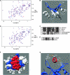
A conserved acidic domain in the ATRIP N terminus interacts with the basic cleft of RPA70N. (A) The 15N-1H-HSQC NMR spectrum of 15N-enriched-RPA70N in the absence (blue) and presence (red) of ATRIP1-107. (B) RPA70N residues perturbed (blue) upon addition of ATRIP1-107 mapped onto the crystal structure of RPA70N (PDB accession 2B3G). (C) Sequence alignment of the conserved acidic region in the N terminus of five ATRIP orthologues. (D) The 15N-1H-HSQC NMR spectrum of 15N-enriched RPA70N acquired in the absence (blue) and presence (red) of ATRIP54-70. (E) Alignment of the p53 and ATRIP peptides used in homology modeling. (F) Space-filling diagram of RPA70 and ATRIP55-66 (red), with the residues of RPA70N in the ATRIP binding pocket colored blue. (G) Predicted electrostatic interactions between basic RPA70N basic residues K88 and R41 with ATRIP acidic residues D58 and D59.
FIG. 3.
FIG. 3.
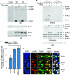
Ddc2-Rfa1 interaction requires the conserved acidic region of Ddc2. (A) Schematic diagram of ATRIP, RPA-binding mutant ATRIP (ATRIPΔN), Ddc2, and Ddc2ΔN. Locations of predicted coiled-coil domains (gray) are indicated. (B) Δddc2 HA-MEC1 yeast containing myc-vector (Vec), myc-Ddc2 (WT), or myc-Ddc2ΔN (ΔN) were exposed to 0 (−) or 60 (+) J/m2 UV and harvested 1 h later. Myc immunoprecipitates from soluble extracts were separated by SDS-PAGE, blotted, and probed with Myc, Rfa1, and HA antibodies. (C) Alignment of conserved acidic region in the N terminus of ATRIP and Ddc2 and schematic of mutations generated in this region of Ddc2. (D) Yeasts containing TAP-Rfa1 and Ddc2 (WT), Ddc2ΔN (ΔN), Ddc2DK (DK), or Ddc2N14 (N14) were damaged with 0.01% MMS (+) or left untreated (−) and harvested 1 h later. Cells were lysed, and TAP-Rfa1 was isolated using immunoglobulin G beads. TAP protein complexes were separated by SDS-PAGE and Western blotted using Myc (Ddc2) and Rfa1 antibodies.
FIG. 4.
FIG. 4.
Ddc2 lacking the N-terminal Rfa1 binding domain is defective in localizing to sites of DNA damage. (A) Δddc2 yeasts transformed with a centromeric plasmid expressing Myc-Ddc2 or Myc-Ddc2ΔN from the DDC2 promoter and harboring a galactose-inducible HO endonuclease were grown to log phase in raffinose-containing media. Galactose (GAL) or glucose (GLU) was added to induce or suppress HO-endonuclease expression. One hour after sugar addition, cells were cross-linked using formaldehyde and harvested. Cells were lysed and sonicated, and Myc-Ddc2 proteins were immunoprecipitated with a myc antibody. Cross-links were reversed, and associated DNA sequences were amplified by PCR using primers specific to regions adjacent to the HO break site (HO-A, HO-B) or to the SMC2 gene (SMC2). Samples were prepared in duplicate. Input samples represent 5% of input into immunoprecipitation reactions. (B) Equal volumes of immunoprecipitation reaction mixtures before (pre) or after (post) isolation of Myc-Ddc2 proteins were separated by SDS-PAGE, blotted, and probed with a myc antibody. (C) Extracts from Δddc2 yeast, or from yeast expressing GFP-Ddc2 (WT) or GFP-Ddc2ΔN (ΔN), were separated by SDS-PAGE and Western blotted using a GFP antibody. (D) Yeasts expressing GFP-Ddc2 (WT) or GFP-Ddc2ΔN (ΔN) and galactose-inducible HO endonuclease were grown to log phase in liquid culture. Glucose (GLU) or galactose (GAL) was added to suppress or induce DNA double-strand-break formation. GFP fluorescence was visualized on a Zeiss Axioplan fluorescent microscope. (E) Quantitation of HO-induced focus formation of GFP-Ddc2 or GFP-Ddc2ΔN 4 h or 6 h after induction of HO endonuclease expression. Error bars represent standard deviations of the results from three experiments.
FIG. 5.
FIG. 5.
Disruption of Ddc2-Rfa1 interaction impairs the DNA damage response. (A and B) Yeasts lacking Ddc2 (Vector) or expressing Ddc2 (WT), Ddc2ΔN (ΔN), Ddc2DK (DK), or Ddc2N14 (N14) were grown to log phase in liquid culture and plated onto rich media containing increasing amounts of HU (A) or MMS (B). Percent viability was calculated as the number of colonies surviving at each dose compared to the number of colonies that survived on plates lacking HU or MMS. Data represent the averages of the results of three experiments. Standard deviations were smaller than symbol width in most cases. (C to E) Yeast strains were arrested in G1 with alpha factor and released into rich media in the presence of 200 mM HU (C and E) or in the indicated concentration of HU (D). Cells were harvested 1 h (C and D) or at the indicated times (E) after G1 release, and trichloroacetic acid was precipitated. Lysates were separated by SDS-PAGE, blotted, and probed with Rad53 or Myc antibodies. (F and G) Δddc2 (V), DDC2 (WT), or ddc2ΔN (ΔN) yeasts were grown to log phase in liquid culture, arrested in G1 with alpha factor, and released into media containing the indicated doses of MMS and harvested 1 h post G1 release (F) or at the indicated various time points after G1 release (G). Cells were lysed, and proteins were separated by SDS-PAGE, blotted, and probed with Rad53 antibody. (G) Membranes containing immobilized proteins were subjected to in situ autophosphorylation to assay Rad53 autophosphorylation activity.
FIG. 6.
FIG. 6.
TopBP1 activates ATR-ATRIP complexes independently of RPA. (A) Wild-type ATR-ATRIP or ATR-ATRIPΔN complexes were isolated from transfected 293T cells and incubated with recombinant wild-type TopBP1 978-1286 (WT) or TopBP1 978-1286 W1145R (WR), Phas1 substrate, and [γ-32P]ATP. Kinase reaction mixtures were separated by SDS-PAGE, stained with Coomassie blue, and exposed to film (32P). A duplicate gel was blotted and probed with anti-ATRIP and anti-ATR antibodies (WB). (B) Wild-type ATR-ATRIP or kinase-dead ATR-ATRIP immune complexes were isolated from transfected 293T cells and incubated with recombinant TopBP1 and/or RPA heterotrimer in the presence of Phas1 substrate and [γ-32P]ATP. Kinase reaction mixtures were separated by SDS-PAGE, stained with Coomassie blue or blotted, and exposed to film (32P) or probed with anti-ATR antibodies (WB). (C and D) 293T cells stably expressing siRNA-resistant ATRIP, ATRIPΔN, or empty vector control were transfected with ATRIP siRNA to deplete endogenous ATRIP. Two days after siRNA transfection, the cells were transfected with GFP-TopB1 978-1286 expression construct. Twenty-four hours later the cells were fixed and stained with antibodies to γH2AX. (C) Representative images collected on a Zeiss Axioplan microscope with the same exposure times. (D) Quantitation of the percentages of the GFP-TopBP1-expressing cells that contained phosphorylated H2AX. Error bars represent standard deviations. The inset presents a Western blot showing the relative expression levels of ATRIP and ATRIPΔN.

References

    1. Amberg, D. C., D. J. Burke, and J. E. Strathern. 2005. Methods in yeast genetics: a Cold Spring Harbor laboratory course manual. Cold Spring Harbor Laboratory Press, Cold Spring Harbor, NY.
    1. Ball, H. L., and D. Cortez. 2005. ATRIP oligomerization is required for ATR-dependent checkpoint signaling. J. Biol. Chem. 280:31390-31396. - PMC - PubMed
    1. Ball, H. L., J. S. Myers, and D. Cortez. 2005. ATRIP binding to RPA-ssDNA promotes ATR-ATRIP localization but is dispensable for Chk1 phosphorylation. Mol. Biol. Cell 16:2372-2381. - PMC - PubMed
    1. Bermudez, V. P., L. A. Lindsey-Boltz, A. J. Cesare, Y. Maniwa, J. D. Griffith, J. Hurwitz, and A. Sancar. 2003. Loading of the human 9-1-1 checkpoint complex onto DNA by the checkpoint clamp loader hRad17-replication factor C complex in vitro. Proc. Natl. Acad. Sci. USA 100:1633-1638. - PMC - PubMed
    1. Binz, S. K., Y. Lao, D. F. Lowry, and M. S. Wold. 2003. The phosphorylation domain of the 32-kDa subunit of replication protein A (RPA) modulates RPA-DNA interactions. Evidence for an intersubunit interaction. J. Biol. Chem. 278:35584-35591. - PubMed

Publication types

MeSH terms