. 2007 Mar 12;176(6):741-7.
doi: 10.1083/jcb.200610027. Epub 2007 Mar 5.

SYCE2 is required for synaptonemal complex assembly, double strand break repair, and homologous recombination

Affiliations

SYCE2 is required for synaptonemal complex assembly, double strand break repair, and homologous recombination

Ewelina Bolcun-Filas et al. J Cell Biol. .

Abstract

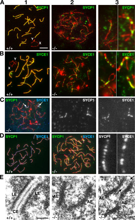
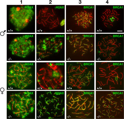
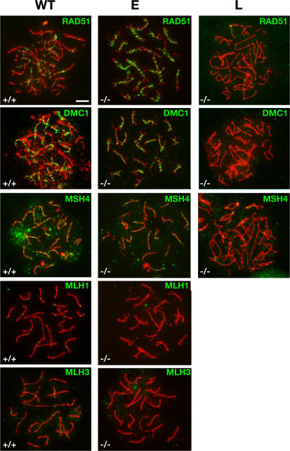
Synapsis is the process by which paired chromosome homologues closely associate in meiosis before crossover. In the synaptonemal complex (SC), axial elements of each homologue connect through molecules of SYCP1 to the central element, which contains the proteins SYCE1 and -2. We have derived mice lacking SYCE2 protein, producing males and females in which meiotic chromosomes align and axes form but do not synapse. Sex chromosomes are unaligned, not forming a sex body. Additionally, markers of DNA breakage and repair are retained on the axes, and crossover is impaired, culminating in both males and females failing to produce gametes. We show that SC formation can initiate at sites of SYCE1/SYCP1 localization but that these points of initiation cannot be extended in the absence of SYCE2. SC assembly is thus dependent on SYCP1, SYCE1, and SYCE2. We provide a model to explain this based on protein-protein interactions.


Figures

Figure 1.
Figure 1.
Syce2 mutants fail to form a SC between homologues, but small regions of synapsis can be observed. Wild-type and mutant meiotic spreads were immunostained with anti-SYCP1 or anti-SYCE1 (green) and anti-SYCP3 (red; AE) antibodies (A–D). Although male +/+ pachytene cells show complete synapsis of autosomes (A1 and B1), mutant cells with well-aligned chromosomes show only small foci of synapsis staining for SYCP1 or SYCE1 on some autosomes (A2 and B2, respectively). A3 and B3 display higher magnification of SYCP1 and SYCE1 localization to regions of AE juxtaposition, respectively. SYCP1 (green) colocalizes with SYCE1 (blue) in the small foci of synapsis (arrows) in male −/− cells (C). C2 shows SYCP1, and C3 shows SYCE1 localization at a higher magnification on the chromosomes indicated by arrows in C1. In female +/+ pachytene cells, synapsis is established between homologous chromosomes as observed by SYCP1 (green) and SYCE1 (blue) staining (D1), but −/− female cells with unpaired AEs show SYCP1 and SYCE1 punctate localization (D2). D3 displays a higher magnification of a single univalent with SYCP1 and SYCE1 signals. Electron microscopic characterization of SYCE2KO male meiotic cells confirmed the existence of small foci of synapsis (E). E1 shows a wild-type SC, whereas E2 and E3 show AEs aligned but not synapsed and the occasional short region of synapsis in the mutant, respectively. Arrow in E3 points to the CE-like structure found in synapsis foci in the mutant. LE, lateral element; X and Y, sex chromosomes. Bars: (A–D) 5 μm; (E) 200 nm.
Figure 2.
Figure 2.
Model for SC assembly. Homodimers of SYCP1 form unstable N-terminal self-associations and do not form C-terminal associations with AE components. These associations in A are stabilized by interactions with SYCE1 in the rudiments of the CE. At this point, the C terminus of the SYCP1 molecule is associated with the AE. These short regions of synapsis are not stably extended until interactions between the three proteins, SYCE2, SYCP1, and SYCE1, are established, forming the completed SC.
Figure 3.
Figure 3.
Altered distribution of γH2AX and BRCA1 in Syce2 mutant mice. Surface-spread nuclei were immunostained with anti-γH2AX or BRCA1 (green) and anti-SYCP3 (red). The top row shows male +/+ cells in leptotene (1), pachytene (2 and 4), and zygotene (3). Row 2 shows a SYCE2KO male leptotene cell (1) and cells with aligned (2 and 3) and unaligned chromosomes (4). Row 3 shows +/+ female cells in late zygotene (1), pachytene (2 and 4), and zygotene (3). Row 4 shows SYCE2KO oocytes with aligned (1 and 3) and unaligned chromosomes (2 and 4). Bar, 5 μm.
Figure 4.
Figure 4.
Distribution of repair and recombination proteins in wild-type and mutant meiosis. SYCP3 signal is represented in red and recombination and repair proteins in green. Columns show wild type (WT) and early SYCE2KO (E) and late SYCE2KO (L) stages. Bar, 5 μm.
Figure 5.
Figure 5.
SYCE2KO spermatocytes do not form an XY body. SYCP3 signal is represented in red; REC8, BRCA1, or γH2AX in green; and CREST staining for centromeres in blue. Arrows in +/+ mark the XY body, whereas −/− unpaired X and Y are marked by arrows and arrowheads, respectively. Insets show higher magnification of the Y chromosome. Bar, 5 μm.

References

    1. Ashley, T., C. Westphal, A. Plug-de Maggio, and D.G. de Rooij. 2004. The mammalian mid-pachytene checkpoint: meiotic arrest in spermatocytes with a mutation in Atm alone or in combination with a Trp53 (p53) or Cdkn1a (p21/cip1) mutation. Cytogenet. Genome Res. 107:256–262. - PubMed
    1. Bergerat, A., B. de Massy, D. Gadelle, P.C. Varoutas, A. Nicolas, and P. Forterre. 1997. An atypical topoisomerase II from Archaea with implications for meiotic recombination. Nature. 386:414–417. - PubMed
    1. Chen, W.V., and P. Soriano. 2003. Gene trap mutagenesis in embryonic stem cells. Methods Enzymol. 365:367–386. - PubMed
    1. Chen, W.V., J. Delrow, P.D. Corrin, J.P. Frazier, and P. Soriano. 2004. Identification and validation of PDGF transcriptional targets by microarray-coupled gene-trap mutagenesis. Nat. Genet. 36:304–312. - PubMed
    1. Costa, Y., R. Speed, R. Ollinger, M. Alsheimer, C.A. Semple, P. Gautier, K. Maratou, I. Novak, C. Hoog, R. Benavente, and H.J. Cooke. 2005. Two novel proteins recruited by synaptonemal complex protein 1 (SYCP1) are at the centre of meiosis. J. Cell Sci. 118:2755–2762. - PubMed

Publication types