. 2007 Mar 12;176(6):757-63.
doi: 10.1083/jcb.200701065. Epub 2007 Mar 5.

Functional genomics identifies a Myb domain-containing protein family required for assembly of CENP-A chromatin

Affiliations

Functional genomics identifies a Myb domain-containing protein family required for assembly of CENP-A chromatin

Paul S Maddox et al. J Cell Biol. .

Abstract

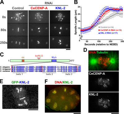
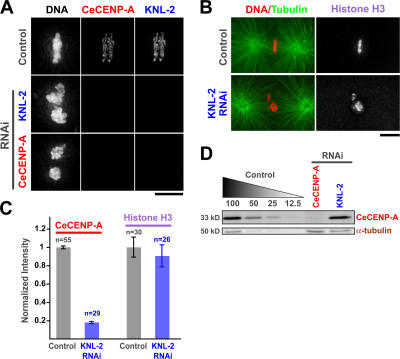
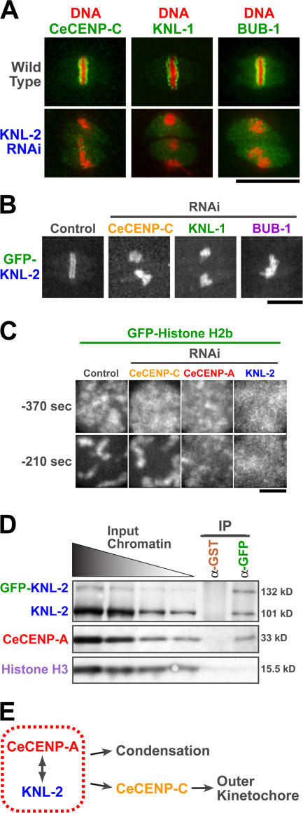
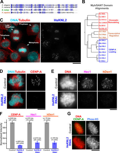
Nucleosomes containing the centromere-specific histone H3 variant centromere protein A (CENP-A) create the chromatin foundation for kinetochore assembly. To understand the mechanisms that selectively target CENP-A to centromeres, we took a functional genomics approach in the nematode Caenorhabditis elegans, in which failure to load CENP-A results in a signature kinetochore-null (KNL) phenotype. We identified a single protein, KNL-2, that is specifically required for CENP-A incorporation into chromatin. KNL-2 and CENP-A localize to centromeres throughout the cell cycle in an interdependent manner and coordinately direct chromosome condensation, kinetochore assembly, and chromosome segregation. The isolation of KNL-2-associated chromatin coenriched CENP-A, indicating their close proximity on DNA. KNL-2 defines a new conserved family of Myb DNA-binding domain-containing proteins. The human homologue of KNL-2 is also specifically required for CENP-A loading and kinetochore assembly but is only transiently present at centromeres after mitotic exit. These results implicate a new protein class in the assembly of centromeric chromatin and suggest that holocentric and monocentric chromosomes share a common mechanism for CENP-A loading.


Figures

Figure 1.
Figure 1.
KNL-2 is a novel Myb domain protein whose depletion results in a CeCENP-A loss of function phenotype. (A) Images of control, CeCENP-A, and KNL-2–depleted embryos expressing GFP-histone H2b (arrow) to mark chromosomes and GFP–γ-tubulin (arrowheads) to mark the spindle poles. Time is given in seconds relative to nuclear envelope breakdown (NEBD). (B) Spindle pole separation kinetics in control, CeCENP-A depletion, and KNL-2 depletion. Error bars represent the SEM with a confidence interval of 0.95. (C) Primary sequence features of KNL-2. The divergent Myb domain (amino acids 624–677) is shown on the bottom for C. elegans, C. briggsae, and C. remanei KNL-2. The predicted helical secondary structure is shown under the sequences, and the conserved tryptophan (W) residues are boxed in red. (D) KNL-2 and CeCENP-A colocalize at kinetochores during mitosis. (E) GFP–KNL-2 exhibits kinetochore localization. (F) KNL-2 localizes to kinetochores in later embryonic divisions. A multicellular embryo in which DNA (red) and KNL-2 (green) are labeled is shown. Bars (A, D, and E), 5 μm; (F) 10 μm.
Figure 2.
Figure 2.
KNL-2 is required for CeCENP-A but not histone H3 targeting to chromatin. (A) CeCENP-A and KNL-2 localization in wild-type, KNL-2–depleted, or CENP-A– depleted embryos. (B) KNL-2 depletion does not prevent histone H3 localization. Note that GFP-histone H2b levels on chromatin were also unaffected (Fig. 1 A). (C) Quantitation of CeCENP-A and histone H3 localization. For CeCENP-A analysis, KNL-2 was depleted in embryos expressing GFP-histone H2b, and the fluorescence intensity ratio of CeCENP-A to GFP-histone H2b was calculated in deconvolved images of fixed embryos. For histone H3, the signal on chromatin was directly quantified. Error bars represent the SEM with a confidence interval of 0.95. (D) KNL-2 depletion does not affect CeCENP-A protein levels. Immunoblot comparing serially diluted control C. elegans extract to CeCENP-A–and KNL-2–depleted C. elegans extracts is shown. α-Tubulin was used as a loading control. Bars, 5 μm.
Figure 3.
Figure 3.
KNL-2 and CeCENP-A make functionally equivalent contributions to kinetochore assembly and chromosome condensation and are physically proximal on chromatin. (A) Like CeCENP-A, KNL-2 is required for the localization of CeCENP-C, KNL-1, and BUB-1 to kinetochores. (B) Depletion of CeCENP-C, KNL-1, or BUB-1 does not prevent GFP–KNL-2 localization to chromatin. (C) CeCENP-A–and KNL-2–depleted embryos exhibit a similar chromosome condensation defect. Representative images of the sperm pronucleus at −370 and −210 s before nuclear envelope breakdown (for quantitation see Fig. S2, available at http://www.jcb.org/cgi/content/full/jcb.200701065/DC1). (D) KNL-2 and CeCENP-A are physically proximal on chromatin. Nuclei purified from C. elegans embryos expressing GFP–KNL-2 were sonicated to generate chromatin fragments of ∼500–1,500 bp. The sonicated chromatin was used as the input for anti-GST (control) and anti-GFP immunoprecipitations. Dilutions of the input chromatin and immunoprecipitates were blotted for KNL-2, CeCENP-A, and histone H3. Chromatin fragments enriched for KNL-2 were coenriched for CeCENP-A but not for histone H3 (Fig. S3). (E) CeCENP-A and KNL-2 coordinately direct kinetochore assembly and chromosome condensation. Bars (A and B), 10 μm; (C) 2 μm.
Figure 4.
Figure 4.
KNL-2 is the founding member of a widely conserved protein family required for CENP-A loading in both invertebrates and vertebrates. (A) The Myb domain of the B. malayi KNL-2 homologue, which was aligned with two of the rhabditid KNL-2 Myb domains, was used to identify a putative human KNL2 homologue (c14orf106). Shading indicates degrees of conservation. (B) Tree of a small subset of the Myb/SANT domain protein superfamily aligned using Myb/SANT domain sequences. SANT domain proteins (red) are implicated in chromatin remodeling activities. The founding canonical Myb protooncogene (A–C refer to three isoforms in humans; gold) is a transcription factor that directly binds specific DNA sequences. A new subfamily (blue) is defined by the KNL-2 Myb domain and includes, in addition to the nematode members, a clear homologue in every sequenced vertebrate. KNL-2 homologues were not evident by sequence in fungi or Drosophila. GenBank/EMBL/DDBJ accession nos. for the putative vertebrate KNL2s are as follows: Hs, human c14orf106; Mm, mouse C79407; Gg, chicken XP_421481; and Xt, frog AL873597.2. (C) Localization of HsKNL2 in HeLa cells. The chosen field includes cells in different mitotic stages and highlights the striking increase in centromere staining in the late anaphase/telophase stage of the cell cycle. (D–F) HsKNL2 depletion dramatically reduces CENP-A localization at centromeres and equivalently reduces targeting of medial (hDsn1) and outer (Hec1) kinetochore proteins. Anticentromere antibody staining was used to identify centromere regions in all cases but is not shown for brevity. The total number of kinetochores (kts) and cells in which fluorescence intensities were measured is indicated for each condition. As expected from the kinetochore assembly defect, chromosome segregation is severely perturbed in HsKNL2-depleted cells (Fig. S4 and Video 10, available at http://www.jcb.org/cgi/content/full/jcb.200701065/DC1). Error bars represent SEM. (G) HsKNL2 depletion does not affect phosphohistone (S10) H3 staining. Bars (C), 10 μm; (D, E, and G) 5 μm.

Comment in

  • Centromeric chromatin gets loaded.
    Carroll CW, Straight AF. Carroll CW, et al. J Cell Biol. 2007 Mar 12;176(6):735-6. doi: 10.1083/jcb.200702020. Epub 2007 Mar 5. J Cell Biol. 2007. PMID: 17339381 Free PMC article.

References

    1. Aasland, R., A.F. Stewart, and T. Gibson. 1996. The SANT domain: a putative DNA-binding domain in the SWI-SNF and ADA complexes, the transcriptional co-repressor N-CoR and TFIIIB. Trends Biochem. Sci. 21:87–88. - PubMed
    1. Buchwitz, B.J., K. Ahmad, L.L. Moore, M.B. Roth, and S. Henikoff. 1999. A histone-H3-like protein in C. elegans. Nature. 401:547–548. - PubMed
    1. Cheeseman, I.M., S. Niessen, S. Anderson, F. Hyndman, J.R. Yates III, K. Oegema, and A. Desai. 2004. A conserved protein network controls assembly of the outer kinetochore and its ability to sustain tension. Genes Dev. 18:2255–2268. - PMC - PubMed
    1. Choo, K.H. 2000. Centromerization. Trends Cell Biol. 10:182–188. - PubMed
    1. Cleveland, D.W., Y. Mao, and K.F. Sullivan. 2003. Centromeres and kinetochores: from epigenetics to mitotic checkpoint signaling. Cell. 112:407–421. - PubMed

Publication types

MeSH terms