. 2007 Mar 19;204(3):571-82.
doi: 10.1084/jem.20061931. Epub 2007 Mar 5.

The fibrin-derived gamma377-395 peptide inhibits microglia activation and suppresses relapsing paralysis in central nervous system autoimmune disease

Affiliations

The fibrin-derived gamma377-395 peptide inhibits microglia activation and suppresses relapsing paralysis in central nervous system autoimmune disease

Ryan A Adams et al. J Exp Med. .

Abstract

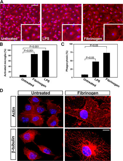
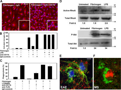
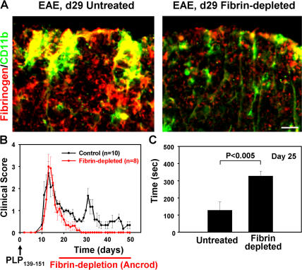
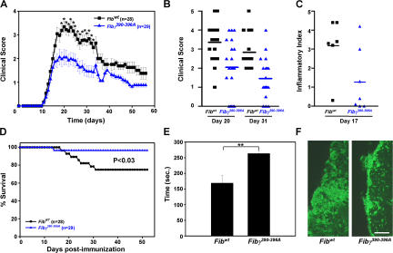
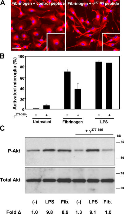
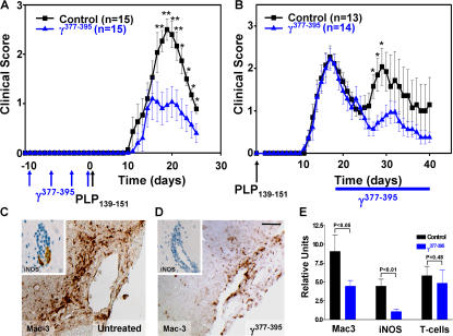
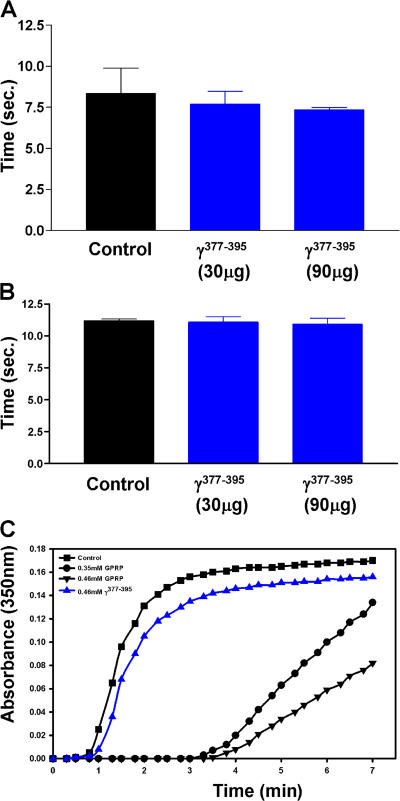
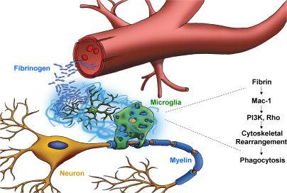
Perivascular microglia activation is a hallmark of inflammatory demyelination in multiple sclerosis (MS), but the mechanisms underlying microglia activation and specific strategies to attenuate their activation remain elusive. Here, we identify fibrinogen as a novel regulator of microglia activation and show that targeting of the interaction of fibrinogen with the microglia integrin receptor Mac-1 (alpha(M)beta(2), CD11b/CD18) is sufficient to suppress experimental autoimmune encephalomyelitis in mice that retain full coagulation function. We show that fibrinogen, which is deposited perivascularly in MS plaques, signals through Mac-1 and induces the differentiation of microglia to phagocytes via activation of Akt and Rho. Genetic disruption of fibrinogen-Mac-1 interaction in fibrinogen-gamma(390-396A) knock-in mice or pharmacologically impeding fibrinogen-Mac-1 interaction through intranasal delivery of a fibrinogen-derived inhibitory peptide (gamma(377-395)) attenuates microglia activation and suppresses relapsing paralysis. Because blocking fibrinogen-Mac-1 interactions affects the proinflammatory but not the procoagulant properties of fibrinogen, targeting the gamma(377-395) fibrinogen epitope could represent a potential therapeutic strategy for MS and other neuroinflammatory diseases associated with blood-brain barrier disruption and microglia activation.


Figures

Figure 1.
Figure 1.
Fibrinogen directly activates microglia resulting in cytoskeletal rearrangements and increased phagocytosis. (A) IsoB4/DAPI immunostained microglia cultured on immobilized fibrinogen have increased cell body size adopting an amoeboid morphology (right). Untreated primary microglia show small cell bodies and thin, bipolar processes (left). LPS-treated cells show activated morphology characterized by cell body swelling (middle). Bar, 26 μm; inset, 21 μm. (B) Quantitation of microglia activation reveals a dramatic increase upon fibrinogen stimulation. (C) Fibrinogen-stimulated microglia showed increased phagocytosis of fluorescent Escherichia coli as compared with untreated microglia. LPS served as a positive control. (D) Deconvolution microscopy of primary microglia revealed significant rearrangements of the cytoskeleton upon treatment with fibrinogen. Microglia were stained with antibodies to actin and β-tubulin in red, and the nucleus was stained blue with DAPI. Bar, 4.4 μm.
Figure 2.
Figure 2.
Fibrinogen directly activates microglia via the Mac-1 integrin receptor. (A) Fibrinogen-induced morphologic activation of microglia is blocked by the addition of a rat anti-CD11b neutralizing antibody (M1/70). Rat IgG (control) did not change the effects of fibrinogen in microglia activation. Bar, 39 μm; inset, 17 μm. (B) Quantitation of microglia activation reveals that the Mac-1 neutralizing antibody blocks fibrinogen-induced activation but not LPS activation of microglia. (C) Increased microglia phagocytosis upon fibrinogen stimulation is blocked by the addition of a PI3K inhibitor (LY294002) and diminished in the presence of a CD11b neutralizing antibody. A control anti-TLR4 or IgG showed no reduction in fibrinogen-stimulated phagocytosis. (D) Western blots show increased active RhoA and Akt activation upon fibrinogen stimulation of microglia. In both assays, LPS served as a positive control. (E) Confocal microscopy demonstrates the spatial interaction between CD11b+ cells (green) and fibrin (red) in spinal cords from PLP139-151–immunized mice. (F) Confocal double immunofluorescence shows fibrin (green) surrounding an activated microglia (red) in a human early demyelinating plaque of acute MS. Bars: E, 6.7 μm; F, 15 μm.
Figure 3.
Figure 3.
Fibrin depletion using ancrod reverses relapsing paralysis and reduces microglia activation in EAE. (A) Immunofluorescence of control (left) and fibrin-depleted (right) spinal cord with antibodies against fibrinogen (red) and CD11b (green). Activated CD11b+ cells colocalize with fibrin deposition in the control spinal cord (yellow). Bar, 8.4 μm. (B) Clinical scores of PLP139-151–immunized mice. Fibrin depletion begins after the first paralytic episode. Fibrin-depleted mice (red circles; n = 8) did not develop first or second relapses as compared with control mice (black circles; n = 10). (C) Rotarod motor strength and coordination test. Fibrin-depleted mice outperformed immunized control untreated mice on day 25 after immunization. Data are represented as the mean clinical score and are mean ± SEM.
Figure 4.
Figure 4.
Fibγ390-396A mice have reduced clinical scores and microglia activation in EAE. (A) Clinical scores of MOG35-55–immunized mice. Fibγ390-396A mice (blue triangles; n = 29) show statistically significant lower clinical scores (*, P < 0.01) than FibγWT control mice (black squares; n = 28). (B) Individual clinical scores from Fibγ390-396A and FibγWT mice on days 20 (average clinical score, 3.3 ± 0.22 for FibγWT [n = 19] vs. 2.1 ± 0.32 for Fibγ390-396A [n = 20]; P < 0.01) and 31 after immunization (average clinical score, 2.8 ± 0.25 for FibγWT [n = 15] vs. 1.5 ± 0.23 for Fibγ390-396A [n = 20]; P < 0.001). (C) Histological analysis of spinal cord sections stained with hematoxylin/eosin revealed increased inflammation in the FibγWT mice versus the Fibγ390-396A mice. (D) Survival curve of Fibγ390-396A and FibγWT mice after MOG35-55 immunization. Fibγ390-396A had a greater survival rate than the FibγWT mice. (E) Rotarod analysis of Fibγ390-396A and FibγWT mice on day 13. Fibγ390-396A significantly outperformed their wild-type counterparts in a behavioral test designed to assess motor skill function. n = 7 mice per group. **, P < 0.05. (F) Increased activation of IsoB4+ cells in FibγWT mice as compared with Fibγ390-396A mice. Bar, 58 μm. Data are represented as the mean clinical score and are mean ± SEM.
Figure 5.
Figure 5.
The fibrin-derived γ377-395 peptide inhibits microglia activation. (A) Microglia activation upon fibrinogen stimulation is attenuated in the presence of γ377-395. Bar, 33 μm; inset, 29 μm. (B) Quantitation of microglia activation reveals that γ377-395 diminishes fibrinogen-stimulated microglia activation but has no effect on LPS activation. (C) Treatment of microglia with γ377-395 blocks fibrinogen-induced Akt activation in vitro. LPS activation of Akt is unaffected by γ377-395 treatment.
Figure 6.
Figure 6.
The fibrin-derived γ377-395 peptide suppresses EAE. (A) Clinical scores from γ377-395 peptide–vaccinated mice. Mice were immunized with γ377-395 peptide before EAE induction with PLP139-151 peptide. Vaccinated mice (blue triangles; n = 15) had significantly reduced clinical scores as compared with control mice (black squares; n = 15). (B) Clinical scores from PLP139-151–immunized mice where the γ377-395 peptide was administered every day intranasally after the peak of the initial paralytic episode. γ377-395–treated mice (blue triangles; n = 14) did not show a relapse at approximately day 30 as compared with the controls (black squares; n = 13). *, P < 0.05; **, P < 0.01. Data are represented as the mean clinical score and are mean ± SEM. Immunostaining for Mac-3 and iNOS (inset) on day 30 after immunization shows dramatic reduction of microglia activation in the γ377-395 peptide–treated mice after EAE induction (D) when compared with control (C). Bar, 20 μm; inset, 10 μm. (E) Quantitation shows a twofold reduction in Mac-3 (P < 0.05) and a 4.2-fold reduction in iNOS (P < 0.01) after γ377-395 peptide treatment, whereas there are no major differences in T cell infiltration.
Figure 7.
Figure 7.
The fibrin-derived γ377-395 peptide does not affect in vivo coagulation or fibrin polymerization. In vivo clotting time (A) and prothrombin times (B) were assayed in the presence of 30 μg γ377-395 peptide, the dose administered in vivo daily, and 90 μg γ377-395 peptide. Neither dose affected blood clotting times. (C) In vitro fibrin polymerization was examined in the presence of the γ377-395 peptide. GPRP, a known inhibitor of fibrin formation, significantly attenuated fibrin formation, whereas the γ377-395 peptide had no effect on fibrin polymerization.
Figure 8.
Figure 8.
Proposed model for the role of fibrin-induced activation of microglia in inflammatory demyelination. BBB disruption permits the leakage of fibrinogen, the high affinity ligand for Mac-1, in the CNS parenchyma. Fibrinogen is converted to fibrin and functions as the spatial signal to induce local activation of microglia via activation of the Mac-1 integrin receptor and induction of signaling pathways downstream of Mac-1, such as Akt and Rho, resulting in phagocytosis that could determine the extent of tissue damage in inflammatory demyelination.

References

    1. Lassmann, H., W. Bruck, and C. Lucchinetti. 2001. Heterogeneity of multiple sclerosis pathogenesis: implications for diagnosis and therapy. Trends Mol. Med. 7:115–121. - PubMed
    1. Platten, M., and L. Steinman. 2005. Multiple sclerosis: trapped in deadly glue. Nat. Med. 11:252–253. - PubMed
    1. Heppner, F.L., M. Greter, D. Marino, J. Falsig, G. Raivich, N. Hovelmeyer, A. Waisman, T. Rulicke, M. Prinz, J. Priller, et al. 2005. Experimental autoimmune encephalomyelitis repressed by microglial paralysis. Nat. Med. 11:146–152. - PubMed
    1. Jack, C., F. Ruffini, A. Bar-Or, and J.P. Antel. 2005. Microglia and multiple sclerosis. J. Neurosci. Res. 81:363–373. - PubMed
    1. Minagar, A., and J.S. Alexander. 2003. Blood-brain barrier disruption in multiple sclerosis. Mult. Scler. 9:540–549. - PubMed

Publication types

MeSH terms