. 2007 Jun 15;109(12):5230-3.
doi: 10.1182/blood-2007-02-072983. Epub 2007 Mar 5.

Friend of GATA-1-independent transcriptional repression: a novel mode of GATA-1 function

Affiliations

Friend of GATA-1-independent transcriptional repression: a novel mode of GATA-1 function

Kirby D Johnson et al. Blood. .

Abstract

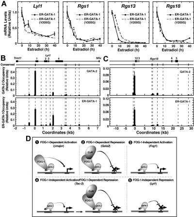
The GATA-1-interacting protein Friend Of GATA-1 (FOG-1) is essential for the proper transcriptional activation and repression of numerous GATA-1 target genes. Although FOG-1-independent activation by GATA-1 has been described, all known examples of GATA-1-mediated repression are FOG-1 dependent. In the GATA-1-null G1E cell line, estrogen receptor ligand binding domain (ER) chimeras of either wild-type GATA-1 or a FOG-1-binding defective mutant of GATA-1 repressed several genes similarly upon activation with beta-estradiol. Repression also occurred in a FOG-1-null cell line expressing ER-GATA-1 and during ex vivo erythropoiesis. At the Lyl1 and Rgs18 loci, we found highly restricted occupancy by GATA-1 and GATA-2, indicating that these genes are direct targets of GATA factor regulation. The identification of genes repressed by GATA-1 independent of FOG-1 defines a novel mode of GATA-1-mediated transcriptional regulation.


Figures

Figure 1
Figure 1
Novel mode of GATA-1 function: FOG-1–independent repression. Real-time RT-PCR analysis of mRNA levels in (A) G1E cells expressing either wild-type ER–GATA-1(WT) or ER–GATA-1(V205G) and (B) FOG-1−/− cells expressing wild-type ER–GATA-1. Transcript levels from cells treated with 1 μM β-estradiol for 24 hours were compared with untreated controls. Transcript levels were normalized to glyceraldehyde-3-phosphate dehydrogenase (GAPDH) transcript levels and, for each clone, the mean value for untreated samples was set to 1 (mean ± standard error [SE] from 3 independent experiments). (C) Real-time RT-PCR analysis of mRNA levels during ex vivo human erythropoiesis. CD34+ cells isolated from peripheral blood were cultured for up to 14 days. RNA was isolated at days 3, 7, 10, and 14 from 2 independent samples. Transcript levels were normalized to 18S RNA levels and the relative level of transcript from day 3 for each sample was set to 1.
Figure 2
Figure 2
Highly restricted GATA factor occupancy at endogenous Lyl1 and Rgs18 domains. (A) Comparison of transcript levels in G1E cells expressing wild-type ER–GATA-1 or ER–GATA-1(V205G) following β-estradiol treatment (1 μM) for various times up to 40 hours. Transcript levels were normalized to GAPDH transcript levels and, for each clone, the mean value for untreated samples was set to 1 (mean ± SE from 3 independent experiments). GATA factor occupancy was measured at (B) the Lyl1 locus and (C) the Rgs18 locus by quantitative real-time ChIP analysis in G1E–ER–GATA-1 cells. For GATA-2 and GATA-1, ChIPs were conducted using untreated or β-estradiol–treated (1 μM, 24 h) cells, respectively (mean ± SD, 2 to 4 independent ChIP experiments). Mean preimmune control signals did not exceed 0.0026 for Lyl1 or 0.0015 for Rgs18 and are not shown. The positions of conserved (mouse to man) WGATAR, NGATAR, and WGATAN sites in each locus are shown above the graph. All nonconserved WGATAR motifs in the Lyl1 locus were also analyzed by ChIP. Coordinates are based upon distance from the conserved WGATAR motif (TTATCA) in the Lyl1 promoter and the transcriptional start site of the Rgs18 locus. (D) Multiple modes of GATA-1–mediated transcriptional regulation. An example of a gene regulated via each mode is indicated in parentheses.

References

    1. Tsang AP, Visvader JE, Turner CA, et al. FOG, a multitype zinc finger protein, acts as a cofactor for transcription factor GATA-1 in erythroid and megakaryocytic differentiation. Cell. 1997;90:109–119. - PubMed
    1. Crispino JD, Lodish MB, MacKay JP, Orkin SH. Use of altered specificity mutants to probe a specific protein-protein interaction in differentiation: the GATA-1:FOG complex. Mol Cell. 1999;3:219–228. - PubMed
    1. Tsang AP, Fujiwara Y, Hom DB, Orkin SH. Failure of megakaryopoiesis and arrested erythropoiesis in mice lacking the GATA-1 transcriptional cofactor FOG. Genes Dev. 1998;12:1176–1188. - PMC - PubMed
    1. Nichols KE, Crispino JD, Poncz M, et al. Familial dyserythropoietic anaemia and thrombocytopenia due to an inherited mutation in GATA1. Nat Genet. 2000;24:266–270. - PMC - PubMed
    1. Weiss MJ, Yu C, Orkin SH. Erythroid-cell-specific properties of transcription factor GATA-1 revealed by phenotypic rescue of a gene-targeted cell line. Mol Cell Biol. 1997;17:1642–1651. - PMC - PubMed

Publication types