. 2007 Mar 15;178(6):3856-64.
doi: 10.4049/jimmunol.178.6.3856.

A cardiovascular drug rescues mice from lethal sepsis by selectively attenuating a late-acting proinflammatory mediator, high mobility group box 1

Affiliations

A cardiovascular drug rescues mice from lethal sepsis by selectively attenuating a late-acting proinflammatory mediator, high mobility group box 1

Wei Li et al. J Immunol. .

Abstract

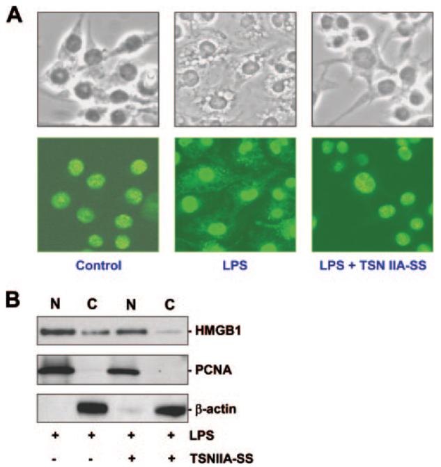
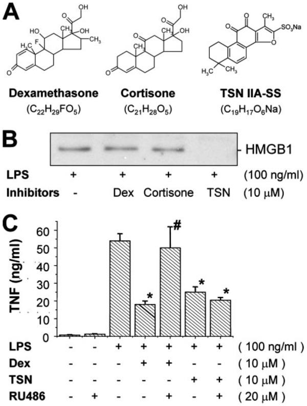
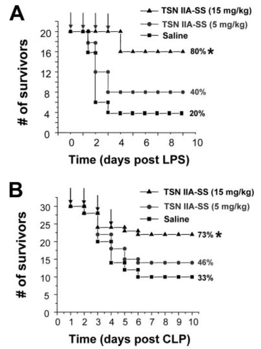
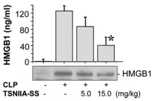
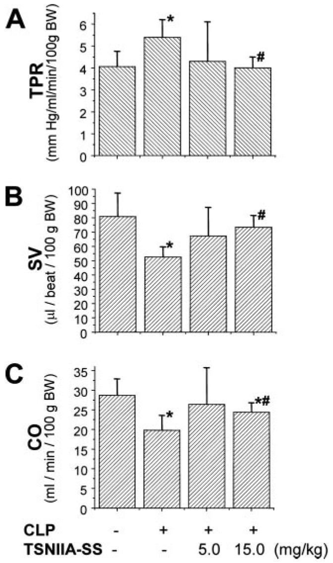
The pathogenesis of sepsis is mediated in part by bacterial endotoxin, which stimulates macrophages/monocytes to sequentially release early (e.g., TNF, IL-1, and IFN-gamma) and late (e.g., high mobility group box 1 (HMGB1) protein) proinflammatory cytokines. The recent discovery of HMGB1 as a late mediator of lethal sepsis has prompted investigation for development of new experimental therapeutics. We found that many steroidal drugs (such as dexamethasone and cortisone) and nonsteroidal anti-inflammatory drugs (such as aspirin, ibuprofen, and indomethacin) failed to influence endotoxin-induced HMGB1 release even at superpharmacological concentrations (up to 10-25 microM). However, several steroid-like pigments (tanshinone I, tanshinone IIA, and cryptotanshinone) of a popular Chinese herb, Danshen (Salvia miltiorrhiza), dose dependently attenuated endotoxin-induced HMGB1 release in macrophage/monocyte cultures. A water-soluble tanshinone IIA sodium sulfonate derivative (TSNIIA-SS), which has been widely used as a Chinese medicine for patients with cardiovascular disorders, selectively abrogated endotoxin-induced HMGB1 cytoplasmic translocation and release in a glucocorticoid receptor-independent manner. Administration of TSNIIA-SS significantly protected mice against lethal endotoxemia and rescued mice from lethal sepsis even when the first dose was given 24 h after the onset of sepsis. The therapeutic effects were partly attributable to attenuation of systemic accumulation of HMGB1 (but not TNF and NO) and improvement of cardiovascular physiologic parameters (e.g., decrease in total peripheral vascular resistance and increase in cardiac stroke volume) in septic animals. Taken together, these data re-enforce the pathogenic role of HMGB1 in lethal sepsis, and support a therapeutic potential for TSNIIA-SS in the treatment of human sepsis.


Figures

FIGURE 1
FIGURE 1
Danshen extract (A) and components (tanshinone I, tanshinone IIA, and cryptotanshinone) (B and C) attenuated endotoxin-induced HMGB1 release. Murine macrophage-like RAW 264.7 cells were stimulated with LPS in the absence or presence of herbal extract (A), and tanshinone I (TSN I), tanshinone IIA (TSN IIA), or cryptotanshinone (C-TSN) (B and C) for 16 h, and levels of HMGB1 in the culture medium were determined by Western blot analysis. A representative Western blot of three independent experiments with similar results is shown (bottom panel). *, p < 0.05 vs controls (+LPS alone).
FIGURE 2
FIGURE 2
TSNIIA-SS specifically abrogated endotoxin-induced HMGB1 release. Murine macrophage-like RAW 264.7 cells, primary murine peritoneal macrophages, or human PBMC were stimulated with LPS in the absence or presence of TSNIIA-SS at indicated concentrations. At 16 h after LPS stimulation, levels of HMGB1 (A and B), NO (A), or TNF (B and C) in the culture medium were determined by Western blot analysis (A and B), Griess reaction (A), ELISA (B), or cytokine array (C). Note that at concentrations that completely abrogated LPS-induced HMGB1 release, TSNIIA-SS did not completely block LPS-induced release of NO (A), TNF (B), or IL-1α, platelet factor 4 (PF-4), IL-12p70, and MCP-5 (C). A representative cytokine array (C) of two independent experiments with similar results is shown.
FIGURE 3
FIGURE 3
Delayed administration of TSNIIA-SS still significantly attenuated endotoxin-induced HMGB1 release. Murine macrophage-like RAW 264.7 cells were stimulated with LPS, and TSNIIA-SS (25 μM (A) or 50 μM (B)) was added at 0, 2, 6, and 12 h after LPS stimulation. Levels of HMGB1 levels in the culture medium were determined at 16 h after LPS stimulation and expressed (in arbitrary unit, AU) as the mean ± SD of two independent experiments. A representative Western blot is shown at bottom of both experiments. *, p < 0.05 vs controls (+LPS alone).
FIGURE 4
FIGURE 4
TSNIIA-SS blocked endotoxin-induced cytoplasmic HMGB1 translocation. Macrophage cultures were stimulated with LPS in the absence or presence of TSNIIA-SS, and assayed for HMGB1 cytoplasmic translocation by immunohistochemistry (A) or cell fractionation (Western blot technique) (B) at 16 h after LPS stimulation. Note in A that HMGB1 was predominantly localized in the nuclear region of unstimulated macrophages (control) (left column), in both cytoplasmic and nuclear regions of LPS-stimulated macrophages (middle column). TSNIIA-SS (100 μM) preserved HMGB1 in the nuclear regions (LPS + TNSIIA-SS) (right column). Following LPS stimulation, cytoplasmic (C) and nuclear (N) fractions were isolated and assayed for levels of HMGB1, a proliferating cell nuclear Ag protein, or cytoplasmic (β-actin) protein using Western blot analysis. Equal loading of samples was confirmed by Western blot analysis of fractions with cytoplasmic (β-actin) or proliferating cell nuclear Ag protein markers. Blots are representative of two independent experiments with similar results.
FIGURE 5
FIGURE 5
TSNIIA-SS attenuates LPS-induced HMGB1 release in a glucocorticoid receptor-independent mechanism. Murine macrophage-like RAW 264.7 cells were stimulated with LPS in the absence or presence of dexamethasone, cortisone, or TSNIIA-SS alone (A), or in combination with a glucocorticoid receptor antagonist, RU486. At 16 h after stimulation, levels of HMGB1 (B) or TNF (C) in the culture medium were determined by Western blot analysis and ELISA, respectively. The representative Western blot of two independent experiments with similar results is shown. *, p < 0.05 vs “+ LPS alone” group; #, p < 0.05 vs “+ LPS + Dex” group.
FIGURE 6
FIGURE 6
TSNIIA-SS dose dependently protects mice against lethal endotoxemia (A) and lethal sepsis (B). BALB/c mice were subjected to lethal endotoxemia (LPS, 15 mg/kg, i.p.), or sepsis (induced by CLP). At +0.5, +24, +48, and +72 h after the onset of endotoxemia or +24, +48, +72, and +96 h after the onset of sepsis, animals were i.p. administered with saline (0.2 ml/mouse) or TSNIIA-SS (0.2 ml/mouse at indicated doses), and animal survival was monitored for up to 2 wk. The Kaplan-Meier method was used to compare the differences in mortality rates between groups. *, p < 0.05 vs saline.
FIGURE 7
FIGURE 7
TSNIIA-SS attenuates sepsis-induced systemic HMGB1 accumulation. BALB/c mice were subjected to lethal sepsis by CLP, and i.p. administered with control saline (0.2 ml/mouse) or TSNIIA-SS (at indicated doses) at +24 and +48 h after CLP. At 52 h after the onset of sepsis, serum HMGB1 levels were determined and expressed as the mean ± SD (n = 10 mice). *, p < 0.05 (by ANOVA or Tukey test).
FIGURE 8
FIGURE 8
TSNIIA-SS prevents sepsis-induced cardiovascular dysfunction. Male Sprague-Dawley rats (290–310 g) were subjected to lethal sepsis by CLP, and TSNIIA-SS was administered via the femoral venous catheter using a Harvard Pump (Harvard Apparatus) at 5 h after CLP. At 20 h after CLP or sham operation, total peripheral resistance (TPR) (A), stroke volume (SV) (B), and cardiac output (CO) (C) were determined using radioactive microspheres as previously described (30). Data are expressed as the mean ± SD (n = 6 mice/group) and compared by one-way ANOVA and the Tukey test. *, p < 0.05 vs sham-operated animals (“−”); #, p < 0.05 vs CLP animals treated with vehicle (+CLP).

References

    1. Oberholzer A, Oberholzer C, Moldawer LL. Sepsis syndromes: understanding the role of innate and acquired immunity. Shock. 2001;16:83–96. - PubMed
    1. Dinarello CA, Thompson RC. Blocking IL-1: interleukin 1 receptor antagonist in vivo and in vitro. Immunol. Today. 1991;12:404–410. - PubMed
    1. Heinzel FP. The role of IFN-γ in the pathology of experimental endotoxemia. J. Immunol. 1990;145:2920–2924. - PubMed
    1. Dinapoli MR, Calderon CL, Lopez DM. The altered tumoricidal capacity of macrophages isolated from tumor-bearing mice is related to reduce expression of the inducible nitric oxide synthase gene. J. Exp. Med. 1996;183:1323–1329. - PMC - PubMed
    1. Calandra T, Echtenacher B, Roy DL, Pugin J, Metz CN, Hultner L, Heumann D, Mannel D, Bucala R, Glauser MP. Protection from septic shock by neutralization of macrophage migration inhibitory factor. Nat. Med. 2000;6:164–170. - PubMed

Publication types

MeSH terms