. 2007 Mar 15;178(6):3865-73.
doi: 10.4049/jimmunol.178.6.3865.

VCAM-1 activation of endothelial cell protein tyrosine phosphatase 1B

Affiliations

VCAM-1 activation of endothelial cell protein tyrosine phosphatase 1B

Tracy L Deem et al. J Immunol. .

Abstract

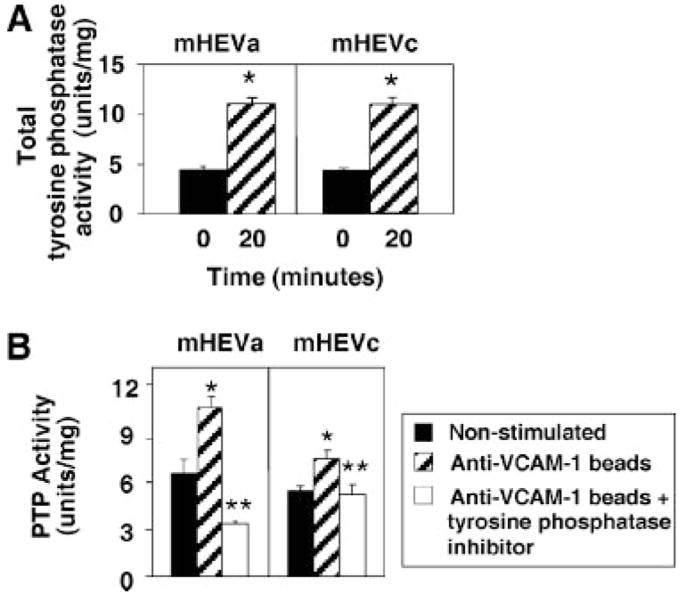
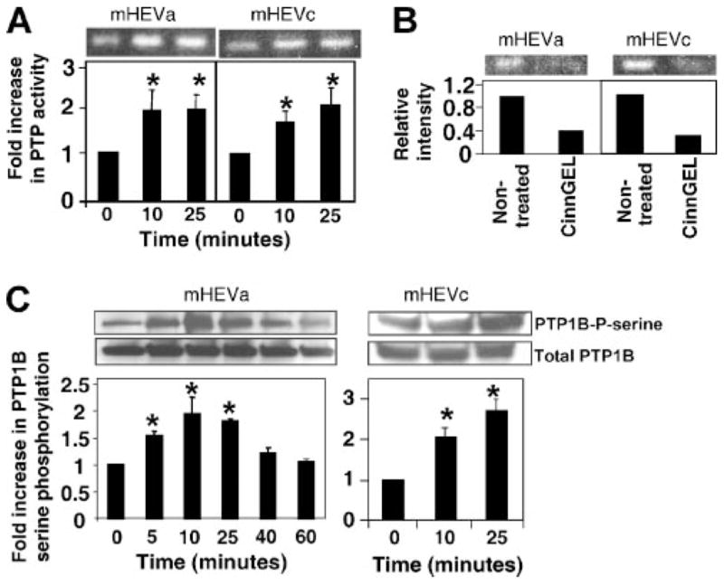
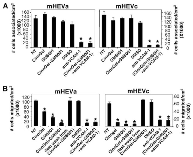
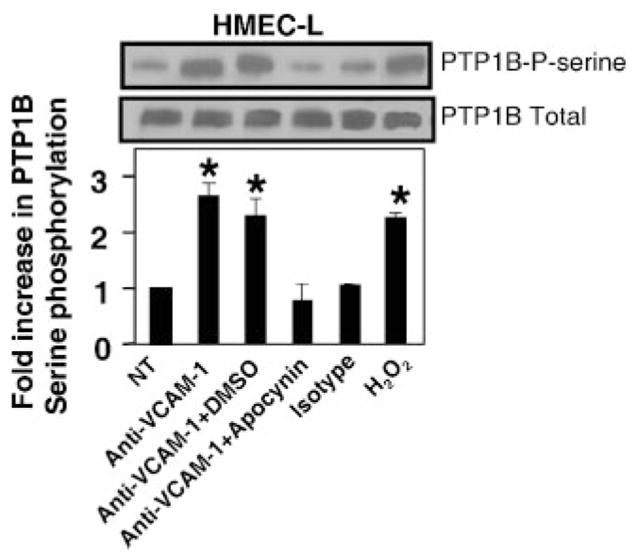
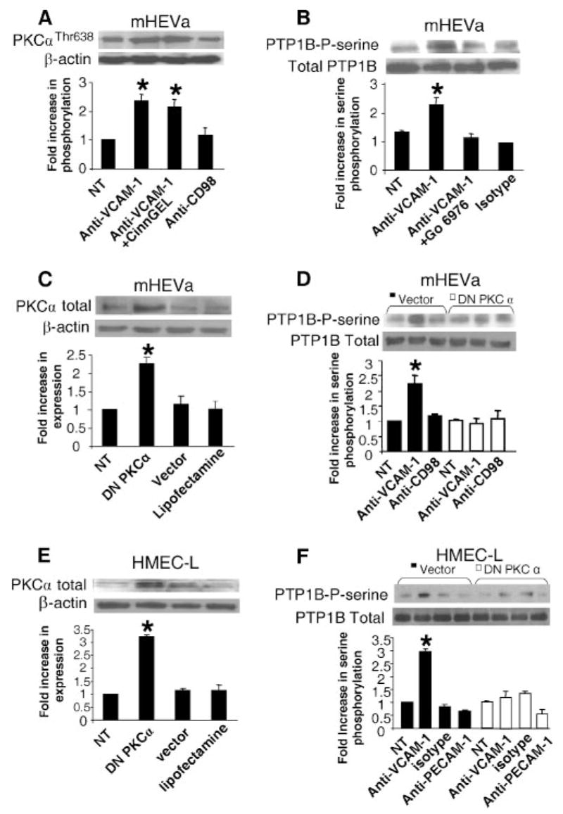
Lymphocytes migrate from the blood into tissue by binding to and migrating across endothelial cells. One of the endothelial cell adhesion molecules that mediate lymphocyte binding is VCAM-1. We have reported that binding to VCAM-1 activates endothelial cell NADPH oxidase for the generation of reactive oxygen species (ROS). The ROS oxidize and stimulate an increase in protein kinase C (PKC)alpha activity. Furthermore, these signals are required for VCAM-1-dependent lymphocyte migration. In this report, we identify a role for protein tyrosine phosphatase 1B (PTP1B) in the VCAM-1 signaling pathway. In primary cultures of endothelial cells and endothelial cell lines, Ab cross-linking of VCAM-1 stimulated an increase in serine phosphorylation of PTP1B, the active form of PTP1B. Ab cross-linking of VCAM-1 also increased activity of PTP1B. This activation of PTP1B was downstream of NADPH oxidase and PKCalpha in the VCAM-1 signaling pathway as determined with pharmacological inhibitors and antisense approaches. In addition, during VCAM-1 signaling, ROS did not oxidize endothelial cell PTP1B. Instead PTP1B was activated by serine phosphorylation. Importantly, inhibition of PTP1B activity blocked VCAM-1-dependent lymphocyte migration across endothelial cells. In summary, VCAM-1 activates endothelial cell NADPH oxidase to generate ROS, resulting in oxidative activation of PKCalpha and then serine phosphorylation of PTP1B. This PTP1B activity is necessary for VCAM-1-dependent transendothelial lymphocyte migration. These data show, for the first time, a function for PTP1B in VCAM-1-dependent lymphocyte migration.


Conflict of interest statement

Disclosures

The authors have no financial conflict of interest.

Figures

FIGURE 1
FIGURE 1
Total tyrosine phosphatase activity in endothelial cells is increased by VCAM-1 binding. mHEV cell lines were stimulated with anti-VCAM-1 Ab-coated beads for 20 min in the absence (A) or presence (B) of the phosphatase inhibitor mixture (1/100) (Sigma-Aldrich). Tyrosine phosphatase activity is the phosphatase inhibitor-blocked free phosphate measured by the colorimetric PTP assay. Background in nonstimulated cells is likely the result of free phosphate in the cells during cell metabolism as the assay measures free phosphate. Units per milligram were calculated as the OD units for free phosphate per milligram of cell protein. Data presented are the mean ± SD from two experiments. *, p < 0.05 compared with nonstimulated mHEV cells.
FIGURE 2
FIGURE 2
VCAM-1 increases activity of a 50 kDa PTP. A, mHEVa and mHEVc cells were incubated with anti-VCAM-1 Ab-coated beads for 0, 10, and 25 min. The cells were lysed, and equal cell protein was analyzed by the PTP in-gel activity assay followed by autoradiography. Shown is the 50 kDa band. Data are from five experiments. B, mHEVa and mHEVc cells were lysed and equal protein was separated by SDS-PAGE. The gel was divided and incubated in the presence or absence of 10 μM CinnGel in the activation buffer plus 4 mM DTT for the PTP in-gel assay. C, mHEV cells were stimulated with anti-VCAM-1-coated beads. Cells were washed with PBS and lysed. PTP1B was immunoprecipitated, separated by SDS-PAGE, and analyzed by Western blot using anti-phosphoserine Ab. The membrane was stripped and probed with anti-total PTP1B Ab to determine protein loading. Densitometry analysis was performed using Image J software from the National Institutes of Health. Data presented are the mean ± SD from three to five experiments. *, p < 0.05 compared with mHEV cells at 0 min.
FIGURE 3
FIGURE 3
Inhibition of PTP1B blocks lymphocyte migration but not adhesion under laminar flow conditions. Confluent monolayers of mHEVa and mHEVc cells on slides were treated for 60 min with 10 μM CinnGEL-2-ME, 10 μM GM6001 or the solvent control, 0.1% DMSO. The culture was washed five times to remove excess inhibitor. Spleen cells (3 × 10⁶) were added on top of the mHEV monolayer (3.5 cm2). The spleen cells were allowed to briefly settle to mediate cell contact with the endothelial cell monolayer as determined by microscopy and then exposed to 2 dyne/cm2 laminar flow for 3 min for cell association (A) or 15 min for migration analysis (B). Cells were washed and fixed in 3% paraformaldehyde for 1 h. Migrated spleen cells are phase dark by phase contrast microscopy (37), whereas nonmigrated spleen cells are phase light (37). The spleen cells that migrate are >88% lymphocytes (12). CinnGel-2-ME and GM6001 had no affect on cell viability, as determined by trypan blue exclusion (data not shown). Data in each panel are from two to three experiments. *, p < 0.05 compared with nontreated or DMSO controls.
FIGURE 4
FIGURE 4
VCAM-1 signaling through ROS activates PTPs by a mechanism independent of oxidation of the active site. A, mHEVa cells were pretreated with the NADPH oxidase inhibitor apocynin or the solvent control DMSO. The cells were stimulated with anti-VCAM-1-coated beads or the binding control (anti-CD98-coated beads). The cells were washed with PBS and lysed. PTP1B was immunoprecipitated, separated by SDS-PAGE, and analyzed by Western blot using anti-phosphoserine Ab. The membrane was stripped and probed with anti-PTP1B Ab. B, mHEVa cells were stimulated for 7 min with H2O2. C, Purified PTP1B was incubated for 5 min with H2O2. D, mHEVa cells were stimulated for 0, 10, and 25 min with anti-VCAM-1 Ab-coated beads. The cells were washed and scraped into PTP lysis buffer ± IAA. BD, PTP activity was examined by the PTP1B in-gel assay followed by autoradiography. AC, Data presented are the mean ± SD from three experiments. *, p < 0.05 compared with nonstimulated cells.
FIGURE 5
FIGURE 5
In HMEC-L cells, VCAM-1 signaling through NADPH oxidase increases serine phosphorylation of PTP1B. HMEC-L cells were treated with 1 ng/ml TNF-α for 24 h to induce VCAM-1 expression. The confluent HMEC-L cells were washed and then pretreated with 4 mM apocynin or the solvent control 0.1% DMSO for 20 min. There was no effect of apocynin on cell viability (data not shown). The HMEC-L cells were stimulated with 27 μg/ml mouse anti-human VCAM-1 Ab or an isotype-matched Ab plus 15 μg/ml goat anti-mouse IgG Ab for 10 min. HMEC-L cells were also stimulated with 1 μM H2O2 for 10 min. Cells were washed with PBS and lysed. PTP1B was immunoprecipitated, separated by SDS-PAGE, and analyzed by Western blot using anti-phosphoserine Ab. The membrane was stripped and probed with anti-PTP1B Ab to determine protein loading. Densitometry analysis was performed using Image J software from the National Institutes of Health. Data presented are the mean ± SD of two experiments. *, p < 0.05 compared with nontreated cells.
FIGURE 6
FIGURE 6
PTP1B functions downstream of PKCα during VCAM-1 signaling. Nontreated mHEVa cells were pretreated with 10 μM CinnGEL-2ME (A) or 2.3 nM Go-6976 for 30 min (B). The cells were washed five times and then were stimulated with anti-VCAM-1-coated beads or control Ab-coated beads for 10 min. The cells were washed with PBS and scraped into SDS-PAGE loading buffer. Cell lysates were separated by SDS-PAGE followed by Western blot. A, Phosphorylated PKCα was detected with a rabbit-anti-mouse phospho-Thr (638)-PKCα Ab (1/1000) and an HRP-conjugated donkey anti-rabbit Ab (1/4000) followed by detection with ECL. The membrane was stripped and equal protein loading was verified using a mouse anti-β-actin Ab (1/5000). B, PTP1B was immunoprecipitated and Western blots were performed with anti-phosphoserine and then with anti-total PTP1B Abs. CF, mHEVa cells or HMEC-L cells were LipofectAMINE transfected with DN PKCα. C and E, Western blots for total PKCα demonstrate increases in PKCα expression after transfection. D and F, The cells were stimulated with anti-VCAM-1-coated beads or control Ab-coated beads (isotype Ab, anti-CD98, or anti-PECAM-1) for 10 min and washed with PBS. PTP1B was immunoprecipitated. The immunoprecipitates were separated by SDS-PAGE followed by Western blot using a rabbit anti-phosphoserine and HRP-conjugated donkey anti-rabbit Ab followed by detection with ECL. The membrane was stripped and equal protein loading was verified using an anti-total PTP1B Ab. The inhibitors and DN PKCα did not affect cell viability (data not shown). Data presented are the mean ± SD from two experiments. *, p < 0.05 compared with non-treated mHEV cells.

References

    1. Chin JE, Hatfield CA, Winterrowd GE, Brashler JR, Vonderfecht SL, Fidler SF, Griffin RL, Kolbasa KP, Krzesicki RF, Sly LM, et al. Airway recruitment of leukocytes in mice is dependent on α4-integrins and vascular cell adhesion molecule-1. Am J Physiol. 1997;272:L219–L229. - PubMed
    1. Baron JL, Madri JA, Ruddle NH, Hashim G, Janeway CA., Jr Surface expression of α4 integrin by CD4 T cells is required for their entry into brain parenchyma. J Exp Med. 1993;177:57–68. - PMC - PubMed
    1. Tummala PE, Chen XL, Sundell CL, Laursen JB, Hammes CP, Alexander RW, Harrison DG, Medford RM. Angiotensin II induces vascular cell adhesion molecule-1 expression in rat vasculature: a potential link between the renin-angiotensin system and atherosclerosis. Circulation. 1999;100:1223–1229. - PubMed
    1. Wright PS, Cooper JR, Kropp KE, Busch SJ. Induction of vascular cell adhesion molecule-1 expression by IL-4 in human aortic smooth muscle cells is not associated with increased nuclear NF-κB levels. J Cell Physiol. 1999;180:381–389. - PubMed
    1. Balogh P, Aydar Y, Tew JG, Szakal AK. Appearance and phenotype of murine follicular dendritic cells expressing VCAM-1. Anat Rec. 2002;268:160–168. - PubMed

Publication types

MeSH terms

Substances