. 2007 Apr;150(7):839-50.
doi: 10.1038/sj.bjp.0707161. Epub 2007 Mar 5.

Erythropoietin involves the phosphatidylinositol 3-kinase pathway, 14-3-3 protein and FOXO3a nuclear trafficking to preserve endothelial cell integrity

Affiliations

Erythropoietin involves the phosphatidylinositol 3-kinase pathway, 14-3-3 protein and FOXO3a nuclear trafficking to preserve endothelial cell integrity

Z Z Chong et al. Br J Pharmacol. 2007 Apr.

Abstract

Background and purpose: Clinical indications for erythropoietin (EPO) in the vascular system reach far beyond the treatment of anemia, but the development of EPO as a non-toxic agent rests heavily upon the cellular pathways controlled by EPO that require elucidation.

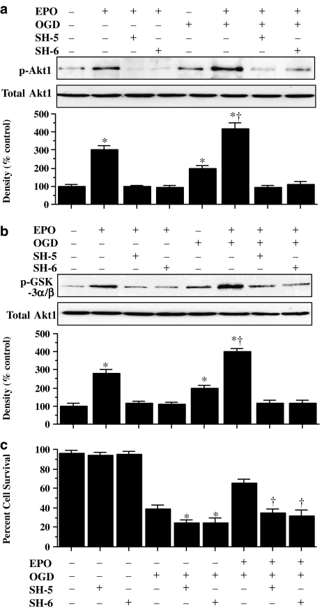
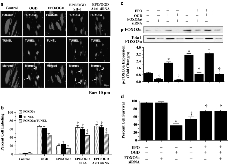
Experimental approach: We modulated gene activity and examined cellular trafficking of critical pathways during oxidative stress that may work in concert with EPO to protect primary cerebral endothelial cells (ECs) during oxidative stress, namely protein kinase B (Akt1), 14-3-3 protein, the Forkhead transcription factor FOXO3a.

Key results: Here, we show that preservation of ECs by EPO during oxygen-glucose deprivation (OGD) required the initial activation of the phosphatidylinositol 3-kinase (PI-3K) pathway through Akt1, since specific pharmacological blockade of Akt1 activity or gene silencing of Akt1 prevented EC protection by EPO. EPO subsequently involved a series of anti-apoptotic pathways to activate STAT3, STAT5, and ERK 1/2. Furthermore, EPO maintained the inhibitory phosphorylation and integrity of the 'pro-apoptotic' transcription factor FOXO3a, promoted the binding of FOXO3a to 14-3-3 protein and regulated the intracellular trafficking of FOXO3a. Additionally, gene silencing of FOXO3a during OGD significantly increased EC survival, but did not synergistically improve cytoprotection by EPO, illustrating that EPO relied upon the blockade of the FOXO3a pathway.

Conclusions and implications: Our work defines a novel cytoprotective pathway in ECs that involves PI-3 K, STAT3, STAT5, ERK 1/2, 14-3-3 protein and FOXO3a, which can be targeted for the development of EPO as a clinically effective and safe agent in the vascular system.


Figures

Figure 1
Figure 1
EPO has a window of concentration and temporal administration for therapeutic effects during OGD. (a) OGD leads to progressive EC injury with increased exposure time (*P< 0.01 vs untreated control). (b) Representative images of ECs pretreated with EPO (10 ng ml−1). (c) Quantitation of EC survival with EPO (0.01–1000 ng ml−1) 1 h before exposure to OGD. Cell survival was assessed 24 h later (*P< 0.01 vs OGD). (d) ECs were treated with EPO (10 ng ml−1) at 2, 4, 6 and 12 h post-OGD exposure and cell survival was assessed 24 h later (*P< 0.01 vs OGD). (e and f) ECs were pretreated with EPO (10 ng ml−1) 1 h before exposure to OGD and DNA fragmentation was determined 24 h later using the TUNEL assay; representative images in (e) and quantitation of results in (f) (*P< 0.01 vs OGD). In all cases, control = untreated ECs.
Figure 2
Figure 2
Cytoprotection by EPO requires the phosphorylation and activation of Akt1. (a and b) EC protein extracts (50 μg lane−1) were immunoblotted with antiphosphorylated-Akt1 (p-Akt1) (a) or with antiphosphorylated-GSK-3α/β (p-GSK-3α/β) (b) to assess Akt1 activity. Exposure to EPO (10 ng ml−1) or OGD significantly increased p-Akt1 and p-GSK-3α/β expression. Application of the specific Akt1 inhibitors SH-5 (20 μmol l−1) or SH-6 (20 μmol l−1) were sufficient to block the expression of p-Akt1 and p-GSK-3α/β in the presence of EPO during OGD (*P<0.01 vs control; P<0.01 vs OGD). (c) At concentrations that block phosphorylation and activation of Akt1 during OGD, SH-5 (20 μM) or SH-6 (20 μM) applied 1 h before OGD significantly reduced the cytoprotective capacity of EPO (10 ng ml−1) (*P<0.01 vs OGD alone; P<0.01 vs EPO with OGD). In all cases, control = untreated ECs.
Figure 3
Figure 3
Gene silencing of Akt1 abolishes cytoprotection by EPO. (ac) EC protein extracts (50 μg lane−1) were immunoblotted with antitotal Akt1 (a), antiphosphorylated-Akt1 (p-Akt1) (b) or antiphosphorylated-GSK-3α/β (p-GSK-3α/β) (c) to measure Akt1 activity. Exposure to EPO (10 ng ml−1) or OGD significantly increased p-Akt1 and p-GSK-3α/β expression. Transfection with Akt1 siRNA significantly reduced expression of total Akt1 (a), p-Akt1 during EPO (10 ng ml−1) (b) and p-GSK-3α/β during EPO (10 ng ml−1), OGD alone, or combined EPO (10 ng ml−1) with OGD (c) (*P<0.01 vs control untreated ECs; P<0.01 vs corresponding band without siRNA). For (a), negative control with multiple siRNAs prevented total Akt1 expression, but a positive control lacking specific Akt1 siRNA did not alter total Akt1 expression. (d) Gene silencing with Akt1 siRNA significantly prevented EPO (10 ng ml−1) from blocking EC membrane injury assessed by trypan blue staining and genomic DNA degradation assessed by TUNEL (*P<0.01 vs OGD alone; P<0.01 vs EPO/OGD without siRNA). Akt1 siRNA alone was not toxic.
Figure 4
Figure 4
In the presence of OGD, EPO increases the activity and phosphorylation of STAT3, STAT5 and ERK 1/2 in ECs. (ac) EC protein extracts (50 μg lane−1) were immunoblotted with antiphosphorylated-STAT3 (p-STAT3) (a), antiphosphorylated-STAT5 (p-STAT5) (b) or antiphosphorylated-ERK 1/2 (p-ERK 1/2) (c) to assess STAT3, STAT5 and ERK 1/2 activities 6 h following OGD. Exposure to EPO (10 ng ml−1) either alone or during OGD significantly increased p-STAT3, p-STAT5 and p-ERK 1/2 expression (*P<0.01 vs control). In all cases, control = untreated ECs.
Figure 5
Figure 5
EPO maintains the inhibitory phosphorylation of FOXO3a during OGD. (a and b) EC protein extracts (50 μg lane−1) were immunoblotted with antiphosphorylated-FOXO3a (p-FOXO3a, Ser253) (a) or antitotal FOXO3a (b) at 6 and 12 h following EPO (10 ng ml−1), OGD alone, or combined EPO (10 ng ml−1) with OGD. OGD led to the loss of p-FOXO3a (a) and the loss of total FOXO3a (b) at 12 h, but exposure to EPO (10 ng ml−1) maintained p-FOXO3a (a) and total FOXO3a (b) at 6 and 12 h following OGD (*P<0.01 vs OGD at 6 h). (c) At concentrations that block phosphorylation and activation of Akt1 during OGD, SH-5 (20 μM) or SH-6 (20 μM) applied 1 h before EPO (10 ng ml−1) or EPO (10 ng ml−1) combined with OGD significantly prevented the capacity of EPO to maintain the phosphorylation of p-FOXO3a at 6 h following OGD (*P<0.01 vs OGD alone; P<0.01 vs EPO with OGD). In all cases, control = untreated ECs.
Figure 6
Figure 6
Through Akt1, EPO uses 14-3-3 protein to bind to FOXO3a and sequester FOXO3a in the cytoplasm during OGD. (a) EC protein extracts were immunoprecipitated 6 h after EPO or OGD exposure with anti-14-3-3 antibody and immunoblotted with FOXO3a. Lysates were from wild type and Akt1 siRNA transfected cells with EPO (10 ng ml−1) or combined EPO (10 ng ml−1) with OGD. Transfection with Akt1 siRNA significantly reduced expression of the FOXO3a/14-3-3 complex during EPO alone or during EPO combined with OGD. (b) EPO (10 ng ml−1) or combined EPO (10 ng ml−1) with OGD was followed at 6 h with immunofluorescent staining for FOXO3a (Texas-red). Nuclei of ECs were counterstained with DAPI. In merged images, cells with EPO alone or combined EPO and OGD with white arrows show EC nuclei with minimal FOXO3a staining (blue/white) and green arrows show EC cytoplasm with significant FOXO3a staining (red) in contrast to cells with OGD alone or Akt1 siRNA transfection with combined EPO and OGD with minimal FOXO3a staining (gray), demonstrating the inability of EPO to sequester FOXO3a in the cytoplasm during Akt1 gene silencing. (c) EPO prevents FOXO3a translocation to the nucleus during OGD, but this ability of EPO is lost during gene silencing of Akt1 (*P<0.01 vs OGD alone or Akt1 siRNA). Control = untreated ECs.
Figure 7
Figure 7
EPO modulates intracellular trafficking of FOXO3a to block nuclear DNA degradation and EPO cytoprotection parallels EC survival levels during FOXO3a gene silencing. (a) Immunofluorescent double staining for FOXO3a and TUNEL was performed at 6 h after OGD. EPO (10 ng ml−1) during OGD prevents nuclear DNA degradation and FOXO3a nuclear translocation in the same ECs with no overlap of staining in merged images. In contrast, white arrows in merged images show both nuclear FOXO3a and TUNEL staining (yellow) in ECs with OGD alone, with combined EPO/OGD and inhibition of Akt1 activity (SH-6, 20 μM), or with combined EPO/OGD and Akt1 siRNA gene silencing, illustrating that EPO requires Akt1 to prevent FOXO3a nuclear translocation that leads to apoptotic DNA degradation. (b) EPO (10 ng ml−1) during OGD prevented FOXO3a and TUNEL nuclear staining in the same ECs, but this ability of EPO is lost during combined EPO/OGD with inhibition of Akt1 activity (SH-6, 20 μM) or with combined EPO/OGD and Akt1 siRNA (*P<0.01 vs OGD; P<0.01 vs EPO/OGD). (c) EC protein extracts (50 μg lane−1) were immunoblotted with antiphosphorylated-FOXO3a (p-FOXO3a, Ser253) at 6 h following with EPO (10 ng ml−1), OGD alone or combined EPO (10 ng ml−1) with OGD in lysates from wild type and FOXO3a siRNA transfected cells. Transfection with FOXO3a siRNA significantly reduced expression of p-FOXO3a and total FOXO3a during EPO alone, OGD alone and combined EPO with OGD (*P<0.01 vs untreated control; P<0.01 vs corresponding band without siRNA). (d) Gene silencing with FOXO3a siRNA significantly increased survival during OGD, but lead to similar survival level during combined EPO with OGD without a synergistic increase, suggesting that EPO required the prevention of FOXO3a activity for cytoprotection (*P<0.01 vs untreated control; P<0.01 vs OGD alone).

Comment in

  • EPO tecting the endothelium.
    Peterson TE, Katusic ZS. Peterson TE, et al. Br J Pharmacol. 2007 Apr;150(7):823-5. doi: 10.1038/sj.bjp.0707162. Epub 2007 Mar 12. Br J Pharmacol. 2007. PMID: 17351666 Free PMC article.

References

    1. Abbott NJ, Hughes CC, Revest PA, Greenwood J. Development and characterisation of a rat brain capillary endothelial culture: towards an in vitro blood–brain barrier. J Cell Sci. 1992;103:23–37. - PubMed
    1. Avasarala JR, Konduru SS. Recombinant erythropoietin down-regulates IL-6 and CXCR4 genes in TNF-alpha-treated primary cultures of human microvascular endothelial cells: implications for multiple sclerosis. J Mol Neurosci. 2005;25:183–189. - PubMed
    1. Bahlmann FH, Song R, Boehm SM, Mengel M, von Wasielewski R, Lindschau C, et al. Low-dose therapy with the long-acting erythropoietin analogue darbepoetin alpha persistently activates endothelial Akt and attenuates progressive organ failure. Circulation. 2004;110:1006–1012. - PubMed
    1. Bierer R, Peceny MC, Hartenberger CH, Ohls RK. Erythropoietin concentrations and naurodevelopmental outcome in preterm infants. Pediatrics. 2006;118:e635–e640. - PubMed
    1. Bullard AJ, Govewalla P, Yellon DM. Erythropoietin protects the myocardium against reperfusion injury in vitro and in vivo. Basic Res Cardiol. 2005;100:397–403. - PubMed

Publication types

MeSH terms