. 2007 Apr;5(4):e63.
doi: 10.1371/journal.pbio.0050063.

Cdk5 is involved in BDNF-stimulated dendritic growth in hippocampal neurons

Affiliations

Cdk5 is involved in BDNF-stimulated dendritic growth in hippocampal neurons

Zelda H Cheung et al. PLoS Biol. 2007 Apr.

Abstract

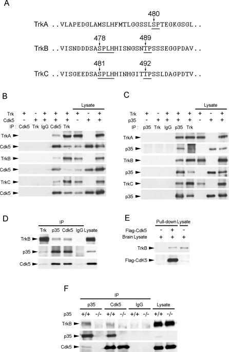
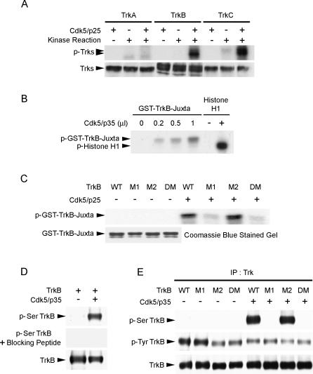
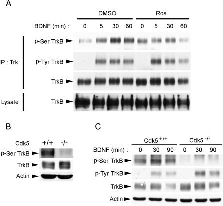
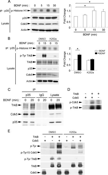
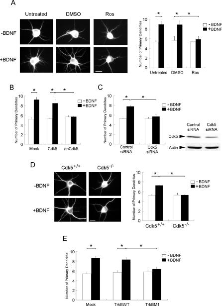
Neurotrophins are key regulators of neuronal survival and differentiation during development. Activation of their cognate receptors, Trk receptors, a family of receptor tyrosine kinases (RTKs), is pivotal for mediating the downstream functions of neurotrophins. Recent studies reveal that cyclin-dependent kinase 5 (Cdk5), a serine/threonine kinase, may modulate RTK signaling through phosphorylation of the receptor. Given the abundant expression of both Cdk5 and Trk receptors in the nervous system, and their mutual involvement in the regulation of neuronal architecture and synaptic functions, it is of interest to investigate if Cdk5 may also modulate Trk signaling. In the current study, we report the identification of TrkB as a Cdk5 substrate. Cdk5 phosphorylates TrkB at Ser478 at the intracellular juxtamembrane region of TrkB. Interestingly, attenuation of Cdk5 activity or overexpression of a TrkB mutant lacking the Cdk5 phosphorylation site essentially abolishes brain-derived neurotrophic factor (BDNF)-triggered dendritic growth in primary hippocampal neurons. In addition, we found that Cdk5 is involved in BDNF-induced activation of Rho GTPase Cdc42, which is essential for BDNF-triggered dendritic growth. Our observations therefore reveal an unanticipated role of Cdk5 in TrkB-mediated regulation of dendritic growth through modulation of BDNF-induced Cdc42 activation.


Conflict of interest statement

Competing interests. The authors have declared that no competing interests exist.

Figures

Figure 1
Figure 1. TrkB Interacted with Cdk5 and p35
(A) TrkA, TrkB, and TrkC all contain proline-directed serine/threonine residues in the juxtamembrane region of the receptors (indicated by arrows). Nonetheless, only TrkB and TrkC contain Cdk5 consensus sites S/TPXK/H/R. (B) Cell lysates from HEK293T cells overexpressing Cdk5 and TrkA, TrkB, or TrkC were immunoprecipitated (IP) with Cdk5 antibody and immunoblotted with pan-Trk antibody. TrkA, TrkB, and TrkC were all observed to associate with Cdk5. (C) Cell lysates from HEK293T cells overexpressing p35 and TrkA, TrkB, or TrkC were immunoprecipitated with p35 antibody and immunoblotted with pan-Trk antibody. TrkA, TrkB, and TrkC were all observed to associate with p35. (D) Brain lysate from P7 rat brain was immunoprecipitated with pan-Trk, p35, or Cdk5 antibody and immunoblotted with p35, Cdk5, and TrkB antibodies. Rabbit normal IgG was used as a control. TrkB was observed to associate with both p35 and Cdk5 in P7 rat brain. (E) The membrane fraction of adult brain lysates was incubated with or without Flag-tagged Cdk5. Flag-tagged Cdk5 pulled down TrkB from the membrane fraction of adult brain lysates. (F) Brain lysates from P7 p35+/+ or p35−/− mouse brains were immunoprecipitated with p35 and Cdk5 antibodies and immunoblotted with p35, Cdk5, and TrkB antibodies. Rabbit normal IgG served as a control. Association between Cdk5 and TrkB was abolished in p35−/− brain, indicating that p35 was required for the association between Cdk5 and TrkB.
Figure 2
Figure 2. Cdk5 Phosphorylated TrkB at Ser478
(A) Lysates from COS7 cells overexpressing TrkA, TrkB, and TrkC were immunoprecipitated with pan-Trk antibody and incubated with Cdk5/p25 in an in vitro kinase assay. TrkB and TrkC, but not TrkA, were phosphorylated by Cdk5/p25. (B) GST-TrkB-juxtamembrane fusion protein was incubated with increasing amount of Cdk5/p35 and subjected to an in vitro kinase assay. Histone H1 served as control to verify the activity of the Cdk5 kinase. The TrkB-juxtamembrane region was phosphorylated by Cdk5/p35 in a dose-dependent manner. (C) Purified WT GST-TrkB-juxtamembrane fusion protein and mutants (M1, M2, and DM) were incubated with Cdk5/p25 in an in vitro kinase assay. While WT and M2 were strongly phosphorylated by Cdk5/p25, phosphorylation of M1 and DM were markedly attenuated. Quality of the purified GST and GST-fusion proteins used in the GST pull-down assay was verified by Coomassie blue staining. (D) Characterization of p-Ser TrkB antibody raised against phosphorylated Ser478 of TrkB. TrkB was overexpressed with or without p35/Cdk5 in HEK293T cells. Preincubation of purified p-Ser478 TrkB antibody with blocking peptide completely abolished detection of Ser478 phosphorylation of TrkB. (E) Full-length TrkB WT, M1, M2, and DM were overexpressed with or without Cdk5/p35 in HEK293T cells. In the absence of Cdk5/p35, Ser478-phosphorylated TrkB (p-Ser TrkB) was not detected. Overexpression of Cdk5/p35 resulted in phosphorylation of TrkB WT at Ser478, but phosphorylation at Ser478 was essentially abolished when TrkB M1 and DM were overexpressed. IP, immunoprecipitation.
Figure 3
Figure 3. Ser478 Phosphorylation of TrkB Required Cdk5 Activity In Vivo
(A) BDNF stimulation resulted in an increase in p-Ser478 TrkB (p-Ser TrkB) levels in cortical neurons. Treatment with Cdk5 selective inhibitor Ros (25 μM) inhibited the BDNF-induced increase in p-Ser478 TrkB, although Ros treatment also resulted in a slight increase in basal p-Ser478 TrkB. (B) cdk5 +/+ and cdk5 −/− brain lysates were immunoblotted against TrkB, phospho-TrkB at Ser478, and β-actin as loading control. p-Ser478 TrkB was almost completely absent in cdk5 −/− brain, indicating the importance of Cdk5 in the phosphorylation of TrkB at Ser478 in vivo. (C) Cortical neurons isolated from cdk5 +/+ and cdk5 −/− brain were treated with BDNF for different periods. Interestingly, while BDNF enhanced TrkB Ser478 phosphorylation in cdk5 +/+ cortical neurons, TrkB Ser478 phosphorylation was not detected in cdk5 −/− neurons, nor did BDNF stimulation enhance Ser478 phosphorylation, indicating that BDNF-stimulated increase in TrkB Ser478 phosphorylation requires Cdk5 activity.
Figure 4
Figure 4. BDNF Enhanced Cdk5 Activity
(A) Cortical neurons were stimulated with BDNF for different time intervals. Lysates were immunoprecipitated (IP) with p35 antibody and subjected to in vitro kinase assay using histone H1 as substrate. BDNF stimulation for 15 min resulted in a marked increase in Cdk5 activity in cortical neurons. Quantification of the changes in phospho-Histone H1 level following BDNF stimulation was normalized to the value obtained from untreated cultures (time 0) and is shown in the histogram. *, p < 0.05. (B) Addition of Trk inhibitor K252a abolished BDNF-induced increase in Cdk5 activity. Cortical neurons were pretreated with vehicle control (DMSO) or K252a for 30 min before stimulation with BDNF for 15 min. Lysates were immunoprecipitated with p35 antibody and subjected to in vitro kinase assay using histone H1 as substrate. We found that K252a pretreatment markedly reduced the increase in Cdk5 activity triggered by BDNF stimulation, indicating that the induction of Cdk5 activity was dependent on TrkB activation. Quantification of the changes in phospho-Histone H1 level following BDNF stimulation in the presence or absence of K252a treatment was normalized to the value obtained from untreated cultures (time 0) and is shown in the histogram. *, p < 0.05. (C) Cortical neurons were treated with BDNF for 20 min. Lysates were immunoprecipitated with p35 antibody and immunoblotted with TrkB, p35, or Cdk5 antibody. While association between Cdk5 and p35 was not affected by BDNF stimulation, association between p35 and TrkB increased following 20 min of BDNF stimulation. (D) Recombinant TrkB was incubated with GST-Cdk5 in an in vitro kinase assay. TrkB was found to phosphorylate GST-Cdk5 (middle lane). (E) GST-Cdk5 and recombinant TrkB were pretreated with vehicle (DMSO) or K252a for 10 min, subjected to in vitro kinase assay, and immunoblotted with antibodies against phospho-tyrosine (p-Tyr) and the Tyr15 phosphorylated form of Cdk5 (pTyr15 Cdk5). Cdk5 was phosphorylated by TrkB at Tyr15. Addition of K252a abolished phosphorylation of Cdk5 by TrkB, further verifying that Cdk5 was phosphorylated by TrkB.
Figure 5
Figure 5. Attenuation of Cdk5 Activity Abolished BDNF-Induced Increase in Primary Dendrites in Hippocampal Neurons
(A) Hippocampal neurons were stimulated with BDNF for 3 d in the presence or absence of Ros (10 μM). Interestingly, while BDNF treatment markedly enhanced the number of primary dendrites, treatment with Ros abrogated the increase. (B) Hippocampal neurons were transfected with Cdk5 or DN Cdk5. Twenty-four hours after transfection, cells were exposed to BDNF for 3 d. Overexpression of DN Cdk5 abolished the BDNF-induced increase in primary dendrites. (C) Hippocampal neurons were transfected with Cdk5 siRNA or control siRNA. Twenty-four hours after transfection, cells were exposed to BDNF for 3 d. Transfection with Cdk5 siRNA attenuated Cdk5 expression in hippocampal neurons. More importantly, BDNF-induced increase in primary dendrites was abrogated in Cdk5 siRNA–transfected cells. (D) Hippocampal neurons isolated from cdk5 +/+ and cdk5 −/− brains were treated with BDNF for 3 d. BDNF treatment failed to enhance primary dendrites in Cdk5−/− neurons. (E) Hippocampal neurons were transfected with TrkB WT or TrkB M1. Twenty-four hours after transfection, cells were exposed to BDNF for 3 d. Overexpression of TrkB M1 markedly reduced the BDNF-induced increase in primary dendrites. Scale bar = 10 μm. *, p < 0.05.
Figure 6
Figure 6. Cdk5-Mediated Phosphorylation of TrkB Affected BDNF-Induced Dendritic Growth through Attenuation of Cdc42 Activity
(A) Hippocampal neurons were transfected with the WT or DN form of Rac1, RhoA, or Cdc42. Twenty-four hours after transfection, cells were exposed to BDNF for 3 d. Overexpression of DN Cdc42 markedly reduced BDNF-induced increase in primary dendrites compared to overexpression of WT Cdc42, indicating that Cdc42 may contribute to the BDNF-dependent induction of dendritic growth in hippocampal neurons. (B) Cortical neurons were pretreated with Cdk5 selective inhibitor Ros or vehicle (DMSO) for 30 min prior to treatment with BDNF for different time intervals. Ros pretreatment markedly reduced BDNF-induced increase in Cdc42 activity following 15 and 30 min of BDNF treatment, indicating that Cdk5 activity was involved in BDNF-triggered Cdc42 activation. Quantification of the changes in Cdc42 activity following BDNF stimulation with or without Ros pretreatment was normalized to the value obtained for the DMSO-treated group at time 0 and is shown in the histogram. *, p < 0.05. (C) TrkB WT or TrkB M1 mutant were co-transfected with WT or CA Cdc42 in hippocampal neurons. Twenty-four hours after transfection, cells were exposed to BDNF for 3 d. Overexpression of CA Cdc42 reversed the abrogation of BDNF-induced increase in primary dendrites following overexpression of TrkB M1. *, p < 0.05. (D) Hippocampal neurons isolated from cdk5 +/+ and cdk5 −/− brains were transfected with WT or CA Cdc42. Twenty-four hours after transfection, cells were exposed to BDNF for 3 d. Overexpression of CA Cdc42 rescued the lack of dendritic growth following BDNF treatment in Cdk5−/− hippocampal neurons. *, p < 0.05.

References

    1. Huang EJ, Reichardt LF. Trk receptors: Roles in neuronal signal transduction. Annu Rev Biochem. 2003;72:609–642. - PubMed
    1. Segal RA. Selectivity in neurotrophin signaling: Theme and variations. Annu Rev Neurosci. 2003;26:299–330. - PubMed
    1. Fu AK, Fu WY, Cheung J, Tsim KW, Ip FC, et al. Cdk5 is involved in neuregulin-induced AChR expression at the neuromuscular junction. Nat Neurosci. 2001;4:374–381. - PubMed
    1. Li BS, Ma W, Jaffe H, Zheng Y, Takahashi S, et al. Cyclin-dependent kinase-5 is involved in neuregulin-dependent activation of phosphatidylinositol 3-kinase and Akt activity mediating neuronal survival. J Biol Chem. 2003;278:35702–35709. - PubMed
    1. Cheung ZH, Fu AK, Ip NY. Synaptic roles of Cdk5: Implications in higher cognitive functions and neurodegenerative diseases. Neuron. 2006;50:13–18. - PubMed

Publication types

Substances